Accurate and efficient identification and segmentation of gases play an important role in industrial processes and public safety. Although Optical Gas Imaging (OGI) has proven effective in acquiring images and videos of gas leaks, accurately determining the leakage points and their diffusion areas in real-world conditions remains a significant challenge. To address this, we propose a novel gas segmentation annotation methodology. This approach applies differencing between gas leakage scenes and corresponding static background to delineate precise leakage regions, which are subsequently annotated manually. The resulting dataset, consisting of 926 static background-gas image pairs extracted from 38 comprehensive video sequences, provides a robust foundation for advancing gas segmentation research. Based on this dataset, we present a novel semantic segmentation model specifically designed for the unique characteristics of gas leakage scenarios. Our model integrates a gas contrast attention mechanism to capitalize on static background information, resulting in improved segmentation precision. Comparative evaluations demonstrate that the proposed model achieves state-of-the-art performance on our dataset, outperforming widely adopted semantic segmentation models. All code and the dataset will be made publicly available at https://github.com/ProAlize/CGNet.
This paper presents a high-precision miniature force sensor based on fiber Bragg grating (FBG) for measuring axial contact force (CF) between a medical instrument tip and tissue during minimally invasive surgery (MIS) palpation. The newly designed FBG force sensor, with a reduced diameter of 1.5 mm and a total length of 12 mm, is easier to integrate into miniature medical instruments compared to existing 3 mm diameter sensors. The sensor consists of a tubular elastomer structure and two optical fibers. A unique staggered inclined notch design on the elastomer surface enhances sensitivity to axial forces and mitigates lateral force-induced strains. The central axial optical fiber has FBG1 inscribed to detect strains from axial forces and temperature changes, while the adjacent fiber has FBG2 to measure only temperature-induced strains. Calibration and temperature compensation experiments confirmed the sensor's accuracy in measuring CF and decoupling temperature effects. The sensor has an axial force sensitivity range of 0 to 1.4 N with a sensitivity of 270.809 pm/N and a dynamic measurement error of less than 0.2 g. The post-temperature compensation error is less than 0.08 N. Simulation palpation experiments, using silicone embedded with hard blocks to mimic pathological tissues, validated the sensor's effectiveness in detecting tissue abnormalities and distinguishing the hardness of different blocks during MIS palpation. A comparative experiment was conducted with a traditional helical-structured sensor to assess lateral force resistance, confirming that the designed sensor exhibits excellent lateral force resistance.
This work introduces a novel fiber Bragg grating (FBG)-based tactile sensor specifically developed for real-time force monitoring at the tips of flexible ureteroscopes. With a diameter of only 1.5 mm, the sensor features a dual-FBG configuration that effectively separates temperature effects from force signals, integrated with an innovative elastomer structure based on staggered parallelogram elements. Finite element analyses comparing traditional spiral and parallel groove designs indicate that the new configuration not only enhances axial sensitivity through optimized deformation characteristics but also significantly improves resistance to transverse forces via superior stress distribution and structural stability. In the sensor, a suspended lateral FBG is employed for thermal compensation, while an axially constrained FBG is dedicated to force detection. Calibration using a segmented approach yielded dual-range sensitivities of approximately 283.85 pm/N for the 0–0.5 N range and 258.57 pm/N for the 0.5–1 N range, with a maximum error of 0.07 N. Ex vivo ureteroscopy simulations further demonstrated the sensor’s capability to detect tissue–instrument interactions and to discriminate contact events effectively. This miniaturized solution offers a promising approach to achieving precise force feedback in endoscopic procedures while conforming to the dimensional constraints of standard ureteroscopes.
In the treatment of atrial fibrillation (AF), ablation stands as the foremost therapeutic strategy. Crucial to the success of ablation is the creation of transmural injuries in the cardiac tissue. The integration of a force sensor at the distal end of the catheter holds the potential to accurately quantify the extent of ablation-induced damage, thereby significantly enhancing the success rate of the procedure. This article introduces a novel isotropic three-axis force sensor, leveraging fiber Bragg grating (FBG) technology. A unique segment-by-segment sensitivity differentiation technique is implemented to decouple three-axis forces. This innovation empowers the sensor with adaptable stiffness adjustments in all spatial directions and robust elastomeric integration. Moreover, the utilization of a single optical fiber substantially enhances the overall flexibility of the catheter. The sensor's structural integrity is rigorously validated numerically and experimentally. Calibration of the sensor is performed using both linear and nonlinear models. Results demonstrate that the designed sensors meet technical specifications, achieving a resolution of less than 1 g. In terms of accuracy, the nonlinear model exhibits superior performance. Root-mean-square errors for sensors operating within lateral [-1 N, 1 N] and axial [0 N, 2 N] ranges are all within 1.2% of the full scale.
Accurate measurement of contact forces between medical instrument tips and tissues is a critical challenge in minimally invasive surgery (MIS), where the lack of tactile feedback can compromise surgical precision and safety. To address this, this article presents a novel miniature high-resolution 3-D force sensor based on fiber Bragg grating (FBG) technology. Compared to traditional methods that use multiple optical fibers and complex structures to decouple forces, this sensor employs a cantilever beam structure for biaxial lateral force sensing, while a hollow cylindrical structure is utilized for axial force sensing. This unique combination allows for the independent adjustment of sensitivity in each segment. Finite-element analysis is used to optimize the structure to approximate isotropy. Experiments demonstrate that the sensor achieves an axial force Z resolution of 3.36 mN and a dynamic measurement error of 2.97% within a range of 0-2 N. Meanwhile, lateral force resolutions for X- and Y-directions within a range of -0.5 to 0.5 N are 4.37 and 3.95 mN, respectively, with dynamic measurement errors of 3.79% and 5.12%. Additionally, porcine kidneys are adopted for ex vivo experiments to simulate tumors, validating the feasibility of the designed FBG force sensor in MIS palpation. The simulated vessel identification experiment further validated the ability to detect vessels at different depths within organs, highlighting the potential of the designed sensor for applications in MIS.
Underwater optical imaging plays a crucial role in maritime safety, enabling reliable navigation, efficient search and rescue operations, precise target recognition, and robust military reconnaissance. However, conventional underwater imaging methods often suffer from severe backscattering noise, limited detection range, and reduced image clarity—challenges that are exacerbated in turbid waters. To address these issues, Underwater Laser Range-Gated Imaging has emerged as a promising solution. By selectively capturing photons within a controlled temporal gate, this technique effectively suppresses backscattering noise-enhancing image clarity, contrast, and detection range. Nevertheless, residual noise within the imaging slice can still degrade image quality, particularly in challenging underwater conditions. In this study, we propose an enhanced U-Net neural network designed to mitigate noise interference in underwater laser range-gated images, improving target recognition performance. Built upon the U-Net architecture with added residual connections, our network combines a VGG16-based perceptual loss with Mean Squared Error (MSE) as the loss function, effectively capturing high-level semantic features while preserving critical target details during reconstruction. Trained on a semi-synthetic grayscale dataset containing synthetically degraded images paired with their reference counterparts, the proposed approach demonstrates improved performance compared to several existing underwater image restoration methods in our experimental evaluations. Through comprehensive qualitative and quantitative evaluations, underwater target detection experiments, and real-world oceanic validations, our method demonstrates significant potential for advancing maritime safety and related applications.
Tactile image sensors are efficient and useful in the robotics and healthcare fields due to their advantage of being impervious to electromagnetic fields. The functionality and applications of the sensors have been extensively researched. Furthermore, the pattern with the arrangement of sizes, colors, shapes and densities is like the “heart” of tactile image sensors, which immediately determines the results of detecting position and force. However, the effect of patterns on the resolution of tactile sensors has not been fully investigated yet. Hence, we experimentally studied the effect of different patterns on the resolution of tactile image sensors due to difficulties in theoretical analysis. Optical fibers and cameras were used to integrate flexible sensors with different features, which are divided into 61 dots. The sensors are put through their trials in various positions and forces, and the data is processed using deep learning methods. The sensors with four different patterns were all successful in distinguishing pressure, shear, and torque. More crucially, the root mean square error (RMSE) values were used to compare the impact of the various patterns on the sensors. All sensors with four types of patterns exhibited outstanding resolution for positions, the normal force, shear forces and torsion, which should be due to the optimization of the neural network and sensor structure. The seven position and force parameters of these four patterned sensors have the same order of magnitude of RMSEs, which suggests that the pattern complexity has negligible influence on the tactile image sensor resolution for these factors in our sensor structure. Moreover, we have demonstrated the outstanding resolution of the sensor with a pure color pattern. This work not only guides the pattern designs of forthcoming tactile image sensors but also enables the mass manufacture of flexible sensors for intelligent robots.
Laser range-gated underwater imaging technology, by removing most of the backscattering noise, can effectively increase image contrast and extend the detection range. The optical signal captured by a range-gated imaging system primarily comprises reflected light from the object and backscattered light from the surrounding water. Consequently, surfaces with low reflectivity or highly turbid water environments substantially constrain the applicability of the range-gated imaging system. To enhance the detection capability of underwater laser range-gated imaging, this paper proposes the incorporation of underwater polarized light imaging technology as an enhancement method. Based on polarization differences, backscattered light and reflected light from an object can be distinguished. Experimental results indicate that, compared to images obtained using a conventional range-gated laser imaging system, those captured with a polarization-enhanced system exhibit an increase of up to 47% for the corresponding Enhancement Measure Evaluation (EME) index. The proposed approach, which integrates polarization imaging with range-gated laser imaging, has the potential to broaden the applicability of underwater laser imaging scenarios, such as deep-sea exploration and military applications.
Amorphous TiTaNbZr multi-element alloy (Multi-principal elements alloys, MPCA) films were prepared by magnetron sputtering. The impact of 6 MeV Au ion irradiation with different temperatures (room temperature, 623 K, and 773 K) on the microstructure and mechanical properties of amorphous films was investigated. The findings indicate that when Au ion irradiation is administered at room temperature, the transformation of initially amorphous films into crystalline structures occurs. Furthermore, an increase in irradiation dose leads to enhanced crystallinity and growth in grain size. This is mainly due to cascade collisions between ion irradiation and alloy atoms, which promote atomic rearrangement, crystal nucleation, and growth. With the increase of irradiation temperature, there is no discernible defect structure under the irradiation condition at 623 K. Meanwhile, due to T < 0.3T(m) (T-m approximate to 2443 K), the crystallization behavior is mainly related to the Au ion energy deposition. The inelastic collision predominates at the initial stage. The crystal 20 nm away below the surface has a single-phase body-centered cube structure, and the elastic collision increases with the depth to form a biphase BCC structure. Similar grain size also increases with the increase of irradiation depth. Additionally, the hardness of the film remains unchanged at high temperatures and high radiation doses; its expansion rate is only 2 % due to the growth of grain boundaries and the self-healing mechanism. These results show that TiTaNbZr MPCA still has strong self-healing ability under high temperatures coupled with heavy ion irradiation, indicating excellent radiation resistance.
The widespread use of various chemical gases in industrial processes necessitates effective measures to prevent their leakage during transportation and storage, given their high toxicity. Thermal infrared-based computer vision detection techniques provide a straightforward approach to identify gas leakage areas. However, the development of high-quality algorithms has been challenging due to the low texture in thermal images and the lack of open-source datasets. In this paper, we present the RGB-Thermal Cross Attention Network (RT-CAN), which employs an RGB-assisted two-stream network architecture to integrate texture information from RGB images and gas area information from thermal images. Additionally, to facilitate the research of invisible gas detection, we introduce Gas-DB, an extensive open-source gas detection database including about 1.3K well-annotated RGB-thermal images with eight variant collection scenes. Experimental results demonstrate that our method successfully leverages the advantages of both modalities, achieving state-of-the-art (SOTA) performance among RGB-thermal methods, surpassing single-stream SOTA models in terms of accuracy, Intersection of Union (IoU), and F2 metrics by 4.86%, 5.65%, and 4.88%, respectively. The code and data can be found at https://github.com/logic112358/RT-CAN.
This paper develops a high-precision miniature three-dimensional Fiber Bragg Grating (FBG) force sensor designed for detecting and locating hard masses within tissues during Minimally Invasive Surgery (MIS) and robotic-assisted MIS palpation processes. The prototype consists of three FBG-etched optical fibers and an integrated elastomer. The fibers are suspended and parallel to each other within the elastomer, allowing each FBG to be directly compressed or stretched under stress. A novel model, combining segment-by-segment sensitivity and differential decoupling, is adopted for decoupling three-dimensional forces. This approach significantly reduces the crosstalk error caused by spatial forces, avoids the complexity of assembling multiple fibers in previous designs, and reduces the sensor's size, facilitating its integration into MIS procedures. Calibration experiments are conducted to determine the sensor's force sensitivity coefficients, achieving resolutions of 4.35 mN, 3.99 mN, and 3.30 mN in the X, Y, and Z directions, respectively. Through discrete and drag palpation tests on silicone with different types of hard inclusions, the results demonstrate that the proposed prototype has the ability to effectively identify the size, hardness, and depth of hard objects from the contact force information. Additionally, ex vivo porcine kidney palpation experiments are conducted to further verify the feasibility of locating hard masses during MIS palpation.
Urban water supply and drainage systems are a crucial component of urban infrastructure, directly affecting residents' livelihoods and industrial production. The normal operation of the water supply and the drainage pipeline is of great significance for conserving water resources and preventing water pollution. However, due to characteristics such as deep burial, diverse materials, and extensive lengths, the detection of defects becomes exceptionally complex. Traditional detection methods used in practical applications, such as ground excavation and destructive testing, typically require the shutdown of water pipelines. This process is time-consuming and labor-intensive, often resulting in significant economic losses. This paper proposes an effective technique for detecting defects in the water supply and the drainage pipeline. The method involves capturing images of the inner walls of water supply conduits and subsequently utilizing an artificial intelligence large-scale model approach (grounded language-image pre-training, GLIP) and a You Only Look Once version 5 (YOLOv5) model to detect defects within them. The experimental results show that GLIP demonstrates impressive detection performance in zero-shot scenarios, while YOLOv5 performs well on existing datasets. By combining these two models, we were able to achieve a balance between fast, flexible detection and high precision, making our approach both practical and efficient for real-world applications.
Human skin can accurately sense subtle changes of both normal and shear forces. However, tactile sensors applied to robots are challenging in decoupling 3D forces due to the inability to develop adaptive models for complex soft materials. Therefore, a new soft tactile sensor has been designed in this paper to detect shear and normal forces, including a soft probe and image acquisition device. First, to capture the deformation of the sensor, colored silicone squares were embedded in the soft probe. Capcamera movement of the colored squares under external forces. The image dataset collected at different 3D forces is then input into a deep learning model. Finally, a custom miniature image device is acquired and embedded in the soft probe to miniaturize the sensor. Computing results obtained from experimental datasets show that the proposed method can accurately decouple the 3D forces. Robots can grap vulnerable objects with sensors prepared at the robot’s tip. The tactile sensors studied in this paper are expected to be applied in robotics fields such as adaptive grasping, dexterous manipulation and human-computer interaction.
Hand electromyogram (EMG) signals, instrumental in tasks like movement recognition, rehabilitation monitoring, disease diagnosis, and human-computer collaboration, are typically obtained via high-density EMG electrodes on the dorsal hand. Conventional polyimide (PI)-based flexible high-density electrodes, despite their popularity, suffer from high modulus and poor stretchability. These factors lead to conformability issues, particularly on complex human body surfaces such as the dorsal hand skin, causing motion artifacts, signal degradation, and in severe cases, signal loss due to electrode detachment.This paper presents a thermoplastic polyurethane (TPU)-based high-density stretchable sEMG electrode for reliable EMG signal capture on complex surfaces. Performance comparisons between the proposed stretchable electrode (SE) and the traditional Pl-based flexible sEMG electrode (FE), focusing on time-domain waveforms and signal-to-noise ratio (SNR) under identical fixation conditions, indicate that SE maintains stable EMG signal acquisition across various hand postures. In contrast, the flexible electrode demonstrates signal weakness or loss under large deformations. Further, SNR analysis highlights SE's superior performance during high-intensity movements causing substantial skin deformation. Thus, the proposed SE emerges as a more convenient solution for stable and reliable hand EMG signal monitoring.
Objective: To accurately achieve distal contact force, a novel temperature-compensated sensor is developed and integrated into an atrial fibrillation (AF) ablation catheter. Methods: A dual elastomer-based dual FBGs structure is used to differentiate the strain on the two FBGs to achieve temperature compensation, and the design is optimized and validated by finite element simulation. Results: The designed sensor has a sensitivity of 90.5 pm/N, resolution of 0.01 N, and root–mean–square error (RMSE) of 0.02 N and 0.04 N for dynamic force loading and temperature compensation, respectively, and can stably measure distal contact forces with temperature disturbances. Conclusion: Due to the advantages, i.e., simple structure, easy assembly, low cost, and good robustness, the proposed sensor is suitable for industrial mass production.
Infrared imaging technology is a useful tool for detecting gas leaks due to its significant advantages in detection spacial range, efficiency, and visualization. However, the original infrared images of gas leak traces often suffer from low contrast due to a small thermal radiation intensity difference between the gas plume and the scene. So it is difficult to directly separate them in the grayscale histogram distribution. Traditional enhancement methods lack targeted optimizations for traces of leaking gas. Therefore, enhancing the trace of gas leaks is crucial to the performance of gas leak detection systems based on infrared imaging. In this paper, we propose an image enhancement method based on guided filtering, the visibility restoration algorithm. The proposed method comprises three stages: image decomposition, base and detail layer enhancement, and image fusion output. We selected three infrared images of gas leakage taken in different scenes for experiments. There are obvious differences in the original images taken from the three scenes. We compare the image enhancement result of Histogram Equalization(HE), Contrast Limited Adaptive Histogram Equalization (CLAHE) and our proposed method. The image enhanced by our proposed has the highest values of quantitative indicators: average gradient(AG) and Enhancement Measure Evaluation(EME); which are three times higher than the original image. By visual observation, the gas leak plume in the images enhanced by our proposed method are obvious significantly.
Infrared imaging systems have been widely applied in gas leak detection. However, The existing gas detection methods have many limitations and are difficult to apply in real-world scenarios. At the same time, there are very few methods that combine gas detection and semantic segmentation with deep learning. In this study, a novel approach for gas detection using image semantic segmentation in deep learning is proposed. This method presents a new multi-scale semantic segmentation model named PUNet, based on PSPNet and U-Net, for automatic segmentation of infrared gas leakage images. Meanwhile, to solve the problems of single scene and fixed leakage location in the gas leakage image dataset, we added more self-collected infrared gas leakage images to the existing dataset. The experimental findings demonstrate that PUNet has higher accuracy than traditional foreground segmentation algorithm and outperforms the conventional U-Net model in segmenting gas leakage images, and exhibits enhanced efficacy in handling multi-scale gas leakage scenarios.
由于水体对电磁波的吸收与散射,水下光学成像存在"看不远"和"看不清"的问题,而水下激光距离选通成像技术可以提高水下光学成像距离和图像对比度.该文介绍了以水下激光距离选通成像技术为基础的水下远距离目标智能识别系统研究.实验结果显示,成像距离超过 7 倍衰减长度.该研究结合深度学习算法,在功率受限的硬件条件下,实现了目标的准实时检测,检测速度达 0.8 f/s.水下激光距离选通成像技术与深度学习算法的结合,有望实现水下光学成像"看得远"和"看得清"的同时,实现"看得快"和"看得准".
 Yuming Dong (董玉明)合作论文数Shenzhen Institute of Advanced Technology, Chinese Academy of Sciences;University of Chinese Academy of Sciences11
 Guohua Jiao (焦国华)合作论文数Faculty of Computer Science and Artificial Intelligence, Shenzhen University of Advanced Technology;Shenzhen Institutes of Advanced Technology, Chinese Academy of Sciences;University of Chinese Academy of Sciences11
 Jianwei wu合作论文数7
 Yang Tianyu合作论文数7
 Chen Si-Hai合作论文数6CHEN Shuaibao (陈帅宝)合作论文数6Huanhuan Liu合作论文数6Liangpei Chen (陈良培)合作论文数4Qi Zhang合作论文数4
 Wei Chen (陈伟)合作论文数Institute of Semiconductors, Chinese Academy of Sciences4
 Zhiyuan Liu (刘志远)合作论文数Shenzhen Institute of Advanced Technology, Chinese Academy of Sciences;University of Chinese Academy of Sciences4Xiaoxi Nie (聂晓曦)合作论文数4Yuxiang Lin合作论文数3
 Zhengshuai Cao合作论文数3Qi Zhao合作论文数3Yuyang Lou合作论文数3Hanlong Zhang合作论文数3
 Hao Liu (刘昊)合作论文数School of Mechanical and Electrical Engineering, China University of Mining and Technology3
 Qiong Tian合作论文数Xi'an Jiaotong Univ, Xi'an Jiaotong Univ.3
 Xiaogang Xiong (熊小刚)合作论文数School of Intelligence Science and Engineering, Harbin Institute of Technology, Shenzhen3Jue Wang合作论文数2TAN Yuguang (谈宇光)合作论文数2Qiran Li (李其然)合作论文数Center for Optoelectronic Technology & Engineering, Shenzhen Insititues of Advanced Technology, Chinese Academy of Sciences;University of Chinese Academy of Sciences2
 Wei He (何伟)合作论文数School of Microelectronics and Communication Engineering, Chongqing University2
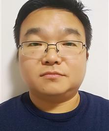 Zhenyu Lu (陆振宇)合作论文数School of Artificial Intelligence, Nanjing University of Information Science & Technology2Peng Liu (刘鹏)合作论文数State Key Laboratory of High Field Laser Physics and CAS Center for Excellence in Ultra-intense Laser Science, Shanghai Institute of Optics and Fine Mechanics (SIOM), Chinese Academy of Sciences (CAS), Shanghai 201800, China
2Center of Materials Science and Optoelectronics Engineering, University of Chinese Academy of Sciences, Beijing 100049, China2
Zhenyu Lu (陆振宇)合作论文数School of Artificial Intelligence, Nanjing University of Information Science & Technology2Peng Liu (刘鹏)合作论文数State Key Laboratory of High Field Laser Physics and CAS Center for Excellence in Ultra-intense Laser Science, Shanghai Institute of Optics and Fine Mechanics (SIOM), Chinese Academy of Sciences (CAS), Shanghai 201800, China
2Center of Materials Science and Optoelectronics Engineering, University of Chinese Academy of Sciences, Beijing 100049, China2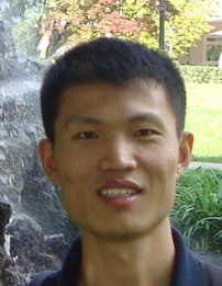 Tianyu Yang (楊田雨)合作论文数Alibaba DAMO Academy2
 Wenming Li (李文明)合作论文数School of Energy and Environment, Southeast University1
 Ruofan Yang合作论文数1Hang Zhao合作论文数1Tong Wei合作论文数1He Wei (何为)合作论文数1Jin Shanghai合作论文数1Guo Pu合作论文数1Lu Wu合作论文数1Xin Wu合作论文数1Jing Zhou合作论文数1Qingsong Li (李青松)合作论文数1Yan Liang合作论文数Sch Resources Environm & Mat, Guangxi Univ1
 Kun Zhang (张坤)合作论文数Institute of Mechanics, Chinese Academy of Sciences;University of Chinese Academy of Sciences1
 Bin Liu (刘彬)合作论文数Power Metallurgy Research Institute, Central South University;State Key Laboratory of Powder Metallurgy, Central South University1
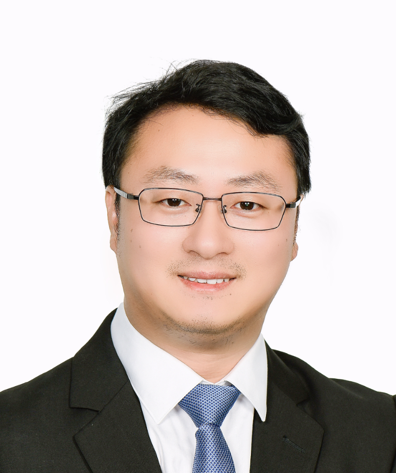 Jia Li (李甲)合作论文数College Of Mechanical And Vehicle Engineering, Hunan University1




