This chapter contains sections titled: Normal blood flow Arterial stenoses Stenosis as part of a larger arterial circuit Bypass grafts Conclusions References
This is the story of how a young surgeon, Donald Eugene Strandness Jr (Gene) was instrumental in the development of the Doppler ultrasonic flow meter, which evolved into the duplex scanner — perhaps the most versatile instrument in the modern vascular lab. He was born in Bowman, North Dakota, in 1928, and attended high school in Olympia, washington, where he was a football player and a star gymnast. He graduated from Pacific Lutheran University in 1946, studied medicine at the University of washington (Uw), and in 1950 entered the general surgical residency programme. At the time of the Korean war, Gene was drafted out of his residency; met his 2-year service obligation to the United States Air Force; and in 1959 returned to Seattle, where he hoped to join in the extensive research underway on the gastrointestinal system under the direction of Professor Henry Harkins. Instead, Dr Harkins urged him to change directions and join a small group at the Seattle VA Hospital who were investigating arterial disease. This group included John Bell, Hub Radke, and J.E. Jesseph. Strandness, swallowing his initial disappointment at having to give up gastrointestinal research, quickly embraced the vascular challenge. The 1950s were a particularly exciting time in the history of vascular surgery. Improved sutures, grafts, and anaesthesia made it possible for the first time to perform major arterial surgery, such as resection of abdominal aortic aneurysms, endarterectomy of the carotid bifurcation, and bypass of iliac, femoral, and popliteal arteries. In preparation for major arterial surgery, the need for imaging was keenly felt. Physiological studies to select patients for surgery took a backseat to arteriograms and physical examination — in part because pulse palpation and patient testimony were the only methods readily available for measuring preoperative functional impairment or postoperative success. Invasive methods for studying blood flow were limited to electromagnetic flowmetry, which was performed in the operating room with the patient anaesthetized. No effort was made to duplicate normal physiological conditions. Prior to the 1960s, a few surgeons and internists maintained rudimentary vascular labs where systolic blood pressure and blood flow were measured plethysmographically.
Introduction: Percent diameter reduction provides an imperfect assessment of the risk for stroke from carotid atheroembolism. Stroke associated with atherosclerotic carotid stenosis commonly results from plaque disruption brought about by hemodynamic shear stress and Bernoulli forces. The aim of the present study was to predict the effect of incomplete intracranial collateralization through the circle of Willis (COW) on disruptive hemodynamic forces acting on carotid plaques.Methods: A simple circuit model of the major pathways and collaterals that form and supply the COW was developed. We modeled the intra-and extracranial arterial circuits from standard anatomic references, and the pressure-flow relationships within these conduits from standard fluid mechanics. The pressure drop caused by (laminar and turbulent) flow along the internal carotid artery path was then computed. Carotid circulation to the brain was classified as being with or without collateral connections through the COW, and the extracranial carotid circuit as being with or without severe stenosis. The pressure drop was computed for each scenario. Finally, a linear circuit model was used to compute brain blood flow in the presence/absence of a disconnected COW.Results:, Pressure drop across a carotid artery stenosis increased as the flow rate within the carotid conduit increased. Poststenotic turbulence from a sudden expansion distal to the stenosis resulted in an additional pressure drop. Despite the stenosis, mean brain blood flow was sustained at 4.15 mL/s bilaterally. In the presence of an intact (collateralized) COW, this was achieved by enhanced flow in the contralateral (normal) carotid artery. However, in a disconnected COW, this was achieved by sustained systolic and enhanced diastolic flow through the stenosed artery. For a similar degree of stenosis, flow and velocity across the plaque was much higher when the COW was disconnected compared with an intact COW. Furthermore, the pressure drop across a similar stenosis was significantly higher with a disconnected COW compared with an intact COW.Conclusions: Incomplete intracranial collateralization through the COW results in increased flow rates and velocities, and therefore large pressure drops across a carotid artery stenosis. This exerts large disruptive shear stress on the plaque compared with patients with an intact COW. Percent diameter reduction provides an inaccurate assessment of risk for atheroembolic stroke. An assessment of carotid flow rates, flow velocities, and the intracranial collateral circulation may add independent information to refine the estimation of stroke risk in patients with asymptomatic carotid atherosclerosis. (J Vase Surg 2011;54:1461-71.)
The venous system is, in many respects, more complex than the arterial system and a thorough understanding of venous anatomy, pathophysiology, and available diagnostic tests is required in the management of acute and chronic venous disorders. The venous system develops through several stages, which may be associated with a number of development anomalies. A thorough knowledge of lower extremity venous anatomy, anatomic variants, and the recently updated nomenclature is required of all venous practitioners. Effective venous return from the lower extremities requires the interaction of the heart, a pressure gradient, the peripheral muscle pumps of the leg, and competent venous valves. In the absence of pathology, this system functions to reduce venous pressure from approximately 100 mm Hg to a mean of 22 mm Hg within a few steps. The severe manifestations of chronic venous insufficiency result from ambulatory venous hypertension, or a failure to reduce venous pressure with exercise. Although the precise mechanism remains unclear, venous hypertension is thought to induce the associated skin changes through a number of inflammatory mechanisms. Several diagnostic tests are available for the evaluation of acute and chronic venous disease. Although venous duplex ultrasonography has become the standard for detection of acute deep venous thrombosis, adjuvant modalities such as contrast, computed tomographic, and magnetic resonance venography have an increasing role. Duplex ultrasonography is also the most useful test for detecting and localizing chronic venous obstruction and valvular incompetence. However, it provides relatively little quantitative hemodynamic information and is often combined with measurements of hemodynamic severity determined by a number of plethysmographic methods. Finally, critical assessment of venous treatment modalities requires an understanding of the objective clinical outcome and quality of life instruments available.
PURPOSE The mechanism of venous pressure decrease during exercise still remains unclear. To explore the components involved with the regulation of ambulatory venous pressure, we reinvestigated the pattern of pressure decrease during tiptoe exercise with a newly developed mathematic model. METHODS Seventeen healthy limbs of 16 volunteers (normal group) and 35 limbs of 33 patients with signs and symptoms of chronic venous insufficiency were studied. Seventeen limbs had superficial venous incompetence (SVI), and 18 showed deep venous incompetence with or without concomitant superficial venous reflux. All subjects were examined with ambulatory venous pressure measurements. As parameters obtained from serial phasic changes in pressure during tiptoe movements, the pressure reduction fraction per step (decreasing component) and the pressure increase per step (increasing component) were calculated with application of the mathematic hemodynamic model and assessed comparatively in the three groups with different types of reflux (normal, SVI, and deep venous incompetence group). RESULTS The pressure increase per step was significantly different in each of the three groups (P <.0001, with analysis of variance), whereas no apparent difference was seen in the mean pressure reduction fraction per step among the groups. With addition of the inflation of ankle cuff, the value of pressure increase in limbs with SVI was significantly reduced (P =.0004, with Wilcoxon signed rank test), although no changes were seen in the pressure reduction fraction in each group. CONCLUSION Our results indicate that the pressure reduction fraction, representing calf muscle pump function, is independent of the existence or site of valve incompetence. On the other hand, the pressure increase, corresponding to the degree of reflux during exercise, correlates strongly with the severity of venous insufficiency. The theoretic model can separate the two components responsible for ambulatory venous pressure changes, calf muscle pump function and venous reflux, and provide better understanding of venous hemodynamics.
Little is known about the clinical outcome of patients who develop ipsilateral transient ischemic attacks (TIAs) or amaurosis fugax (AF) following carotid endarterectomy (CEA). This review was undertaken to define the natural history of this group of patients and to formulate a rational approach for the evaluation and treatment of their symptoms. From April 1976 to December 1996, 1,121 CEAs were performed at the authors' institution. They reviewed the short- and long-term outcomes of 33 patients (34 CEAs) who developed ipsilateral AF or TIAs as their initial postoperative neurologic event following surgery. Mean follow-up after CEA was 77.0 ± 57.4 months (range 3-220 months), and 49.9 ±52.2 months after occurrence of the initial episode of TIA or AF. Eleven patients suffered from an early initial ipsilateral TIA or AF within 30 days of CEA, and 23 patients developed late initial ipsilateral TIA or AF during later follow-up. Five secondary neurologic events (15%) occurred after initial ipsilateral TIA or AF. Two patients suffered ipsilateral strokes (one fatal and one nonfatal), one patient developed a recurrent ipsilateral TIA, another a contralateral stroke, and a fifth patient suffered nonhemispheric symptoms following postoperative ipsilateral AF or TIAs. Following the occurrence of initial ipsilateral TIA or AF, 1-, 2-, and 3-year ipsilateral stroke-free rates were 96%, 96%, and 89%, respectively. Recurrent carotid stenosis (>50% diameter reduction) as determined by color flow duplex scanning was present in 10 of 34 (29%) arteries at the time of the initial ipsilateral TIA or AF but correlated poorly with the development of secondary neurologic symptoms. Secondary CEA was performed on six arteries, two for ulcerated carotid lesions < 50%, two for recurrent lesions with 50-79%, and two for recurrent lesions with 80-99% stenosis. All six patients remained free of symptoms following their secondary operation. These results suggest that the majority of patients who suffer initial ipsilateral TIAs or AF following CEA enjoy a favorable neurologic prognosis, are not associated with recurrent carotid stenosis, and enjoy a natural history similar to patients who develop TIA or AF with normal or minimal to mild internal carotid artery disease. The authors conclude that most patients with postoperative AF or TIA can be managed safely with medical therapy and routine clinical follow-up and that few require surgical treatment.
OBJECTIVE Many new patients evaluated by vascular surgeons are referred by internal medicine physicians (IMPs). Objectives shared by vascular surgeons and IMPs include early identification of peripheral arterial disease (PAD), improved referral relationships, and reduction of health care costs. The approach to PAD by IMPs and identification of deficiencies that might contribute to suboptimal care form the basis for this report. METHODS An anonymous survey was mailed to all IMPs (n = 843) in the central and southern parts of Illinois. Questions concerned IMP demographics, approach to diagnostic testing, referral patterns, perception of adequacy of education of PAD, and how often parts of the history and physical examination for PAD would be performed on the initial office visit of a hypothetical 65-year-old male with hypertension (each answer measured as 0%-25%, 25%-50%, 50%-75%, and 75%-100% of the time completed). RESULTS There was a response from 360 IMPs: 230 IMPs (27.3%) returned the questionnaire, and 130 IMPs (15.4%) declined to participate. Practice locations for IMPs returning the questionnaire included rural (36%), suburban (22%), and urban (40%). Practice types included academic (7%), solo private (29%), group private (53%), and other (14%). A history of cardiac disease was obtained most of the time by 92% of IMPs (75%-100% answer category). Histories for pulmonary disease, diabetes mellitus, stroke, and smoking were obtained most of the time with similar frequencies (85%, 86%, 73%, and 96%, respectively). In contrast, only 37% obtained a history for claudication, and 26% obtained a history for foot ulceration 75% to 100% of the time (P <.05, all comparisons). Examination of the heart (95%) and lungs (96%) occurred most of the time (75%-100% answer category) compared with each part of the pulse examination (range, 34%-60%; P <.05, all comparisons) and aortic aneurysm palpation (39%; P <.05). If pedal pulses were absent, examination by IMPs with Doppler scan and ankle-arm indices were mostly distributed in the 0% to 25% answer category (79% and 79%, respectively). After suspecting PAD, most IMPs obtained diagnostic tests first compared with specialist referral: carotid disease (91% vs 9%), aortic aneurysm (91% vs 9%), and lower extremity PAD (86% vs 14%). Initial referral patterns were made to vascular surgeons (49%), general surgeons (33%), cardiothoracic surgeons (13%), cardiologists (4%), and radiologists (1%). Most IMPs believed medical school (70%) and residency (73%) provided adequate training for PAD diagnosis. CONCLUSIONS Deficiencies may exist in the identification of PAD by IMPs that could adversely affect diagnosis, time to referral, health care costs, and ultimately, patient outcome. Improvements in medical school education and IMP training in the diagnosis of PAD are needed.
Life is short, and the Art long; the occasion fleeting; experience fallacious, and judgment difficult.Hippocrates, Aphorisms As we enter the third millennium since the birth of Christ, I thought it appropriate to review the history of venous disease. What have we learned and when, and where can we expect to go in this rapidly changing world? How does progress in our understanding of the pathophysiology, diagnosis, and treatment of venous disease compare with that of other disciplines? Have we moved or are we moving apace with world progress? Are we on the threshold of major advances, or can we expect more of the same? As someone said, “Predicting is difficult, especially about the future.” I remember at the close of World War II in the late 1940s poring over Popular Mechanics and Popular Science , intrigued by the predictions of the way the world was going to be by the end of the century. Most of the predictions were far off. Every family was going to own a multipurpose vehicle, in which you could drive on dry land or fly through the heavens. Just imagine a sky filled with private planes, the traffic being equivalent to a Los Angeles freeway, dodging here and there! Strangely enough, little was said about the massive jumbo jets that now constitute the main vehicles for long-distance travel. There were giant UNIVAC computers, but no one in the lay literature predicted the impact of the transistors and the miniaturization of electronics that followed. Who envisioned the personal computer, far more powerful than those of refrigerator-size during the 1960s and 1970s? Where was the Internet, the worldwide web with its 800 million pages? True, Dick Tracy introduced the “wrist radio,” but did not foresee the explosion of cellular phones, global positioning devices, and all the other things that depend on satellite technology. What happened to the 4-day workweek and to all the leisure time that household and business machines were supposed to provide? We now expect more, work twice as hard, and have—if anything—less free time. So, it is with considerable audacity that anyone undertakes to predict the future. It is far easier to examine the past. When varicose veins or venous ulcers developed in the first human is unknown. Other mammals, apparently, do not have these diseases, despite some of them having very long legs with correspondingly high hydrostatic pressures (giraffes, for example). Perhaps, the stage was set when our remote ancestors came down from the trees and adopted an upright bipedal existence. Thus, Australopithecus afarensis , the famous “Lucy,” may have been a candidate some 3 to 4 million years ago. A more likely candidate is Homo sapiens , the “Cro-Magnon Man,” who appeared 36,000 years ago and lives today as modern man. Short lives, much squatting, and a lot of physical activity may, however, have prevented venous problems in primitive society. Venous disease, therefore, probably appeared sometime after the dawn of civilization. For the earliest reference to varicose veins, we have to go back more than three millennia, to the civilization on the Nile. The reference appears in the Ebers papyrus, which was written in 1550 BC. It was said to have been found between the legs of a mummy in the Theban necropolis. The papyrus itself was copied from a series of books, one of which dates back to the First Dynasty (3400 BC ). Apparently, the ancient Egyptians had some bad experiences with varicose veins, because the surgical therapy of these serpentine windings was advised against for fear that potentially fatal hemorrhage could occur.1Bryan CP. The Papyrus Ebers. Oldest medical book in the world.in: ECA Associates, Chesapeake, VA1990: 1-167Google Scholar Further documentation of venous disease comes from gross and microscopic examination of a mummy found in the Valley of the Kings. Estimated to be from the New Kingdom (1580-1085 BC ), the mummy had a shallow 3-cm ulcer located just above the lateral malleolus. On the basis of histologic studies, which showed hemosiderin deposition, it was concluded that the ulcer was most compatible with a venous origin.2Haneveld GT. An Egyptian mummy of the New Kingdom with an ulceration of the leg.Arch Chir Neerl. 1974; 26: 103-107PubMed Google Scholar Hippocrates, the “Father of Medicine,” was the most famous and most influential physician (ιατρσς) in the millennium before the birth of Christ. Born on the small island of Cos around 460 BC , he lived in the “Golden Age” of Greece, in the time of Pericles, Socrates, and Plato, and died about 377 BC.3Major RH. A history of medicine. Vol 1. Charles C. Thomas Publishers, Springfield1954: 1-563Google Scholar He cautioned against “... wetting all sorts of ulcers except with wine... for, the dry is nearer to the sound, and the wet to the unsound, since an ulcer is wet, but a sound part is dry. And it is better to leave the part without a bandage unless a [poultice] be applied.” He goes on to say: “... it is not expedient to stand; more especially if the ulcer be situated in the leg; but neither, also, is it proper to sit or walk. But quiet and rest are particularly expedient.” A spare diet, water, and gentle purging of the bowels were also recommended. He advocated frequent cleaning of the ulcer, debridement of circular ulcers, and removal of granulation tissue.4Adams F. The genuine works of Hippocrates.in: Williams and Wilkins, Baltimore1939: 1-374Google Scholar The author also notes that “... when a varix occasions an overflow of blood in the part,” the ulcer is not disposed to heal. He continues, “When a varix is on the fore part of the leg and is very superficial, or below the flesh, and the leg is black and seems to stand in need of having the blood evacuated from it, such swellings are not, by any means to be cut open; for, generally, large ulcers are the consequence of the incisions... but the varix itself is to be punctured in many places, as circumstances may indicate.” He thought that Scythians developed varicose veins because they spent too much time on horseback with their legs hanging down. He also noted that “The bald are not subject to varicose veins; but should they occur, the hairs are reproduced.” Hippocrates had no conception of the heart’s function and did not distinguish between arteries and veins, both of which were thought to carry air. He believed that diseases had natural causes and were not the result of divine or demoniac intervention. The Pythagorean doctrine of the four humors (blood, phlegm, yellow bile, and black bile) was fundamental to Hippocratic medicine.3Major RH. A history of medicine. Vol 1. Charles C. Thomas Publishers, Springfield1954: 1-563Google Scholar, 5Majno G. The healing hand: man and wound in the ancient world.in: Harvard University Press, Cambridge (MA)1975: 1-571Google Scholar An excess or deficit of one of the humors was thought to be responsible for disease. As irrational as this theory seems to us today, it remained the basis for medical practice for 2000 years! In the first century AD , Celsus, the famous Roman encyclopedist, advocated the use of plasters and roller bandages for treating chronic, purulent ulcers. He wrote that a surgeon should be youthful, have a strong and steady hand, sharp vision, and an undaunted spirit, be filled with pity, but oblivious to cries of pain.5Majno G. The healing hand: man and wound in the ancient world.in: Harvard University Press, Cambridge (MA)1975: 1-571Google Scholar Roman surgeons had scalpels with blunt handles that could be used for dissecting varicose veins. Varicose veins were treated by avulsion with a blunt hook and cauterization with a red-hot iron.6Quintal D Jackson R. Leg ulcers: a historical perspective.Clin Dermatol. 1990; 8: 4-12Abstract Full Text PDF PubMed Scopus (6) Google Scholar The treatment was performed without anesthesia; one can imagine that this was not a pleasant experience. Plutarch verified this in his description of a varicose vein operation on the Roman tyrant Caius Marius, who died in 86 BC.7Plutarch Plutarch’s lives: Caius Marius.URL:http://classics.mit.edu/Plutarch/c_marius.htmlDate: 1994–1998Google Scholar According to Dryden’s translation Marius... [had]... both his legs full of great tumours, and disliking the deformity, he determined to put himself into the hands of an operator; when, without being tied, he stretched out one of his legs, and silently, without changing countenance, endured most excessive torments in the cutting, never either flinching or complaining; but when the surgeon went to the other, he declined to have it done, saying, “I see the cure is not worth the pain.” Claudius Galen was born in the Greek city of Pergamon in AD 130. Galen authored more than 100 books containing 2.5 million words, and his work dominated medical theory and practice for nearly 1500 years.3Major RH. A history of medicine. Vol 1. Charles C. Thomas Publishers, Springfield1954: 1-563Google Scholar He advocated the use of a blunt hook for the excision of varicose veins and taught that ulcers should be treated by starving, purging, and bleeding to cleanse the body of noxious humors.8Rose SS. Historical development of varicose vein surgery.in: 2nd ed. Varicose veins and telangiectasias, diagnosis and treatment. Quality Medical Publishing, St Louis1999: 150-174Google Scholar Galen cites the case of a man with a chronic ulcer of the leg who was treated initially by means of the excision of varicose veins proximal to the ulcer. Although the ulcer healed rapidly, the surgical wound remained open for a year. Venesection carried out for 4 days allowed the wound to heal.5Majno G. The healing hand: man and wound in the ancient world.in: Harvard University Press, Cambridge (MA)1975: 1-571Google Scholar Bleeding, thus, became the method by which wounds were purged of blood, before the blood stagnated and decayed. Galen had a very distorted view of cardiac function. He taught that diastole was the active part of the cardiac cycle, that all the blood is made in the liver, that blood from the right side of the heart passed through tiny holes in the interventricular septum to the left ventricle, and that, rather than circulating, blood ebbed and flowed in an alternating motion to all segments of the body.9Galen On the natural faculties, book 3.URL:http://classics.mit.edu/Galen/natfac.3.three.htmlDate: 1994–1998Google Scholar These misconceptions were finally dispelled by Harvey in the 17th century. Such was the rudimentary state of venous medicine two millennia ago. At least some surgeons excised varicose veins, but this operation probably was rarely performed. By this time, ulcers were differentiated from nonhealing traumatic wounds, and some, apparently, were associated with varicose veins. They were treated by means of washing with vinegar or wine, application of various poultices, and bandaging, not to mention the old standbys, purging and bleeding. With the fall of Rome in AD 476, Europe lapsed into the period of chaos and intellectual stagnation known as the Middle Ages. Were it not for scholars working in the Muslim world, who translated into Arabic most of Greek medicine, these writings would have been lost to Latin Europe. Sadly, in much of medieval Europe 1000 years ago, medicine was associated with magic and disease with the wrath of God. So, it is unlikely that the lessons learned during the Greek and Roman era were applied outside of the perimeter of the Muslim and Byzantine worlds. Byzantine surgeons ligated varicose veins,10Anning ST. The historical aspects.in: 2nd ed. The pathology and surgery of the veins of the lower limb. Churchill Livingstone, Edinburgh1976: 3-17Google Scholar and the use of an external stripper was first reported by Albucasis of Cordova (AD 936-1013).8Rose SS. Historical development of varicose vein surgery.in: 2nd ed. Varicose veins and telangiectasias, diagnosis and treatment. Quality Medical Publishing, St Louis1999: 150-174Google Scholar Avicenna (AD 980-1037), the brilliant Persian physician and philosopher, believed that ulcers drained evil humors and should not be allowed to heal—a perception that persisted into the 19th century.8Rose SS. Historical development of varicose vein surgery.in: 2nd ed. Varicose veins and telangiectasias, diagnosis and treatment. Quality Medical Publishing, St Louis1999: 150-174Google Scholar During the second half of the Middle Ages, the “Black Death,” which reached Europe in 1347, killed an estimated 25 million people, or about one fourth of the population. Guy de Chauliac (1298-1368), “the greatest surgeon of the Middle Ages,” worked with the victims and contracted the disease himself, but lived to write a masterly description of the plague.3Major RH. A history of medicine. Vol 1. Charles C. Thomas Publishers, Springfield1954: 1-563Google Scholar For treating leg ulcers, Guy used compression dressings of linen, to which he applied an adhesive plaster made by boiling together lead oxide, olive oil, and water.6Quintal D Jackson R. Leg ulcers: a historical perspective.Clin Dermatol. 1990; 8: 4-12Abstract Full Text PDF PubMed Scopus (6) Google Scholar This might be considered a precursor of the Unna boot. Artists of the early Renaissance, led by the incomparable genius Leonardo da Vinci (1452-1519), dissected the human body to ensure anatomical accuracy. Leonardo’s sketches of the veins were not only an artistic triumph, but were also remarkably true to life. Venous valves were discovered by Canano in 1547 and were first clearly illustrated by Hieronymus Fabricius ab Aquapendente (1533-1619), professor of anatomy and surgery at Padua. He noted that valves prevented retrograde flow, but thought they were involved in controlling the ebb and flow of blood, according to Galen’s precepts.3Major RH. A history of medicine. Vol 1. Charles C. Thomas Publishers, Springfield1954: 1-563Google Scholar, 11Browse NL Burnand KG Irvine AT Wilson NM. Milestones, pebbles, and grains of sand.in: 2nd ed. Diseases of the veins. Arnold, London1999: 1-22Google Scholar Ambroise Paré (1510-1590), the most renowned surgeon of the 16th century, while acting as the surgeon to Henri II, was taken prisoner in 1553 by the Spanish and, being unable to raise the ransom, was offered his freedom if he could cure the leg ulcer, which had been present 6 or 7 years, of his captor, Lord Vaudeville.12Menzoian JO Lest we forget: the contributions of Andreas Vesalius and Ambroise Paré to my surgical practice.Am J Surg. 1999; 178: 85-91Abstract Full Text PDF PubMed Scopus (6) Google Scholar The ulcer was fed by a large varicose vein. Paré proposed a rigid diet, purgatives, bleeding, and bed rest. After local debridement, topical treatment, and cleaning, he massaged the leg from the foot upward and applied a lead plate. Although Lord Vaudeville insisted that he stay until the ulcer was completely healed, Paré, knowing this would take a long time, negotiated to be released when healing was 50% accomplished. Reflecting the influence of Galen, Paré thought that varicose veins were caused by the accumulation of melancholy blood in men and that “suppressed menstrual evacuation” was responsible in pregnant women. He cautioned against meddling with chronic varicose veins, for if they were cured, the melancholy blood might reflux to the “noble parts,” where they might cause cancer, madness, or suffocation.10Anning ST. The historical aspects.in: 2nd ed. The pathology and surgery of the veins of the lower limb. Churchill Livingstone, Edinburgh1976: 3-17Google Scholar Jean Fernel (1497-1558), the greatest physiologist of the century, also explained varicose veins and ulcers according to Galenic concepts, but anticipated developments in the 17th century by teaching that nothing in medicine was outside the laws of nature and by condemning both astrology and magic.3Major RH. A history of medicine. Vol 1. Charles C. Thomas Publishers, Springfield1954: 1-563Google Scholar The 17th century has been called, appropriately, the “Age of Reason.” Francis Bacon (1561-1626) and René Descartes (1596-1650) emphasized inductive reasoning and fostered the concept that all things, including man, are machines and obey the laws of mechanics. The century also boasted some of the true scientific geniuses of all time: Kepler, Galileo, Robert Boyle, and Isaac Newton. The discovery of the circulation of blood by William Harvey (1578-1657) ranks as one of the most significant events in the history of physiology, crushing forever the theories of Galen. In his book, De Motu Cordis (1628), he argues that a ridiculously large quantity of blood would have to be made by the liver and absorbed in a given time if there were no circulation.13Harvey W. Anatomical studies on the motion of the heart and blood. Exercitatio anatomica de motu cordis et sanguinis in animalibus.in: 4th ed. Charles C. Thomas, Springfield1958: 1-150Google Scholar By compressing the visible veins of the forearm, he simply but persuasively demonstrated that the venous valves permitted flow in only one direction, toward the heart. Coupled with his anatomic studies of the heart and blood vessels, these observations permitted only one conclusion: Blood circulates and in one direction only. There was only one catch: How does the blood get from the arteries to the veins? Although Harvey postulated the existence of capillaries, it was 4 years after Harvey’s death (1661) before Marcello Malpighi confirmed their presence by means of a microscopic examination of frogs’ lungs. Richard Wiseman (1622-1676), sergeant surgeon to Charles II, noted the association between varicose veins and ulceration and coined the term “varicose ulcer.”11Browse NL Burnand KG Irvine AT Wilson NM. Milestones, pebbles, and grains of sand.in: 2nd ed. Diseases of the veins. Arnold, London1999: 1-22Google Scholar For treating such ulcers, he advocated a laced stocking made of soft leather (dog skin was said to be best) that closely resembles today’s CircAid appliance.6Quintal D Jackson R. Leg ulcers: a historical perspective.Clin Dermatol. 1990; 8: 4-12Abstract Full Text PDF PubMed Scopus (6) Google Scholar Wiseman was opposed to the surgical treatment of varices unless they were painful, formed a large “tumor,” ulcerated, or bled. Wiseman said if treatment was necessary, the surgeon “ought to begin with Purging and Bleeding, not once or twice, but often repeated.” When these measures failed, the varicose vein should be ligated, then slit open to “cast out the gross blood.” He goes on to say, however, that he had never met a patient who wanted cure by means of ligature.11Browse NL Burnand KG Irvine AT Wilson NM. Milestones, pebbles, and grains of sand.in: 2nd ed. Diseases of the veins. Arnold, London1999: 1-22Google Scholar Leg ulcers were very common in 18th- and early 19th-century England. About one fifth of the lower socioeconomic population was afflicted, and an equal proportion of hospital admissions were for leg ulcers. The average length of hospitalization was 15 weeks.14Loudon IS. Leg ulcers in the eighteenth and early nineteenth centuries.J R Coll Gen Pract. 1981; 31: 263-273PubMed Google Scholar Few ulcers were attributed to venous disease, either because they were uncommon or because the association between venous disease and ulcers either was forgotten or was not recognized. Still influenced by Galen’s theories, many physicians in the 18th century remained convinced that chronic ulcers, especially those of the legs, were caused by the presence of noxious humors and that the ulcers functioned as drains, allowing the humors to leave the body. This posed a dilemma, because if the ulcer were to heal completely, the evil humors might accumulate and damage other organs, perhaps fatally. The alleviation of symptoms was justified, but to effect a cure might be risky. One approach was to apply various irritants to keep the ulcer open and draining. Another, more imaginative option was to create an artificial ulcer on the neck, arm, knee, or along the spine (called “opening an issue”). These surrogate ulcers could be kept open by inserting peas into the cavity. Then, when the alternative site was draining properly, the leg ulcer could be allowed to heal.15Loudon IS. Leg ulcers in the eighteenth and early nineteenth centuries, II: treatment.J R Coll Gen Pract. 1982; 32: 301-309PubMed Google Scholar Before the 19th century, physicians, for the same reason, considered bleeding to be proper treatment.6Quintal D Jackson R. Leg ulcers: a historical perspective.Clin Dermatol. 1990; 8: 4-12Abstract Full Text PDF PubMed Scopus (6) Google Scholar A related concept was the theory, then prevalent, that phlegmasia alba dolens was caused by the accumulation of milk in the legs of pregnant or postpartum women, when the fetus or newborn could not consume all the milk produced (hence the term, “milk leg”). For those seeking to cure extremity ulcers, a vast array of topical agents were in use, some benign, some frankly toxic, and all empirical. Although this approach was popular, it was scathingly criticized for its lack of scientific merit by John Bell (1763-1820) of Edinburgh, who was convinced that enforced bed rest and leg elevation were the keys to healing venous ulcers.15Loudon IS. Leg ulcers in the eighteenth and early nineteenth centuries, II: treatment.J R Coll Gen Pract. 1982; 32: 301-309PubMed Google Scholar Bandaging the leg was a recognized alternative to prolonged bed rest. The most successful and popular technique was that of Thomas Baynton (1761-1820), a Bristol surgeon, who applied adhesive strips to the ulcer and pulled as tight as the patient would tolerate.10Anning ST. The historical aspects.in: 2nd ed. The pathology and surgery of the veins of the lower limb. Churchill Livingstone, Edinburgh1976: 3-17Google Scholar, 11Browse NL Burnand KG Irvine AT Wilson NM. Milestones, pebbles, and grains of sand.in: 2nd ed. Diseases of the veins. Arnold, London1999: 1-22Google Scholar, 15Loudon IS. Leg ulcers in the eighteenth and early nineteenth centuries, II: treatment.J R Coll Gen Pract. 1982; 32: 301-309PubMed Google Scholar During the 19th century, many surgeons accepted the hypothesis that varicose veins were responsible for leg ulcers and believed that interruption of the saphenous vein would hasten ulcer healing. Sir Everard Home, the first president of the Royal College of Surgeons, Sir Benjamin Brodie, and the French surgeon Velpeau popularized several simple procedures that could be performed rapidly. However, the high rate of infections (a significant number of which were fatal), pain during the operation, and less than spectacular results dampened the enthusiasm for surgery.15Loudon IS. Leg ulcers in the eighteenth and early nineteenth centuries, II: treatment.J R Coll Gen Pract. 1982; 32: 301-309PubMed Google Scholar The introduction of anesthesia by Morton on Oct 16, 1846, and antiseptic surgery by Joseph Lister in 1867 had profound and almost immediate influences on surgical practice. Pain was eliminated, and infection, the nemesis of surgery throughout history, could now be minimized. A varicose vein operation was performed with the patient under anesthesia in Finland as early as Feb 16, 1847.16Tammisto T Tammisto C. Finland’s first anaesthesia revealed by newspapers.Acta Anaesthesiol Scand. 1999; 43: 97-99Crossref PubMed Scopus (2) Google Scholar The concept of “varicose ulcer” was challenged by John Gay, who thought that the term was misleading and should be replaced by “venous ulcer.” He pointed out (in a book published in 1866) that ulcers of the “varicose” type could exist without varicose veins and, conversely, that varicose veins can be present for many years without any ulcers or bronzing of the skin.15Loudon IS. Leg ulcers in the eighteenth and early nineteenth centuries, II: treatment.J R Coll Gen Pract. 1982; 32: 301-309PubMed Google Scholar He found, instead, that ulcers and skin changes were invariably associated with (post-thrombotic) disease of the deep veins, of which varicose veins may also be a complication. Gay noted that although ulcers may heal after the ligation of varicose veins, on follow-up examination both the ulcers and the varicose veins often reoccur. He postulated that healing may have nothing to do with venous ligation, but rather is a consequence of the perioperative period of bed rest. Toward the end of the 19th century, Friedrich Trendelenburg popularized ligation of the saphenous vein in the upper third of the thigh through a 3-cm incision—an operation designed to prevent reflux flow in incompetent saphenous veins, but which was performed well below the saphenofemoral junction. Patients were hospitalized for 5 weeks.8Rose SS. Historical development of varicose vein surgery.in: 2nd ed. Varicose veins and telangiectasias, diagnosis and treatment. Quality Medical Publishing, St Louis1999: 150-174Google Scholar (He boasted that he could do the operation so fast that no anesthesia was required!) Martin, in 1878, introduced a rubber bandage and, around 1883, Unna, the prominent German dermatologist, developed the noncompliant zinc oxide and gelatine paste dressing, which, with minor modifications, remains in use today.6Quintal D Jackson R. Leg ulcers: a historical perspective.Clin Dermatol. 1990; 8: 4-12Abstract Full Text PDF PubMed Scopus (6) Google Scholar, 11Browse NL Burnand KG Irvine AT Wilson NM. Milestones, pebbles, and grains of sand.in: 2nd ed. Diseases of the veins. Arnold, London1999: 1-22Google Scholar, 17Kalan D Unna’s boot.N C Med J. 1989; 50: 103PubMed Google Scholar Sclerotherapy was first tried and then abandoned because of complications. Toward the middle of the century, Rudolf Virchow proposed his familiar “triad” and established the relationship between propagating deep venous thrombosis in the legs and pulmonary emboli. Trendelenburg, Sir Benjamin Brodie, and Georg Perthes, devised their well-known tests for superficial venous incompetence and deep vein obstruction.11Browse NL Burnand KG Irvine AT Wilson NM. Milestones, pebbles, and grains of sand.in: 2nd ed. Diseases of the veins. Arnold, London1999: 1-22Google Scholar The accomplishments of the first half of the 20th century set the stage for the modern era. They include the introduction of vein stripping, perforator ligation, sclerotherapy, heparin, and phlebography, to single out a few, as well as important advances in venous physiology and pathology. John Homans emphasized the importance of flush ligation of the greater saphenous vein at the saphenofemoral junction.18Homans J. The operative treatment of varicose veins and ulcers, based upon a classification of these lesions.Surg Gynecol Obstet. 1916; 22: 143-158Google Scholar He believed that post-thrombotic damage to the deep veins was the probable cause of leg ulcers and that “venous stasis” was the important factor.19Homans J. The aetiology and treatment of varicose ulcers of the leg.Surg Gynecol Obstet. 1917; 24: 300-311Google Scholar Gunnar Bauer’s studies of the natural history of venous disease are frequently referenced even today.20Bauer G. A roentgenological and clinical study of the sequels of thrombosis.Acta Chir Scand. 1942; 86: 1-116Google Scholar Pertinent to our present interest in the subfascial endoscopic approach to perforator vein ligation (SEPS) is the work of Robert Linton,21Linton RR. The communicating veins of the lower leg and the operative technique for their ligation.Ann Durg. 1938; 107: 582-593PubMed Google Scholar who was convinced by his anatomical dissections that incompetent perforators play a major role in the genesis of venous ulcers, leading him to recommend their subfascial ligation, and the work of Frank Cockett,22Cockett FB Jones DE. The ankle blow-out syndrome: a new approach to the varicose ulcer problem.Lancet. 1953; 1: 17Abstract PubMed Scopus (135) Google Scholar who coined the picturesque term “ankle blow-out syndrome” and advocated extrafascial ligation of perforating veins. In the last half of the 20th century, much progress was made in the field of venous disease. Many of the contributions came from members of the American Venous Forum or from their mentors. The SEPS procedure has decreased the incidence of wound complications associated with open procedures. Ingenious operations, such as valvuloplasty and valve transplantation, have been devised to restore valvular competence, and balloon angioplasty and stenting have proved useful in treating certain venous stenoses. Skin substitutes (Apligraf) and growth factors are being investigated as measures of accelerating ulcer healing. Compression therapy now includes nonelastic (CirAid) support, improved gradient elastic stockings, and intermittent compression devices. Duplex scanning has not only facilitated the noninvasive diagnosis of acute and chronic venous disease and made evaluations more accurate, but it has also furnished a powerful tool for studying the natural history of venous thrombosis and the pathophysiology of venous disease. Clinically applicable plethysmographic methods that have proved useful for evaluating the physiologic status of limbs with chronic venous insufficiency were developed. Significant progress has been made in the prevention and treatment of venous thrombosis with the introduction of low molecular-weight heparin and catheter-directed thrombolysis. The coagulation cascade and intrinsic thrombolysis are much better understood, and the mechanisms precipitating venous thrombosis and modulating thrombus development are now being studied at the molecular level. How venous hypertension and venous congestion produce lipodermatosclerosis and ulcers is the subject of ongoing investigations. Finally, efforts have been and are being
Background: Endoluminal grafting of abdominal aortic aneurysms (AAA) has shown promising early results. However, endoleaks present a new and challenging obstacle to successful aneurysm exclusion. We report our experience with primary, persistent endoleaks and provide an algorithm for their diagnosis and management. Methods: Over a 19-month period, 73 patients underwent endoluminal repair of their AAAs using a modular bifurcated endograft as part of a US FDA Investigational Device Exemption trial. Spiral computed tomography (CT) scanning was performed prior to discharge after repair to evaluate for complete aneurysm exclusion. If no endoleak was present on that initial CT scan, color-flow duplex scanning was performed at 1 month, with repeat CT scanning at 6 months and 1 year. If the initial CT scan revealed the presence of an endoleak, repeat CT scanning was performed at 2 weeks, 1 month, and 3 months, or until the endoleak resolved. Any patient with an endoleak that persisted beyond 3 months underwent angiographic evaluation to localize the source of the leak. Results: At 1 month, 62 patients (85%) had successful aneurysm exclusion. The remaining 11 patients (15%) had primary endoleaks, 8 (11%) of which persisted beyond 3 months, prompting angiographic evaluation. In 2 patients the endoleak was related to a graft-graft or graft-arterial junction. One was from the endograft terminus in the common iliac artery and was successfully embolized, along with its outflow lumbar artery. The other required placement of an additional endograft component across a leaking graft-graft junction to successfully exclude the aneurysm. The remaining six endoleaks were due to collateral flow through the aneurysm sac. In 4 cases this was lumbar to lumbar flow fed by hypogastric artery collaterals to the inflow lumbar artery. In the remaining 2 patients the endoleak was found to be due to flow between a lumbar and inferior mesenteric artery. Resolution of the endoleak by coil embolization of the feeding hypogastric artery branch in 1 patient was unsuccessful due to rapid recruitment of another hypogastric branch. Two of the six collateral flow endoleaks have resolved spontaneously without treatment, while the remaining cases have been followed up without evidence of aneurysm expansion. Conclusion: Systematic postoperative surveillance facilitates proper diagnosis and treatment of endoleaks. This involves serial CT scans to detect the presence of endoleaks, followed by angiography to determine their etiology and guide treatment, if clinically indicated.
BACKGROUND:The feasibility of endograft exclusion of abdominal aortic aneurysms (AAA) has been established. However, the technical challenges of graft delivery through tortuous or diseased iliac arteries and the treatment of associated iliac aneurysmal disease have received little attention.METHODS:Over 19 months, 74 patients underwent endoluminal repair of AAA and/or iliac artery aneurysms. Iliac anatomy that required special consideration during endografting was reviewed.RESULTS:Of the 74 patients, 35 (47%) had iliac anatomy that required special attention. Thirteen patients (18%) had aneurysmal involvement of a common iliac artery. Eleven of these patients required endograft extension into the external iliac artery (EIA) and hypogastric coil embolization due to the proximity of the aneurysm to the hypogastric origin. Eleven patients with ectatic, nonaneurysmal iliac arteries required aortic cuffs to achieve a distal seal in these oversized vessels. Iliac artery tortuosity or stenosis were complicating factors in 27 of the 74 patients (36%), requiring the use of brachial guidewire tension in 2 patients to facilitate tracking of the delivery device. Five patients with severely splayed aortic bifurcations required crossed placement of the iliac limbs to prevent kinking of the endograft. Occlusive atherosclerotic disease of the EIA mandated preprocedural dilatation and stenting in 3 patients and postprocedural surgical EIA reconstruction in another 5 patients. Three patients who underwent successful endograft placement required subsequent endovascular repair of traumatized EIAs.CONCLUSIONS:Iliac artery anatomy plays a significant role in the endoluminal treatment of infrarenal abdominal aortic aneurysms, complicating the procedure in up to 47% of patients with otherwise suitable anatomy. A variety of supplemental procedures, both surgical and endovascular, may be required to facilitate endograft placement. A special understanding of these constraints and proper planning is required for optimal therapy.
Current Problems in Surgery keeps readers up-to-date on the latest surgical advances. Each month, this publication examines a single clinical problem or condition commonly seen by general surgeons. Issues also focus on topics in surgical research and emerging ideas in surgical subspecialties. Current Problems in Surgery is ideal for information too urgent to await book publication, yet too important to be summarized in a brief journal article.
Purpose: Local anesthesia has been shown to reduce cardiopulmonary mortality and morbidity rates in patients who undergo selected peripheral vascular procedures. The efforts to treat abdominal aortic aneurysms (AAAs) with endovascular techniques have largely been driven by the desire to reduce the mortality and morbidity rates as compared with those associated with open aneurysm repair. Early results have indicated a modest degree of success in this goal. The purpose of this study was to investigate the feasibility of endovascular repair of AAAs with local anesthesia.Methods: During a 14-month period, 47 patients underwent endovascular repair of infrarenal AAAs with local anesthesia that was supplemented with intravenous sedation. Anesthetic monitoring was selective on the basis of comorbidities. The patient ages ranged from 48 to 93 years (average age, 74.4 +/- 9.8 years). Of the 47 patients, 55% had significant coronary artery disease, 30% had significant chronic obstructive pulmonary disease, and 13% had diabetes. The average anesthesia grade was 3.1, with 30% of the patients having an average anesthesia grade of 4. The mean aortic aneurysm diameter was 5.77 cm (range, 4.5 to 12.0 cm). All the implanted grafts were bifurcated in design.Results: Endovascular repair of the infrarenal AAA was successful for all 47 patients. One patient required the conversion to general anesthesia to facilitate the repair of an injured external iliac artery via a retroperitoneal approach. The operative mortality rate was 0. No patient had a myocardial infarction or had other cardiopulmonary complications develop in the perioperative period. The average operative time was 170 minutes, and the average blood loss was 623 mL (range, 100 to 2500 mL). The fluid requirements averaged 2491 mL. Of the 47 patients, 46 (98%) tolerated oral intake and were ambulatory within 24 hours of graft implantation. The patients were discharged from the hospital an average of 2.13 days after the procedure, with 87% of the patients discharged less than 48 hours after the graft implantation. Furthermore, at least 30% of the patients could have been discharged on the first postoperative day except for study protocol requirements for computed tomographic scanning at 48 hours.Conclusion: This is the first reported series that describes the use of local anesthesia for the endovascular repair of infrarenal AAAs. Our preliminary results indicate that the endovascular treatment of AAAs with local anesthesia is feasible and can be performed safely in a patient population with significant comorbidities. The significant potential advantages include decreased cardiopulmonary morbidity rates, shorter hospital stays, and lower hospital costs. A definitive evaluation of the benefits of local anesthesia will necessitate a direct comparison with other anesthetic techniques.
The ability to reliably gain access to the vascular system is fundamental to the performance of all endovascular diagnostic and therapeutic procedures. Competence with a variety of different access sites and techniques is essential if one is to be able to address the full spectrum of vascular disease and diversity of clinical circumstances. Although open surgical access is sometimes indicated, most endovascular procedures can and should be performed percutaneously, because this is the least invasive technique. Understanding the advantages, risks, and potential pitfalls of the various approaches influences the site of access chosen and whether a percutaneous or open arteriotomy technique is used. Furthermore, this knowledge permits efforts to minimize attendant risks and facilitates recognition of problems when they do occur. The net result is a procedure unspoiled by failure to gain access or avoidable access site complications.
 Kim J. Hodgson合作论文数36Don E. Ramsey合作论文数33Mattos M A合作论文数27Lynne D. Barkmeier合作论文数26D.E. Strandness合作论文数16D J Mozersky合作论文数14De Hokanson合作论文数14Rd Miles合作论文数9Douglas B. Hood合作论文数8PS VANBEMMELEN合作论文数7Mark A. Mattos合作论文数7WE FAUGHT合作论文数6Dermot Moore合作论文数6John Henretta合作论文数6Jb Russell合作论文数6De Strandness合作论文数5Londrey G L合作论文数5
 Ma Ashraf Mansour合作论文数5
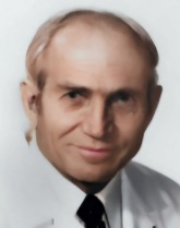 Gene Strandness合作论文数University of Washington School of Medicine5E A Wolf合作论文数4Donald P. Spadone合作论文数4Waleed Hajjar合作论文数4Scott N. Hurlbert合作论文数3Rw Barnes合作论文数3
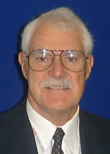 Robert B. Rutherford合作论文数University of Colorado School of Medicine3
 Dennis F Bandyk合作论文数3Ann Lambeth合作论文数3Yaron Sternbach合作论文数3Thomas L. Criblez合作论文数3
 Ashraf Mansour合作论文数3Shigetsugu Ohgi合作论文数2Richard D. Miles合作论文数2Anne M. Jones合作论文数2Dw Baker合作论文数2PJ CALL合作论文数2Paul Kolm合作论文数2Douglas M G合作论文数2Hokanson D E合作论文数2Bada H S合作论文数2D.Eugene Strandness合作论文数2Robert W. Hobson合作论文数2
 Kirk W. Beach合作论文数Department of Bioengineering, College of Engineering, University of Washington2Robert B. Mclafferty合作论文数2M.-P. Colgan合作论文数2
 Denis s Quill合作论文数2Robert B Mclafferty合作论文数2James B. Russell合作论文数2Brian L Thiele合作论文数2




