Severe prolonged migrainous symptoms and prolonged partial status epilepticus are characteristic features of the MELAS syndrome (mitochondrial encephalomyopathy, lactic acidosis, and stroke-like episodes). Maternal transmission previously found in myoclonus epilepsy and ragged-red fibers (MERRF), another mitochondrial disease, is suggested in this disorder as well.
The doctor of today must adopt the 'cellular way of thinking' in the evaluation of diseases. This ultrastructural outlook provides him with much indispensable information that also serves a practical purpose. A diseased cell organelle is at the basis of every clinical sign and any attempt of therapy must be aimed at that specific point of lesion. We intend, in the light of a long experience, to propose to clinicians a new way of thinking in which a precise correlation between symptoms and submicroscopic changes of the cell is considered. Many different examples amply justify this proposal. Electron microscopy can contribute by enabling identification of structural subcellular modifications suitable for the finest differential diagnosis, more and more complete understanding of pathogenic pathways of various diseases, the establishment of guidelines for precise pharmacological interventions at the molecular level.
In four patients, calf muscular hypertrophy developed after the onset of sciatica. Hypertrophic muscles were weak and showed electromyographic signs of denervation. In all cases, calf muscle biopsy showed striking hypertrophy of type 1 and, especially, type 2 muscle fibers. This hypertrophy was associated with other signs indicating a neurogenic lesion. Muscle hypertrophy is a rare finding in neurogenic lesions. Stretch and exercise of muscle are probably the causative factors.
 E Lugaresi合作论文数2Fabrizio Rasi合作论文数2Renzo Laschi合作论文数2Paolo Martinelli合作论文数2W. Neri合作论文数1
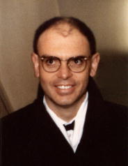 Pasquale Montagna合作论文数University of Bologna1
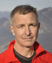 Massimo Zeviani合作论文数Department of Neurosciences, University of Padova;Veneto Institute for Molecular Medicine1Rossella Medori合作论文数Janssen-Cilag EMEA1
 Roberto Gallassi合作论文数1
 Fabio Cirignotta合作论文数S. Orsola-Malpighi Hospital, University of Bologna1




