The aim of this study is to investigate additional genetic alterations in papillary thyroid carcinomas (PTCs) with known RET/PTC rearrangements. We applied array-based comparative genomic hybridization (array CGH) to 33 PTC (20 PTC from adults, 13 post-Chernobyl PTC from children) with known RET/PTC status. Principal component analysis and hierarchical cluster analysis identified cases with similar aberration patterns. Significant deviations between tumour-groups were obtained by statistical testing (Fisher's exact test in combination with Benjamini–Hochberg FDR-controlling procedure). FISH analysis on FFPE sections was applied to validate the array CGH data. Deletions were found more frequently in RET/PTC-positive and RET/PTC-negative tumours than amplifications. Specific aberration signatures were identified that discriminated between RET/PTC-positive and RET/PTC-negative cases (aberrations on chromosomes 1p, 3q, 4p, 7p, 9p/q, 10q, 12q, 13q and 21q). In addition, childhood and adult RET/PTC-positive cases differ significantly for a deletion on the distal part of chromosome 1p. There are additional alterations in RET/PTC-positive tumours, which may act as modifiers of RET activation. In contrast, alterations in RET/PTC-negative tumours indicate alternative routes of tumour development. The data presented serve as a starting point for further studies on gene expression and function of genes identified in this study.
Evaluation of 20 cases of radiation-induced childhood papillary thyroid carcinoma using fluorescence in situ hybridization demonstrated the presence of clonal translocations affecting the RET locus. Semiquantitative reverse transcription-PCR indicated overexpression of the RET tyrosine kinase (TK) domain in four cases. In two cases, the RET rearrangements PTC6 and PTC7 were identified and assigned to balanced translocations t(7;10)(q32;q11.2) and t(1;10)(p13;q11.2), respectively. In one case with a balanced translocation t(10;14)(q11.2;q22.1), 5' rapid amplification of cDNA ends revealed a novel type of RET oncogenic activation (PTC8), arising from a fusion of the 5' part of the kinectin (KTN1) gene to the TK domain of the RET gene. The presence of coiled-coil domains in the resulting ktn1/ret fusion protein suggests ligand-independent dimerization and thus constitutive activation of the ret TK domain.
Thyroid carcinoma incidence is increased significantly after ionizing irradiation; however, the possible mechanisms have not yet been identified. To provide clues for an understanding of the radiation-induced transformation of thyroid epithelium, we analyzed the karyotypes of 56 childhood thyroid tumors that appeared in Belarus after the Chernobyl nuclear accident in 1986. We also studied eight secondary thyroid tumors that developed after radiotherapy. Metaphase preparations obtained from primary cultures were analyzed by G-banding. Clonal structural aberrations were found in 13 of 56 Belarussian cases and in 6 of 8 secondary tumors that developed after radiotherapy. Furthermore, we detected multiple chromosomal aberrations as well as complex rearrangements in some of these tumors and performed a detailed analysis of marker chromosomes from a single case using spectral karyotyping and comparative genomic hybridization in a childhood tumor from Belarus with a near-triploid karyotype. Both comparative genomic hybridization and spectral karyotyping analysis revealed structural alterations affecting identical chromosomes 1, 2, 9, and 13, among others. In addition to the known hot spots of alterations in papillary thyroid carcinomas on chromosomes 1q and 10q, a comprehensive breakpoint analysis in the pooled data set revealed novel breakpoints on chromosomes 4q, 5q, 6p, 12q, 13q, and 14q. The chromosomal aberrations in these tumors may provide suitable starting points for the positional cloning of genes involved in radiation-induced tumorigenesis.
A novel type of RET rearrangement, PTC5, was detected in papillary thyroid carcinomas of two patients exposed to radioactive fallout after Chernobyl. Reverse transcription-PCR and rapid amplification of 5'-cDNA ends revealed a fusion of the ret tyrosine kinase (TK) domain with a sequence identical to that described previously as ret-II. Ret-II is a transfection artifact in NIH3T3 cells and has not yet been detected in any human tumor. Overlapping sequences found in the expressed sequence tag databases enabled us to sequence the COOH terminus of the ret-fused gene 5 (RFG5). The combined data made it possible to assemble a full-length rfg5 protein sequence. Computer-assisted analysis of this sequence reveals four putative coiled-coil structures, possibly involved in dimerization, but no membrane-binding sequences. Northern blots show a ubiquitous RFG5 expression in various normal tissues, including the thyroid gland. In addition to the RFG5/RET, we also detected the reciprocal RET/RFG5 transcript in both tumor samples, suggesting that the rearrangement is based on a balanced reciprocal translocation. In agreement with other rearranged TKs, it is concluded that the transforming action of the new fusion protein rfg5/ret in thyroid tumors may be due to an activation of the ret TK by constitutive expression and dimerization potential of the 5'-fused rfg5 protein. Ret immunohistochemistry indicates that the fusion protein is expressed in all cells of PTC5 tumors, suggesting that RFG5/RET rearrangement is an early event in thyroid carcinogenesis.
A high prevalence of RET rearrangements is found in papillary thyroid carcinomas (PTC) of children from Belarus after the Chernobyl reactor accident. The ELE/RET rearrangement (PTC3) is prevailing. Aberrant types of ELE/RET rearrangement have been found with a truncated ELE1 gene: As compared with the common form (PTC3r1) one aberrant type is shorter by one 144 bp exon (PTC3r2) (three cases); in the second atypic form (PTC3r3) the ELE1 part is 18 bp shorter than in PTC3r1. In agreement with the observation that the oncogenic RET is generated by a paracentric inversion at chromosome 10, we found not only ELE/RET, but also RET/ELE transcripts in these tumors. Sequencing of the breakpoint regions at the genomic DNA level revealed DNA modifications that might be relevant for illegitimate recombination after DNA doublestrand breaks. The high prevalence of ELE/RET rearrangements and various subtypes appears to be typical for radiation-induced thyroid carcinomas of children after the Chernobyl reactor accident.
Mutations in the p53 tumour-suppressor gene (exons 5-8) were investigated in 31 Belarussian childhood thyroid tumours (24 cases of papillary thyroid carcinoma, 3 benign tumours and 2 cases each of thyroiditis and goiter); 33 thyroid tumours from juveniles and adults without radiation exposures (25 carcinomas of various histological types, including 11 papillary carcinomas and 8 adenomas) and 6 tumours from adults (4 papillary carcinomas, 1 adenoma, 1 goiter) served as controls. The mutational spectrum of p53 differed greatly between the childhood thyroid carcinomas from Belarus and the control groups. In the control groups of 29 malignant thyroid tumours, 7 different mutations were detected on exons 5-8, none of which occurred among the 15 papillary carcinomas in this group. Five mutations were found in tissue samples of the 24 childhood papillary carcinomas, and they were all the same p53 point mutation (CGA --> CGG) on codon 213 of exon 6. To determine whether this mutation is simply a polymorphism or whether it is specific to the tumour cells, laser-assisted microdissection was applied to collect various areas of tumorous and non-tumorous cells (10-20 cells per sample) from each paraffin-embedded tissue section of 8 of the papillary thyroid carcinomas. Using PCR-SSCP and sequence analysis on these cells, the very same p53 mutation on codon 213 was detected in various microdissected tumour samples of 2 cases, but it was not found in any microdissected non-tumorous sample. The exclusive occurrence of this p53 mutation in selective microdissected samples of tumour cells, even as homozygous mutation in 1 case, reflects a distinct tumour heterogeneity within papillary childhood thyroid carcinomas.
 E P Demidchik合作论文数5
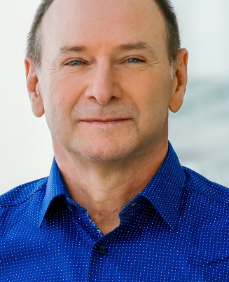 Horst Zitzelsberger合作论文数Research Unit Radiation Cytogenetics, Helmholtz Zentrum München5
 Ludwig Hieber合作论文数3Albrecht M. Kellerer合作论文数3Manfred Bauchinger合作论文数3T Negele合作论文数2J Smida合作论文数2Axel Walch合作论文数2Fritz Spelsberg合作论文数2Konstatinos Salassidis合作论文数2Lars Lehmann合作论文数2Hartmut M Rabes合作论文数2Sabine Klugbauer合作论文数2J Bruch合作论文数1P Lewis合作论文数1Hug Weier合作论文数1Hu Weier合作论文数1M Werner合作论文数1G Minkus合作论文数1G. A. Thomas合作论文数1C. Janish合作论文数1Jingly Fung合作论文数1
 Johannes Christoph Wienberg合作论文数Department of Biology II, Ludwig-Maximilians-University of Munich1Eva Malisch合作论文数1Gerhard Jackl合作论文数1
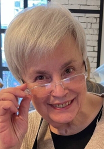 Bogdanova Tetiana合作论文数V.P. Komissarenko Institute of Endocrynology and Metabolism, National Academy of Medical Sciences of Ukraine1
 Kristian Unger合作论文数Helmholtz Zentrum München1
 Herbert Braselmann合作论文数Research Unit Radiation Cytogenetics, Helmholtz Zentrum München1




