Alterations in autonomic function are known to occur in cardiac conditions including sudden cardiac death. Cardiac stimulation via sympathetic neurons can potentially trigger arrhythmias. Dissecting direct neural-cardiac interactions at the cellular level is technically challenging and understudied due to the lack of experimental model systems and methodologies. Here we demonstrate the utility of optical interrogation of sympathetic neurons and their effects on macroscopic cardiac monolayer dynamics to address research targets such as the effects of adrenergic stimulation via the release of neurotransmitters, the effect of neuronal numbers on cardiac wave behaviour and the applicability of optogenetics in mechanistic in vitro studies. We combine photo-uncaging or optogenetic neural stimulation with imaging of cardiac monolayers to measure electrical activity in an automated fashion, illustrating the power and high throughput capability of such interrogations. The methods described highlight the challenges and benefits of co-cultures as experimental model systems.
Aims: Macroscopic cultures of cardiac myocytes are a useful model to study arrhythmogenic mechanisms, but the absence of sympathetic stimulation limits our ability to study one of the main triggers of arrhythmia. In this study, we grow myocytes and cardiac stellate sympathetic neurons (CSSN) in confluent monolayer's to investigate how neurons modulate propagating wave patterns. Methods: Cardiac monolayer's were prepared by plating petri-dishes with neonatal myocytes from Sprague Dawley rat ventricle cells. After 24 hours, co-cultures were produced by adding CSSN isolated from litter mates. Co-cultures were imaged using state-of-the-art dye free optical mapping techniques. Results: SCCN form structural (evidenced by scanning electron microscopy and conventional microscopy) and functional connections with myocytes. We demonstrate that co-cultures are able to support target, spiral and wavelet patterns. Basal unstimulated wave speed of co-cultures was significantly faster than myocyte monocultures (+30%), and the number of co-cultures displaying multiple wave fronts was lower than that found in monocultures (P < 0.05). 10 μM nicotine stimulation of co-cultures showed an immediate increase in beat rate and in cases where the cardiac cultures are in target (analogous to sinus rhythm) acute addition of nicotine may initiate a re-entrant activity or can cause the breakup of the target wave. Conclusion: CSSN affect cardiac conduction velocity and wave patterns in in-vitro co-cultures. This model system may be used for the quantitative investigation of the influence of neural activation on cardiac activity during arrhythmias.
Hypertension is associated with over-activity of cardiac sympathetic neurons resulting in greater transmitter release that is coupled to abnormally high intracellular Ca2+ transients in the spontaneously hypertensive rat (SHR). These neurons also express lower levels of nNOS, the beta1 subunit of soluble guanylate cyclase (sGC), and cGMP compared to normotensive control WKY rats. We tested the hypothesis that dysregulation of the voltage gated Ca2+ channel could provide the trigger for the calcium phenotype seen in pro-hypertensive SHRs that is linked impaired cyclic nucleotide signalling. Cardiac sympathetic stellate ganglions were acutely isolated from 4 week, pro-hypertensive SHR and WKY rats. Whole cell voltage clamp (10 mV steps from -50 to +50) was performed at 37°. The whole cell maximum Ca2+ conductance (in nS/pF) in the SHR was significantly larger when compared to the normotensive WKY (2.48 ± 0.11 SEM, n = 10 vs 2.09 ± 0.13 SEM, n = 10, p = 0.037). Application of Conotoxin GVIA reduced the max conductance by approximately 83% and 78% (0.40 ± 0.05 SEM, n = 6 and 0.46 ± 0.04 SEM, n = 3, p < 0.0001), suggesting a major role for the N-type Ca2+ channel. Increasing the cGMP levels (8-bromo-cGMP) did not affect the WKY (1.96 ± 0.17 SEM, n = 7, p = 0.54), but normalised the SHR conductance to levels seen in WKYs (2.02 ± 0.13 SEM, n = 7, p = 0.0184, all unpaired t-test). There was no change in voltage dependency of the conductances or the half maximum activation (mV) between the groups. In conclusion, an enhanced N-Type Ca2+ channel conductance suggests a channelopathy may be an early hallmark of sympathetic impairment in the SHR that is coupled to impaired cyclic nucleotide regulation.
 David Paterson合作论文数Department of Physiology, Anatomy & Genetics, University of Oxford;Burdon Sanderson Cardiac Science Centre, University of Oxford5
 Guy Stephens合作论文数3Gil Bub合作论文数3
 Hege Ekeberg Larsen合作论文数3
 Rebecca A B Burton合作论文数Medical Sciences Division, Department of Pharmacology, University of Oxford3h e larsen合作论文数2
 Amy Sharkey合作论文数2J Tomek合作论文数2Yi-Ge Huang合作论文数Radcliffe Department of Medicine, University of Oxford2
 Vladyslav V. Vyazovskiy合作论文数Department of Physiology, Anatomy & Genetics, University of Oxford2A. Deli合作论文数1P. Pettingill合作论文数1
 Pantelis Antonoudiou合作论文数Graduate School of Biomedical Sciences, Tufts University School of Medicine1
 Giuseppe Gallone合作论文数Wellcome Sanger Institute;1
 Shenghong He (何盛鸿)合作论文数1
 Michael Lohse合作论文数Sainsbury Wellcome Centre, Faculty of Life Sciences, University College London1Hao Guoliang合作论文数1
 Capel Rebecca A合作论文数Department of Pharmacology, University of Oxford Oxford1
 Angela Vincent合作论文数Medical Sciences Division, Nuffield Department of Clinical Neurosciences, University of Oxford1
 Emilia Entcheva合作论文数Department of Biomedical Engineering, School of Engineering & Applied Science, George Washington University1
 Kahn Martin合作论文数1
 Anjan Nibber合作论文数1
 Bethan Lang合作论文数University of Sydney1
 Huiling Tan合作论文数Nuffield Department of Clinical Neurosciences, University of Oxford1
 Dan Li合作论文数Department of Physiology, Anatomy & Genetics, University of Oxford1Dong Li合作论文数1
 Alex Green合作论文数Medical Sciences Division, Nuffield Department of Surgical Sciences, University of Oxford1
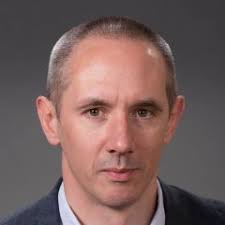 Patrick Waters合作论文数Autoimmune Neurology Diagnostic Laboratory, Nuffield Department of Clinical Neurosciences, Medical Sciences Division, University of Oxford1
 Ashavani Kumar合作论文数Department of Physics, National Institute of Technology Kurukshetra;School of Materials Science and Technology, National Institute of Technology Kurukshetra1
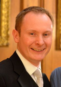 Neil Herring合作论文数Department of Physiology, Anatomy & Genetics, University of Oxford1
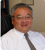 Kiang J Liu合作论文数Department of Preventive Medicine, Feinberg School of Medicine, Northwestern University;Center for Circadian and Sleep Medicine, Feinberg School of Medicine, Northwestern University1




