We accurately modelled acute diarrhea (AUC = 0.90) for rectal cancer patients receiving CRT using AI-contoured small-bowel structures. The treatment arm and small-bowel dose were independently correlated to the diarrhea severity. The explainable model allowed us to quantify the impact of radiation dose, usage of irinotecan, and its combination, with a threshold of V = 160.93 cm yielding an equivalent impact. We will be extending the analysis to the whole trial cohort to improve the statistical power.
Multicentre clinical trials involving a dosimetry component are becoming more prevalent in molecular radiotherapy and are essential to generate the evidence to support individualised approaches to treatment planning and to ensure that sufficient patients are recruited to achieve the statistical significance required. Quality assurance programmes should be considered to support the standardisation required to achieve meaningful results. Trials should be designed to ensure that dosimetry results from image acquisition systems across centres are comparable by incorporating steps to standardise the methodologies used for the quantification of images and dosimetry. Furthermore, it is essential to assess the expertise and resources available at each participating site prior to trial commencement. A quality assurance plan should be drawn up and training provided if necessary. Standardisation of quantification and dosimetry methodologies used in a trial are essential to ensure that results from different centres may be collated. In addition, appropriate uncertainty analysis should be carried out to correct for differences in methodologies between centres. Recommendations are provided to support dosimetry studies based on the experience of several previous and ongoing multicentre trials.
AIMS:Adaptive radiotherapy (ART) is an emerging advanced treatment option for bladder cancer patients. Therapeutic radiographers (RTTs) are central to the successful delivery of this treatment. The purpose of this work was to evaluate the image-guided radiotherapy (IGRT) and ART experience of RTTs before participating in the RAIDER trial. A plan of the day (PoD) quality assurance programme was then implemented. Finally, the post-trial experience of RTTs was evaluated, together with the impact of trial quality assurance participation on their routine practice. MATERIALS AND METHODS:A pre-trial questionnaire to assess the experience of the RTT staff group in IGRT and ART in bladder cancer was sent to each centre. Responses were grouped according to experience. The PoD quality assurance programme was implemented, and the RAIDER trial commenced. During stage 1 of the trial, RTTs reported difficulties in delivering PoD and the quality assurance programme was updated accordingly. A follow-up questionnaire was sent assessing experience in IGRT and ART post-trial. Any changes in routine practice were also recorded. RESULTS:The experience of RTTs in IGRT and ART pre-trial varied. For centres deemed to have RTTs with more experience, the initial PoD quality assurance programme was streamlined. For RTTs without ART experience, the full quality assurance programme was implemented, of which 508 RTTs completed. The quality assurance programme was updated (as the trial recruited) and it was mandated that at least one representative RTT (regardless of pre-trial experience) participated in the update in real-time. The purpose of the updated quality assurance programme was to provide further support to RTTs in delivering a complex treatment. Engagement with the updated quality assurance programme was high, with RTTs in 24/33 centres participating in the real-time online workshop. All 33 UK centres reported all RTTs reviewed the updated training offline. Post-trial, the RTTs' experience in IGRT and ART was increased. CONCLUSION:Overall, 508 RTTs undertook the PoD quality assurance programme. There was a high engagement of RTTs in the PoD quality assurance programme and trial. RTTs increased their experience in IGRT and ART and subsequently updated their practice for bladder cancer and other treatment sites.
Aims: Radiotherapy clinical trials are integral to the development of new treatments to improve the outcomes of patients with cancer. A collaborative study by the National Cancer Research Institute Clinical and Translational Radiotherapy Research Working Group and the National Institute for Health Research was carried out to understand better if and why inefficiencies occur in the set-up of radiotherapy trials in the UK. Materials and methods: Two online surveys collected information on the time taken for UK radiotherapy trials to reach key milestones during set-up and the research support currently being provided to radiotherapy centres to enable efficient clinical trial set-up. Semi-structured interviews with project managers and chief investigators identified better ways of working to improve trial set-up in the future. Results: The timelines for the set-up of 39 UK radiotherapy trials were captured in an online survey showing that the median time from grant approval to trial opening was 600 days (range 169-1172). There were 38 responses from radiotherapy centres to a survey asking about the current support provided for radiotherapy research. Most of these centres have more than one type of staff member dedicated to supporting radiotherapy research. The most frequent barrier to radiotherapy trial set-up identified was lack of physicists' time and lack of time for clinical oncologists to carry out research activities. Four main themes around trial set-up were identified from semi-structured interviews: the importance of communication and building relationships, the previous experience of the chief investigator and clinical trials units, a lack of resources and having the time and personnel required to produce trial documentation and to process trial approval requests. Conclusions: This unique, collaborative project has provided up to date information about the current landscape of trial set-up and research support in the UK and identified several avenues on which to focus future efforts in order to support the excellent radiotherapy trial work carried out across the UK. (C) 2019 The Authors. Published by Elsevier Ltd on behalf of The Royal College of Radiologists.
IntroductionSoft tissue sarcomas of the extremities (STSE) are rare malignancies. We report current UK practice for immobilisation of soft tissue sarcoma of STSE, as part of the initial study set-up within the IMRiS trial, a phase II study of intensity modulated radiotherapy (IMRT) in primary bone and soft tissue sarcoma.MethodsA facility questionnaire (FQ) was circulated to 29 IMRiS centres investigating the variation in immobilisation devices, planning techniques, and imaging protocols. A workshop was held to address concerns raised by centres. It focused on STSE immobilisation and patient set-up. Robustness of patient set-up at each centre was evaluated based on the following criteria: evidence of local set-up audit, calculation of margins based on set-up audit results, imaging frequency, and number of patients treated per centre per annum.ResultsTwenty-seven (93%) questionnaires were returned. 30% (8/27) of responders routinely treated STSE with IMRT. The remaining 70% (19/27) had little or no experience with IMRT for STSE. Vacuum bags were the most frequent immobilisation device (9/27), followed by thermoplastic shells (7/27). Nine centres had audited their local set-up; however, only 4 had calculated margins in response to the results. Ten centres were classified as having high level of robustness.ConclusionsImmobilisation devices and planning techniques for STSE are inconsistent across centres. Robustness of set-up is an important tool to ensure quality of results in a multicentre trial setting with such different levels of experience. The IMRiS trial Quality Assurance programme encourages centres to assess robustness of set-up through local audit and subsequent calculation of treatment margins.Implications for practiceThis is the first study that used robustness criteria to tailor QA support to individual centres.
In the UK, the recent introduction of high-energy proton beam therapy into national clinical practice provides an opportunity for new clinical trials, particularly those comparing proton and photon treatments. However, comparing these different modalities can present many challenges. Although protons may confer an advantage in terms of reduced normal tissue dose, they can also be more sensitive to uncertainty. Uncertainty analysis is fundamental in ensuring that proton plans are both safe and effective in the event of unavoidable discrepancies, such as variations in patient setup and proton beam range. Methods of evaluating and mitigating the effect of these uncertainties can differ from those approaches established for photon therapy treatments, such as the use of expansion margins to assure safety. These differences should be considered when comparing protons and photons. An overview of the effect of uncertainties on proton plans is presented together with an introduction to some of the concepts and terms that should become familiar to those involved in proton therapy trials. This report aims to provide guidance for those engaged in UK clinical trials comparing protons and photons. This guidance is intended to take a pragmatic approach considering the tools that are available to practising centres and represents a consensus across multidisciplinary groups involved in proton therapy in the UK.
To quantify the inter-clinician contouring variability in pediatric abdominal neuroblastoma based on a national pre-trial quality assurance exercise for a multi-center clinical trial. As part of the quality assurance process for the IMAT Neuroblastoma prospective Phase 1/2 trial (ISRCTN10746820), completion of a pre-trial benchmark contouring case was required. Diagnostic MRI, pre-surgical CT, radiotherapy planning CT, and a short clinical history were supplied to each center. Clinicians were requested to delineate the virtual gross tumor volume (GTV) defined as the reconstructed tumor volume seen on the post induction chemotherapy, pre-surgical imaging, as well as any immediately adjacent persistently enlarged lymph nodes, and edited to exclude uninvolved normal organs. Clinical target and planning target volumes (CTV and PTV) were then generated prior to submission and relevant organs at risk (OAR) were also delineated. Contours were reviewed centrally by a trial clinician, who provided feedback in a narrative report with images used for visual clarification. A consensus GTV contour was determined from all submissions using the simultaneous truth and performance level estimation (STAPLE) algorithm at the 95% confidence level. DICE similarity coefficient (DSC) and mean Hausdorff distance were calculated for each submitted contour compared to the STAPLE consensus contour. Clinicians from 12/16 pediatric radiotherapy centers in the United Kingdom submitted contours for review. GTV contours were classified as unacceptable deviations in 3/12 cases and required subsequent amendment prior to acceptance. Narrative detail was provided in 10/12 reviews and identified regions of under-contouring (9/10) and over-contouring (3/10). Under-contouring was identified in the para-aortic nodal region (5/9), retrocrural nodal region (1/9), unspecified regions of residual calcification (2/9), and more generally (2/9). Over-contouring was identified in uninvolved organs (2/3) and more generally (1/3). Substantial variation was present in the submitted GTVs when compared to the consensus contour. Averaged over all clinicians, the DSC was 0.61 ± 0.15 (mean ± 1 S.D.) and mean Hausdorff distance was 4.54 ± 2.25 mm. Good agreement was present for the organs at risk, however. Considerable variability in GTV contouring has been identified between clinicians, primarily in the extent to which nodes and residual calcification had been included. This supports the use of prospective contour review for target volumes in future abdominal neuroblastoma clinical trials. Limited variation in OAR contouring suggests that quality assurance could be streamlined for future trials.
Aims: Accurate delineation of organs at risk (OAR) is vital to the radiotherapy planning process. Inaccuracies in OAR delineation arising from imprecise anatomical definitions may affect plan optimisation and risk inappropriate dose delivery to normal tissues. The aim of this study was to review the provision of OAR contouring guidance in National Institute of Health Research Clinical Research Network (NIHR CRN) portfolio clinical trials. Materials and methods: The National Radiotherapy Quality Trials Assurance (RTTQA) Group carried out a two-round Delphi assessment to determine which OAR descriptions provided optimal guidance. Results: Eighty-four clinical trials involving radiotherapy quality assurance were identified as either in recruitment or in setup within the NIHR CRN portfolio. Fifty-nine trials mandated OAR contouring. In total there were 412 OAR; 171 were uniquely named; 159 OAR had more than one name associated with a single structure, with the greatest nomenclature variation seen for the femoral head +/- neck, the parotid gland, and bowel. The two-round Delphi assessment determined 42 OAR descriptions as providing optimal contouring guidance. Conclusions: This study identified the need for OAR nomenclature and contouring guidance consistency across clinical trials. In response to this study and in conjunction with the Global Quality Assurance of Radiation Therapy Clinical Trials Harmonisation Group, the RTTQA Group is in collaboration with international partners to provide consensus recommendations for OAR delineation in clinical trials. Crown Copyright (C) 2019 Published by Elsevier Ltd on behalf of The Royal College of Radiologists. All rights reserved.
 Catharine Clark合作论文数Department of Medical Physics and Biomedical Engineering, University College London;University College Hospital;National Physical Laboratory17
 Christopher Nutting合作论文数The Royal Marsden Hospital;The Institute of Cancer Research12
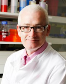 Kevin Harrington合作论文数The Institute of Cancer Research10
 Margaret Bidmead合作论文数8Teresa Guerrero Urbano合作论文数The Institute of Cancer Research7
 Ricardo Simoes合作论文数Department of Gynecology, Federal University of São Paulo7O. Naismith合作论文数6
 Patricia Diez合作论文数6
 Helen McNair合作论文数The Institute of Cancer Research6Sarah Gwynne合作论文数6H. Mayles合作论文数5H. Yang合作论文数5Conibear J合作论文数5
 Peter Hoskin合作论文数Mount Vernon Cancer Centre;Cancer Research UK Manchester Centre, University of Manchester;Christie Hospital;University College Hospital5
 Emiliano Spezi合作论文数Division of Radiation Oncology, Policlinico S. Orsola5
 Sarah Gulliford合作论文数University College Hospital;Department of Medical Physics and Biomedical Engineering, University College London5
 Beatrice M Seddon合作论文数5V. Hansen合作论文数4Y. Tsang合作论文数4
 Maria Hawkins合作论文数Department of Medical Physics and Biomedical Engineering, Faculty of Engineering Sciences, University College London4Maggs Rhydian合作论文数4
 Fiona J Mcdonald合作论文数4V. Khoo合作论文数4
 John Staffurth合作论文数School of Medicine, Cardiff University;Institute of Cancer & Genetics, Cardiff University4
 David Sebag-Montefiore合作论文数University of Leeds4Laura Ciurlionis合作论文数4
 David Paul Dearnaley合作论文数Department of Radiotherapy, The Royal Marsden NHS Foundation Trust;Academic Department of Urology, Institute of Cancer Research4R. Mir合作论文数3
 Kevin Nicholas Franks合作论文数3A. Webster合作论文数3Y. M. Tsang合作论文数3
 Franel le Grange合作论文数3A Miah合作论文数3David Landau合作论文数3Wadsley Jonathan合作论文数3Mayles Philip合作论文数3Wells E合作论文数3
 Alison Tree合作论文数The Royal Marsden NHS Foundation Trust;Convergence Science Centre, Institute of Cancer Research3Forsyth Sharon合作论文数3
 Emma A Hall合作论文数3Baker A合作论文数3Adams Elizabeth J合作论文数3Ray Ruby合作论文数3
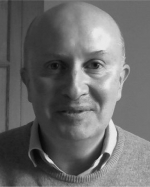 John D. Fenwick合作论文数3
 Raj B.Patel合作论文数Department of Physics, Faculty of Natural Sciences, Imperial College London3T. Guerrero Urbano合作论文数3E. Aird合作论文数3T. Crosby合作论文数3N. Whilde合作论文数2




