The E3 ubiquitin ligase Casitas B-lineage lymphoma (CBL) promotes positive selection and antigen responses in mouse T lymphocytes by ubiquitinating ZAP70. Conversely, mouse CBL and CBL-B mutually redundantly regulate SYK ubiquitination and B cell receptor signaling. Here we studied individuals with somatically homozygous CBL loss-of-function variants in leukocytes. Human CBL is largely redundant for the development and function of human T cells. Conversely, B cell development is altered at the immature stage, with a tenfold increase in transitional cells, enhanced survival of autoreactive clones and impaired tolerance manifested by autoantibody production. B cell maturation is intrinsically impaired by reduced apoptosis and dysregulated B cell receptor signaling. CBL deficiency impairs humoral immunity by limiting memory B cell formation and reducing class switching and somatic hypermutation. Consequently, antigen-specific B cell generation and adaptive immune memory are disrupted, predisposing individuals to infection. Human CBL is critical for B cell development and function but redundant for T cell biology.
Oncostatin M (OSM) is a cytokine with the unique ability to interact with both the OSM receptor (OSMR) and the leukemia inhibitory factor receptor (LIFR). On the other hand, OSMR interacts with IL31RA to form the interleukin-31 receptor. This intricate network of cytokines and receptors makes it difficult to understand the specific function of OSM. While monoallelic loss-of-function (LoF) mutations in OSMR underlie autosomal dominant familial primary localized cutaneous amyloidosis, the in vivo consequences of human OSM deficiency have never been reported so far. Here, we identified 3 young individuals from a consanguineous family presenting with inherited severe bone marrow failure syndromes (IBMFS) characterized by profound anemia, thrombocytopenia, and neutropenia. Genetic analysis revealed a homozygous 1 base-pair insertion in the sequence of OSM associated with the disease. Structural and functional analyses showed that this variant causes a frameshift that replaces the C-terminal portion of OSM, which contains the FxxK motif that interacts with both OSMR and LIFR, with a neopeptide. The lack of detection and signaling of the mutant OSM suggests a LoF mutation. Analysis of zebrafish models further supported the role of the OSM/OSMR signaling in erythroid progenitor proliferation and neutrophil differentiation. Our study provides the previously uncharacterized and unexpectedly limited in vivo consequence of OSM deficiency in humans.
In sickle cell disease (SCD), the β6Glu→Val substitution in the β-globin leads to red blood cell sickling. The transplantation of autologous, genetically modified hematopoietic stem and progenitor cells (HSPCs) is a promising treatment option for patients with SCD. We completed a Phase I/II open-label clinical trial (NCT03964792) for patients with SCD using a lentiviral vector (DREPAGLOBE) expressing a potent anti-sickling β-globin. The primary endpoint was to evaluate the short-term safety and secondary endpoints included the efficacy and the long-term safety. We report on the results after 18 to 36 months of follow-up. No drug-related adverse events or signs of clonal hematopoiesis were observed. Despite similar vector copy numbers in the drug product, gene-marking in peripheral blood mononuclear cells and correction of the clinical phenotype varied from one patient to another. Single-cell transcriptome analyses show that in the patients with poor engraftment, the most immature HSCs display an exacerbated inflammatory signature (via IL-1 or TNF-α and interferon signaling pathways). This signature is accompanied by a lineage bias in the HSCs. Our clinical data indicates that the DREPAGLOBE-based gene therapy (GT) is safe. However, its efficacy is variable and probably depends on the number of infused HSCs and intrinsic, engraftment-impairing inflammatory alterations in HSCs. Trial: NCT03964792.
Patients heterozygous for germline CBL loss-of-function (LOF) variants can develop myeloid malignancy, autoinflammation, or both, if some or all of their leukocytes become homozygous for these variants through somatic loss of heterozygosity (LOH) via uniparental isodisomy. We observed an upregulation of the inflammatory gene expression signature in whole blood from these patients, mimicking monogenic inborn errors underlying autoinflammation. Remarkably, these patients had constitutively activated monocytes that secreted 10 to 100 times more inflammatory cytokines than those of healthy individuals and CBL LOF heterozygotes without LOH. CBL-LOH hematopoietic stem and progenitor cells (HSPCs) outgrew the other cells, accounting for the persistence of peripheral monocytes homozygous for the CBL LOF variant. ERK pathway activation was required for the excessive production of cytokines by both resting and stimulated CBL-LOF monocytes, as shown in monocytic cell lines. Finally, we found that about 1 in 10,000 individuals in the UK Biobank were heterozygous for CBL LOF variants and that these carriers were at high risk of hematological and inflammatory conditions
Hematopoietic stem cell gene therapy (GT) using a γ-retroviral vector (γ-RV) is an effective treatment for Severe Combined Immunodeficiency due to Adenosine Deaminase deficiency. Here, we describe a case of GT-related T-cell acute lymphoblastic leukemia (T-ALL) that developed 4.7 years after treatment. The patient underwent chemotherapy and haploidentical transplantation and is currently in remission. Blast cells contain a single vector insertion activating the LIM-only protein 2 (LMO2) proto-oncogene, confirmed by physical interaction, and low Adenosine Deaminase (ADA) activity resulting from methylation of viral promoter. The insertion is detected years before T-ALL in multiple lineages, suggesting that further hits occurred in a thymic progenitor. Blast cells contain known and novel somatic mutations as well as germline mutations which may have contributed to transformation. Before T-ALL onset, the insertion profile is similar to those of other ADA-deficient patients. The limited incidence of vector-related adverse events in ADA-deficiency compared to other γ-RV GT trials could be explained by differences in transgenes, background disease and patient's specific factors.
FMS-related tyrosine kinase 3 ligand (FLT3L), encoded by FLT3LG, is a hematopoietic factor essential for the development of natural killer (NK) cells, B cells, and dendritic cells (DCs) in mice. We describe three humans homozygous for a loss-of-function FLT3LG variant with a history of various recurrent infections, including severe cutaneous warts. The patients’ bone marrow (BM) was hypoplastic, with low levels of hematopoietic progenitors, particularly myeloid and B cell precursors. Counts of B cells, monocytes, and DCs were low in the patients’ blood, whereas the other blood subsets, including NK cells, were affected only moderately, if at all. The patients had normal counts of Langerhans cells (LCs) and dermal macrophages in the skin but lacked dermal DCs. Thus, FLT3L is required for B cell and DC development in mice and humans. However, unlike its murine counterpart, human FLT3L is required for the development of monocytes but not NK cells.
X-linked chronic granulomatous disease (CGD) is associated with defective phagocytosis, life-threatening infections, and inflammatory complications. We performed a clinical trial of lentivirus-based gene therapy in four patients (NCT02757911). Two patients show stable engraftment and clinical benefits, whereas the other two have progressively lost gene-corrected cells. Single-cell transcriptomic analysis reveals a significantly lower frequency of hematopoietic stem cells (HSCs) in CGD patients, especially in the two patients with defective engraftment. These two present a profound change in HSC status, a high interferon score, and elevated myeloid progenitor frequency. We use elastic-net logistic regression to identify a set of 51 interferon genes and transcription factors that predict the failure of HSC engraftment. In one patient, an aberrant HSC state with elevated CEBPβ expression drives HSC exhaustion, as demonstrated by low repopulation in a xenotransplantation model. Targeted treatments to protect HSCs, coupled to targeted gene expression screening, might improve clinical outcomes in CGD.
BACKGROUND:Allogenic hematopoietic stem cell transplantation (HSCT) and gene therapy (GT) are potentially curative treatments for severe combined immunodeficiency (SCID). Late-onset posttreatment manifestations (such as persistent hepatitis) are not uncommon. OBJECTIVE:We sought to characterize the prevalence and pathophysiology of persistent hepatitis in transplanted SCID patients (SCIDH+) and to evaluate risk factors and treatments. METHODS:We used various techniques (including pathology assessments, metagenomics, single-cell transcriptomics, and cytometry by time of flight) to perform an in-depth study of different tissues from patients in the SCIDH+ group and corresponding asymptomatic similarly transplanted SCID patients without hepatitis (SCIDH-). RESULTS:Eleven patients developed persistent hepatitis (median of 6 years after HSCT or GT). This condition was associated with the chronic detection of enteric viruses (human Aichi virus, norovirus, and sapovirus) in liver and/or stools, which were not found in stools from the SCIDH- group (n = 12). Multiomics analysis identified an expansion of effector memory CD8+ T cells with high type I and II interferon signatures. Hepatitis was associated with absence of myeloablation during conditioning, split chimerism, and defective B-cell function, representing 25% of the 44 patients with SCID having these characteristics. Partially myeloablative retransplantation or GT of patients with this condition (which we have named as "enteric virus infection associated with hepatitis") led to the reconstitution of T- and B-cell immunity and remission of hepatitis in 5 patients, concomitantly with viral clearance. CONCLUSIONS:Enteric virus infection associated with hepatitis is related to chronic enteric viral infection and immune dysregulation and is an important risk for transplanted SCID patients with defective B-cell function.
Background and aims: Germline monoallelic Casitas B-Lineage Lymphoma Proto-Oncogene (CBL) mutations underlie a RASopathy called CBL Syndrome, and predispose to juvenile monomyelocytic leukemia (JMML). Typically, this JMML is driven by loss-of-heterozygosity (LOH) of CBL by uniparental isodisomy (UPD). CBL-driven JMML usually regresses spontaneously, but renders the hematopoietic system homozygous for mutated CBL. Whether and how this genetic transformation affects the adaptive immune system is unclear. Methods: We studied humoral immunity and B cell function in 9 patients with CBL mutation and LOH. They suffered from life-threatening bacterial infections (n = 5), autoinflammation (n = 3) and autoimmunity (n = 2). We quantified immunoglobin levels, humoral responses to vaccines and infections. Furthermore, we studied B cell function in terms of immunoglobulin production and isotype switching in vitro. We characterized the mosaicism for LOH in mature leukocyte subsets by amplicon sequencing. Results: We detect monocytosis (1397+/-322 monocytes/µl) and B cell lymphocytosis (1107+/-122 B cells/µl) in all pediatric patients. Immature transitional B cells are particularly overrepresented compared to age-matched children (409 +/- 169 cells/µl). Five patients have hypergammaglobulinemia (17,2+/-1,6 mg/dl IgG) and two produce antibodies targeting auto-antigens (ANA, LAC, actin, smooth muscle). Sorted B cells of all tested patients produce reduced levels of immunoglobulins in vitro in response to B cell receptor and TLR activation (5to10-fold decreased) but normal levels upon IL-21. We quantified the allele burdens in sorted B-cell subsets. We find that the frequency of B cells lacking LOH is lowest in transitional B cells compared to naïve and memory, where heterozygous cells are 5-fold and 18-fold more frequent, respectively. This suggests that in vivo, maturation and differentiation of LOH B cells is significantly impaired. Conclusions: In summary, patients that recover from CBL-driven JMML have impaired B cell function and anti-bacterial immunity. A B cell intrinsic role of CBL in the maturation and differentiation pathway can be assumed. Keywords: CBL Syndrome, B cells, Bacterial Infection, Inborn error of immunity, IEI
Abstract In a steady state, hematopoietic stem cells (HSC) exhibit very low levels of reactive oxygen species (ROS). Upon stress, HSC get activated and enter into proliferation and differentiation process to ensure blood cell regeneration. Once activated, their levels of ROS increase, as messengers to mediate their proliferation and differentiation programs. However, at the end of the stress episode, ROS levels need to return to normal to avoid HSC exhaustion. It was shown that antioxidants can prevent loss of HSC self-renewal potential in several contexts such as aging or after exposure to low doses of irradiation suggesting that antioxidants can be used to maintain HSC functional properties upon culture-induced stress. Indeed, in humans, HSC are increasingly used for cell and gene therapy approaches, requiring them to be cultured for several days. As expected, we show that a short culture period leads to drastic defects in HSC functional properties. Moreover, a switch of HSC transcriptional program from stemness to differentiation was evidenced in cultured HSC. Interestingly, cultured-HSC treated with 4-hydroxy-2,2,6,6-tetramethylpiperidin-1-oxyl (4-hydroxy-TEMPO or Tempol) exhibited a higher clonogenic potential in secondary colony forming unit cell (CFU-C) assay and higher reconstitution potential in xenograft model, compared to untreated cultured-HSC. By transcriptomic analyses combined with serial CFU-C assays, we show that Tempol, which mimics superoxide dismutase, protects HSC from culture-induced stress partly through VEGFα signaling. Thus, we demonstrate that adding Tempol leads to the protection of HSC functional properties during ex vivo culture.
Sickle cell disease (SCD) and transfusion-dependent β-thalassemia (TDT) are the most prevalent monogenic disorders worldwide. Trial HGB-205 ( NCT02151526 ) aimed at evaluating gene therapy by autologous CD34 + cells transduced ex vivo with lentiviral vector BB305 that encodes the anti-sickling β A-T87Q -globin expressed in the erythroid lineage. HGB-205 is a phase 1/2, open-label, single-arm, non-randomized interventional study of 2-year duration at a single center, followed by observation in long-term follow-up studies LTF-303 ( NCT02633943 ) and LTF-307 ( NCT04628585 ) for TDT and SCD, respectively. Inclusion and exclusion criteria were similar to those for allogeneic transplantation but restricted to patients lacking geno-identical, histocompatible donors. Four patients with TDT and three patients with SCD, ages 13–21 years, were treated after busulfan myeloablation 4.6–7.9 years ago, with a median follow-up of 4.5 years. Key primary endpoints included mortality, engraftment, replication-competent lentivirus and clonal dominance. No adverse events related to the drug product were observed. Clinical remission and remediation of biological hallmarks of the disease have been sustained in two of the three patients with SCD, and frequency of transfusions was reduced in the third. The patients with TDT are all transfusion free with improvement of dyserythropoiesis and iron overload.
Long-term multilineage hematopoietic donor chimerism occurs sporadically in patients who receive a transplanted solid organ enriched in lymphoid tissues such as the intestine or liver. There is currently no evidence for the presence of kidney-resident hematopoietic stem cells in any mammal species. Graft-versus-host-reactive donor T cells promote engraftment of graft-derived hematopoietic stem cells by making space in the bone marrow. Here, we report full (over 99%) multilineage, donor-derived hematopoietic chimerism in a pediatric kidney transplant recipient with syndromic combined immune deficiency that leads to transplant tolerance. Interestingly, we found that the human kidney-derived hematopoietic stem cells took up long-term residence in the recipient's bone marrow and gradually replaced their host counterparts, leading to blood type conversion and full donor chimerism of both lymphoid and myeloid lineages. Thus, our findings highlight the existence of human kidney-derived hematopoietic stem cells with a self-renewal ability able to support multilineage hematopoiesis.
Patients with Wiskott–Aldrich syndrome (WAS) lacking a human leukocyte antigen-matched donor may benefit from gene therapy through the provision of gene-corrected, autologous hematopoietic stem/progenitor cells. Here, we present comprehensive, long-term follow-up results (median follow-up, 7.6 years) (phase I/II trial no. NCT02333760 ) for eight patients with WAS having undergone phase I/II lentiviral vector-based gene therapy trials (nos. NCT01347346 and NCT01347242 ), with a focus on thrombocytopenia and autoimmunity. Primary outcomes of the long-term study were to establish clinical and biological safety, efficacy and tolerability by evaluating the incidence and type of serious adverse events and clinical status and biological parameters including lentiviral genomic integration sites in different cell subpopulations from 3 years to 15 years after gene therapy. Secondary outcomes included monitoring the need for additional treatment and T cell repertoire diversity. An interim analysis shows that the study meets the primary outcome criteria tested given that the gene-corrected cells engrafted stably, and no serious treatment-associated adverse events occurred. Overall, severe infections and eczema resolved. Autoimmune disorders and bleeding episodes were significantly less frequent, despite only partial correction of the platelet compartment. The results suggest that lentiviral gene therapy provides sustained clinical benefits for patients with WAS.
FOXP3 deficiency in mice and in patients with immune dysregulation polyendocrinopathy enteropathy X-linked (IPEX) syndrome results in fatal autoimmunity by altering regulatory T (Treg) cells. CD4+ T cells in patients with IPEX syndrome and Foxp3-deficient mice were analyzed by single-cell cytometry and RNA-sequencing, revealing heterogeneous Treg-like cells, some very similar to normal Treg cells, others more distant. Conventional T cells showed no widespread activation or helper T cell bias, but a monomorphic disease signature affected all CD4+ T cells. This signature proved to be cell extrinsic since it was extinguished in mixed bone marrow chimeric mice and heterozygous mothers of patients with IPEX syndrome. Normal Treg cells exerted dominant suppression, quenching the disease signature and revealing in mutant Treg-like cells a small cluster of genes regulated cell-intrinsically by FOXP3, including key homeostatic regulators. We propose a two-step pathogenesis model: cell-intrinsic downregulation of core FOXP3-dependent genes destabilizes Treg cells, de-repressing systemic mediators that imprint the disease signature on all T cells, furthering Treg cell dysfunction. Accordingly, interleukin-2 treatment improved the Treg-like compartment and survival. FOXP3 deficiency leads to dramatic loss of immune homeostasis. This multicenter collaborative group finds that loss of FOXP3 function only disrupts a few core genes, but this unmasks a degree of systemic inflammation, and it is this environment that then strongly perturbs Treg cells.
The use of chimeric antigen receptor (CAR)-engineered regulatory T cells (Tregs) has emerged as a promising strategy to promote immune tolerance. However, in conventional T cells (Tconvs), CAR expression is often associated with tonic signaling, which can induce CAR-T cell dysfunction. The extent and effects of CAR tonic signaling vary greatly according to the expression intensity and intrinsic properties of the CAR. Here, we show that the 4-1BB CSD-associated tonic signal yields a more dramatic effect in CAR-Tregs than in CAR-Tconvs with respect to activation and proliferation. Compared to CD28 CAR-Tregs, 4-1BB CAR-Tregs exhibit decreased lineage stability and reduced in vivo suppressive capacities. Transient exposure of 4-1BB CAR-Tregs to a Treg stabilizing cocktail, including an mTOR inhibitor and vitamin C, during ex vivo expansion sharply improves their in vivo function and expansion after adoptive transfer. This study demonstrates that the negative effects of 4-1BB tonic signaling in Tregs can be mitigated by transient mTOR inhibition.
Abstract In sickle cell disease (SCD), the β6 Glu→Val substitution leads to sickle hemoglobin (HbS) polymerization and red blood cell (RBC) sickling. Transplantation of autologous, genetically modified hematopoietic stem/progenitor cells (HSPCs) represents a promising therapeutic option for patients lacking a compatible donor. We previously designed a lentiviral vector (βAS3 LV) expressing a potent anti-sickling βAS3 globin and demonstrated its safety and efficacy in SCD patient cells (Weber et al., 2018). In vitro and in vivo preclinical studies demonstrated the safety and efficacy of a gene therapy (GT) protocol based on the efficient transduction of plerixafor-mobilized SCD HSPCs by βAS3 LV. Based on these results a Phase 1/2 clinical trial (NCT03964792) started in January 2020. Three homozygous SCD patients (P1, P2 and P3) were recruited. Before GT, patients suffered from severe SCD symptoms that were not controlled despite of a strict adherence to the treatment. Plerixafor-mobilized HSPCs were transduced achieving a VCN of 0.9±0.2 in liquid culture, 1.6±0.3 in BFU-E and 0.8±0.2 in CFU-GM. The patients were hypertransfused to reach HbS levels <30%. After busulphan-based myeloablative conditioning, they received a drug product 6 to 8.1x 10 6 per kg. Here, for the 3 patients we report the follow-up of 18 (P1), 10 (P2) and 6 (P3) months. No drug-related severe adverse events were observed. Importantly, no sign of clonal hematopoiesis was detected by NGS sequencing performed on the graft before and after transduction. Despite of the similar VCN in the drug product, gene marking in peripheral blood mononuclear cells was variable and ranged between 0.2 and 0.6, as measured at the last follow-up (0.4 in P1, 0.6 in P2 and 0.2 in P3). These results pinpoint the difficulty to estimate the gene marking, self-renewal and engraftment potential of HSCs in the grafts. P1 and P2 clinically benefit from the GT treatment despite they presented non-severe vaso-occlusive crises (VOCs) not requiring hospitalization (two for P1 at month 6 and 14 and one in P2 at month 8 post-GT). Despite the rapid resolution of these episodes, P1 started hydroxyurea at month 10 and stopped it at month 16. P2 initiated a phlebotomy program to treat liver iron overload and decrease viscosity. Intravascular hemolysis improved in P1 and P2, as shown by a decrease in plasma hemoglobin and heme, and an increase in hemopexin (HPX). Interestingly, in P1 a mild worsening occurred at the time of VOC. In P1 and P2, total bilirubin, a marker of extravascular hemolysis, decreased post-GT. Deformability, measured using osmoscan, improved in P1 and to a lesser extent in P2; however, both still exhibited some degree of dehydration compared to normal RBCs (Fig. 1). Adhesion to thrombospondin decreased after GT with an increase during VOC in P1 or prior the VOC in P2 (Fig. 1). In both patients, markers of inflammation (e.g., C reactive protein) and reticulocyte counts decreased upon GT. An in vitro sickling assays was performed for P1 and showed significant improvement, reaching a frequency of sickling cells similar to SCD heterozygous carriers (from 88% before GT to 45% 6 months post-GT). The GT treatment was not effective in P3 due to the modest intake and rapid decline of gene modified cells. In P1 and P2, who were no longer on transfusions, HbAS3 levels correlated with the VCN (2.1 g/dl for P1 and 3.3 g/dl for P2) and the therapeutic Hb accounted for 21.5%, and 28.7% of the total hemoglobins, respectively. For P3, who remained transfusion-dependent, this frequency was <3% due to the low VCN and the presence of the transfusion-derived Hb. Notably, total Hb concentration was only 8.1 g/dl in P2 before the treatment and reached 11.4 g/dl at the last follow-up, suggesting a better survival of RBCs derived from genetically modified HSPCs. Furthermore, in P1 and P2, HbAS3 levels were significantly higher in mature RBCs compared to reticulocytes, indicating a survival advantage of βAS3 globin-expressing cells. Overall, these clinical data indicate a variable efficacy of the DREPAGLOBE GT treatment, which likely depends on the extent of gene marking achieved in HSCs in vivo and on the engraftment capability of genetically modified HSCs in SCD patients' bone marrow. Single-cell RNA-seq analysis of HSPCs and evaluation of the bone marrow niche in SCD patients will aid to define critical parameters for achieving successful outcomes in GT clinical trials for SCD. Figure 1 Figure 1. Disclosures Joseph: bluebird bio: Consultancy. El Nemer: Hemanext: Consultancy. Bartolucci: INNOVHEM: Other: Co-founder; AGIOS: Consultancy; GBT: Consultancy; Bluebird: Consultancy, Research Funding; Jazz Pharma: Other: Lecture fees; Fabre Foundation: Research Funding; Novartis: Consultancy, Membership on an entity's Board of Directors or advisory committees, Other: Lecture fees, Steering committee, Research Funding; F. Hoffmann-La Roche Ltd: Consultancy; Hemanext: Consultancy; Addmedica: Consultancy, Other: Lecture fees, Research Funding; Emmaus: Consultancy. Cavazzana: Smart Immune: Other: co-founder.
Immunodysregulation, polyendocrinopathy, enteropathy, X-linked (IPEX) syndrome is caused by mutations in forkhead box P3 (FOXP3), which lead to the loss of function of regulatory T cells (Tregs) and the development of autoimmune manifestations early in life. The selective induction of a Treg program in autologous CD4+ T cells by FOXP3 gene transfer is a promising approach for curing IPEX. We have established a novel in vivo assay of Treg functionality, based on adoptive transfer of these cells into scurfy mice (an animal model of IPEX) and a combination of cyclophosphamide (Cy) conditioning and interleukin-2 (IL-2) treatment. This model highlighted the possibility of rescuing scurfy disease after the latter's onset. By using this in vivo model and an optimized lentiviral vector expressing human Foxp3 and, as a reporter, a truncated form of the low-affinity nerve growth factor receptor (ΔLNGFR), we demonstrated that the adoptive transfer of FOXP3-transduced scurfy CD4+ T cells enabled the long-term rescue of scurfy autoimmune disease. The efficiency was similar to that seen with wild-type Tregs. After in vivo expansion, the converted CD4FOXP3 cells recapitulated the transcriptomic core signature for Tregs. These findings demonstrate that FOXP3 expression converts CD4+ T cells into functional Tregs capable of controlling severe autoimmune disease.
 Marina Cavazzana合作论文数Research Human Lymphohematopoiesis Laboratory, Imagine Institute;Department of Biotherapy, Necker Hospital, Paris Descartes University;Necker-Enfants Malades Hospital22
 Magnani Alessandra合作论文数21Chantal Lagresle-Peyrou合作论文数20
 Frederic D. Bushman合作论文数Department of Microbiology, Perelman School of Medical, University of Pennsylvania20
 Thomas F E Barth合作论文数Institute of Pathology, University of Ulm20Magrin Elisa合作论文数15
 Salima Hacein-Bey-Abina合作论文数Université Paris Cité15
 Alain Fischer合作论文数Collège de France14Vahid Asnafi合作论文数12Cecile Roudaut合作论文数12
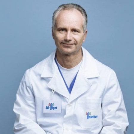 Alessandro Aiuti合作论文数Università Vita-Salute San Raffaele;Pediatric Immunohematology Unit, IRCCS Ospedale San Raffaele;Pediatric Clinical Research Unit, SR-Tiget11
 Stephane Blanche合作论文数Hôpital Necker-Enfants Malades11
 Frederic Rieux-Laucat合作论文数10Michaela Semeraro合作论文数10
 Adrian Thrasher合作论文数Great Ormond Street Institute of Child Health, University College London10
 Capucine Picard合作论文数Centre d’Etude des Déficits Immunitaires, Hôpital Universitaire Necker10Laure Caccavelli合作论文数10
 Anne Galy合作论文数Experimental Cellular Therapy Group SyStemix Incorporated10Adeline Denis合作论文数9Steicy Sobrino合作论文数9
 Charles Berry合作论文数Division of Biostatistics & Bioinformatics, Department of Family Medicine and Public Health, School of Medicine, University of California, San Diego9Frances Male合作论文数9
 Marianne Delville合作论文数Department of Nephrology and Kidney Transplantation, Necker Hospital, and.9
 Julien Zuber合作论文数9Touzot Fabien合作论文数AP HP, Unite Immunol & Hematol Pediat8
 Pierangela Gallina合作论文数8
 Chris Nobles合作论文数Molecular Virology and Microbiology, Baylor College of Medicine8Gabrion Aurélie合作论文数8
 Laura Rudilosso合作论文数8
 Luigi Naldini合作论文数Vita-Salute San Raffaele University;San Raffaele Telethon Institute for Gene Therapy8
 Elizabeth Macintyre合作论文数Université Paris Cité;Istitut Necker Enfants Malades8
 John Everett合作论文数Bushman Lab, Perelman School of Medicine, University of Pennsylvania8
 Eugenio Montini合作论文数8
 Andrea Calabria合作论文数National Research Council Institute for Biomedical Technologies Via Fratelli Cervi8
 Nicolas Cagnard合作论文数Institut Cochin, Université Paris Descartes8Bénédicte Neven合作论文数7Christian Reimann合作论文数7
 Francoise Pflumio合作论文数7Liliane Dal Cortivo合作论文数7Fabrizio Benedicenti合作论文数7
 Jacinta Bustamante合作论文数6S. Charbonnier合作论文数6
 Antonio Rausell合作论文数INSERM UMR 1163, Institut Imagine6
 Giulio Spinozzi合作论文数San Raffaele Telethon Institute for Gene Therapy (TIGET),, San Raffaele Scientific Institute;Department of Informatics, Systems and Communication (DISCo), University of Milano-Bicocca;San Raffaele Scientific Institute, University of Milano-Bicocca6Trinquand Amélie合作论文数6Fulvio Mavilio合作论文数6Alain Israel合作论文数6Felipe Suarez合作论文数Université de Paris6




