Background: Adolescent Idiopathic Scoliosis (AIS) is the most common spinal deformity disorder associated with bad posture and reduced quality of life. The Body Image Disturbance Questionnaire-Scoliosis (BIDQ-S) is a self -report instrument that assesses the concerns of scoliotic patients. This study aimed to translate and evaluate the reproducibility and internal consistency of the BIDQ-S in the Persian-speaking population worldwide suffering from AIS.Methods: The BIDQ-S was translated into Persian by two native-speaking Iranian translators and back-translated into English by two native-English translators. The resulting back-translated English BIDQ-S was then sent to the authors of the English BIDQ-S questionnaire for validation. After translation, it was provided for 41 AIS patients from those who referred to the outpatient clinics of Shafa Yahyaian Hospital from January 2020 to January 2021. Patients were asked to complete the Persian BIDQ-S and Persian Scoliosis Research Society-22 (SRS-22) inventories. Internal consistency and reproducibility were assessed using Cronbach's alpha and interclass correlation coefficients (ICC), respectively. The validity of the questionnaire was evaluated by comparing the scores obtained on the Persian BIDQ-S (P-BIDQ-S) inventory with those obtained on the SRS-22 subscales.Results: The consistency and reliability of the P-BIDQ-S inventory were confirmed by Cronbach's alpha of 0.856 and interclass correlation coefficients of 0.882. The P-BIDQ-S scores directly correlated with the level of education of patients (r=0.21, P=0.041). The correlation coefficient between the P-BIDQ-S inventory and the SRS-22 questionnaire was-0.56 (P=0.001). A significant correlation was also observed between the P-BIDQ-S items and all of the SRS-22 subscales (P<0.05).Conclusion: The P-BIDQ-S inventory maintains adequate reliability, internal consistency, and reproducibility for the evaluation of Persian-speaking AIS patients.
In Adolescent Idiopathic Scoliosis (AIS), correction surgery can correct the maximum movement and balance of the spine. Under certain conditions for two simultaneous curvatures, the procedure, in which correcting one of the curvatures can result in the automatic correction of another curvature, is called selective fusion, attracting spine surgeons' interest because of more movement in the spine. However, the majority of surgeons have not used this technique due to the lack of sufficient information. The current study aimed to totally investigate selective thoracolumbar/lumbar fusion and to provide accurate information on outcomes and complications of surgery for spinal surgeons. This technique can also help spinal surgeons have a better selection of patients' surgical procedures.
Background: Deep vein thrombosis (DVT) is a great postoperative challenge in all orthopaedic surgeries. To the authors’ knowledge, this study is the first to evaluate the efficacy of aspirin administration in the prevention of DVT in patients undergoing lumbar spinal surgery. Methods: In this double-blind parallel randomized clinical trial, a total of 126 candidates (age 40 yr and older) were admitted between June 2021 to December 2021. Patients were randomly assigned to the intervention chemoprophylaxis group (41 patients receiving 325 mg aspirin) and controls. The DVT occurrence was recorded by clinical features (Well’s criteria), Doppler lower limbs ultrasound, and D-dimer levels in all participants at baseline (24 hr before the time of surgery) and 2, 6, and 12 wk after surgery in postoperative visits. Results: The mean age of the participants was 63.72±6.87 yr. Baseline demographic values were similar in both groups (P>0.05). The mean follow-up duration was 6.11±2.33 mo. No cases of DVT or abnormal findings on Doppler ultrasound were observed in either group. The mean duration of hospitalization or intensive care unit (ICU) admission was similar between the two groups. Mean baseline D-dimer levels were significantly higher in the intervention group compared with the controls (P=0.047), while it was similar in both groups 3 mo after the operation (P=0.13). Conclusions: In the current study, no case of DVT was observed in either study group. These data do not support the use of aspirin as an anticoagulant for DVT prophylaxis following regular lumbar spinal surgeries. Level of Evidence: Level II.
Background: Extracorporeal shock wave therapy (ESWT) has been used in the treatment of coccydynia. However, it has not yet been validated due to the dispersion of studies, the low number of patients, and the use of different measuring instruments. This meta-analysis was performed with the aim of collecting homogeneous studies to achieve a reliable result. Methods: The authors searched electronic databases including Google Scholar, Scopus, ScienceDirect, ISI Web of Science, Embase, and PubMed to find studies in which the effect of ESWT on coccydynia using visual analog scale (VAS) scores was evaluated starting in January 1990 through August 2021. All search procedures were performed by two authors. Results: The mean pain changes of 81 patients from four studies in 1 mo, 2 to 4 mo, and 6 to 12 mo after using ESWT were analyzed. Overall mean change of VAS score in 1 mo, 2 to 4 mo, and 6 to 12 mo after applying ESWT was -42.41 (I2=86.96%, 95% confidence interval [CI] of −56.88, −27.94, P=0.001), −41.01 (I2=0.0%, 95% CI of −46.98, −35.04, P=0.001) and −50.13 (I2=82.41%, 95% CI of −67.33; −32.94, P=0.001), respectively. Conclusions: The authors showed that ESWT lessened pain in patients with coccydynia. This effect did not diminish during a year; instead, it further reduced pain in patients. However, more research is needed to verify the results. Level of Evidence: Level I.
Background Shoulder imbalance (SI) is among the most rated manifestations of adolescent idiopathic scoliosis (AIS) pointed to by patients and spine surgeons. It serves as a criterion to assess the outcome of scoliosis surgery and is also a cause of dissatisfaction for the patients postoperatively. Despite the availability of multiple studies on this issue, a comprehensive survey of the risk factors and preventive measures has yet to be elucidated. The present study aimed to highlight the most recent approach to the evaluation and management of SI, as well as medical counseling about the expectations and limitations of the surgery. Methods A systematic literature review using electric databases was conducted, including PubMed, Embase, the Cochrane Library, and Google Scholar, with a well-defined search strategy on SI definition, risk factors, and preventive and surgical recommendations. Results A total of 69 articles were identified; SI > 2 cm was the most used cut-off, and its risk factors included the main thoracic Cobb angle > 80˚, preoperative level shoulder, high left shoulder, and higher Risser grade. The most stated strategies to preclude SI were the sufficient correction of the proximal thoracic curve, and moderate correction of the main thoracic and lumbar curve (LC). Conclusion Shoulder imbalance should be prevented not only for appearance or satisfaction but also for possible complications such as distal adding-on, new LC progression, or trunk shift postoperatively in AIS patients.
A 30-year-old man presented to our clinic with progressive neck pain and dysphagia. He had been operated on for an atlantoaxial instability (os odontoideum) 7 years ago. Imaging studies revealed cervical spondyloptosis of C5-C6 and C7-T1; the neurologic examination was intact. It was decided to correct the deformity through a circumferential approach. Thus C5, C6, and C7 corpectomy, alongside anterior column reconstruction using titanium expandable cage, reinforced by posterior spinal instrumentation from occipital bone to T3 vertebra were resolved; his symptoms resolved completely following an uneventful surgery.
A 45-year-old man presented to our facility with predominantly Sciatica-like leg pain and lower extremity motor weakness, who did not get relief despite undergoing two consecutive lumbar surgeries for suspected lumbar disc herniation. Medical history, physical findings, and a magnetic resonance imaging scan revealed thoracic cord tumor as the underlying disease. Our patient had complete resolution of his back and leg pain following surgical resection of the thoracic Schwannoma. Thoracic cord compression often results in diffuse pain and myelopathic symptoms caused by the irritation of ascending spinothalamic tract, which causes a vague and burning pain that should be differentiated from nerve root lesions and can be the first presentation of a thoracic cord lesion. A B S T R A C T
BACKGROUND:Adolescent idiopathic scoliosis (AIS) is a common type of spinal deformity confronting surgeons. The Scoliosis Research Society Health-Related Quality of Life (SRS-30) Questionnaire has been translated into Persian to evaluate its internal consistency, reliability, validity, and cross-cultural adaptability in the Persian population.METHODS:The translation and cultural adaptation process was based on the American Academy of Orthopaedic Surgeons guidelines. A total of 102 AIS patients referring to our institution were enrolled in this study within March 2014-March 2016. The 36-item Short Form (SF-36) was used for adapting the Persian SRS-30 questionnaire. The convergent validity of the Persian SRS-30 was examined using the Pearson correlation coefficient. Furthermore, its internal consistency and validity were tested using Cronbach α with bootstrapped 95% confidence interval. Interclass Correlation Coefficient (ICC) was used to test and retest reliability.RESULTS:The total correlation coefficient between the Persian SRS-30 and SF-36 was obtained at 0.74 (0.67-0.80), which was statistically significant (P=0.001). The total Cronbach α for the Persian SRS-30 was estimated at 0.84, ranging from 0.51 in satisfaction with management domains to 0.88 in mental health domains. The Persian SRS-30r domains indicated satisfactory test-retest reliability with ICC rage of 0.79-0.87.CONCLUSION:The Persian SRS-30 translation was reliable and valid for the AIS Iranian patients. The internal consistency of this instrument was found to be good and excellent in all domains except satisfaction with management, which was moderate. The authors believe that the Persian version of SRS-30 is simple and easy to use and now it can be applied in clinical settings for future outcome studies in Iran.
Background: Various studies have shown the effects of morbid obesity on the adverse consequences of various surgeries, especially postoperative infections. However, some studies have shown that the complications of spinal surgery in obese and non-obese patients are not significantly different. Objectives: This study investigated and compared the duration of surgery, length of hospital stay, and complications after common spinal surgeries by orthopedic spine fellowship in obese and non-obese patients in a specialized spine center in Iran. Methods: All patients who underwent decompression with or without lumbar fusion were included in this retrospective study. These patients were classified into two groups: non-obese (BMI <30 kg/m2) and obese (BMI ≥30 kg/m2). The data related to type and levels of surgery, 30-day hospital complications, length of hospital stay, rate of postoperative wound infection, blood loss, and need for transfusion were all extracted and compared between the two groups. Results: A total of 148 patients (74%) were in the non-obese group and 52 patients (26%) in the obese group. The number of patients that need packed cells was significantly higher in the obese group (51.8% vs 32.6%) (P=0.01). Otherwise, there were not a significant difference between type of treatment (fusion or only decompression) (P=0.78), interbody fusion (P=0.26), osteotomy (P=0.56), duration of surgery (P=0.25), length of hospital stay (P=0.72), mean amount of blood loss (P=0.09), and postoperative complications (P=0.68) between the two groups. Conclusion: Our results suggest that duration of surgery, length of hospital stay, and postoperative complications are not associated with the BMI of the patients.
A 45-year-old man presented to our facility with predominantly Sciatica-like leg pain and lower extremity motor weakness, who did not get relief despite undergoing two consecutive lumbar surgeries for suspected lumbar disc herniation. Medical history, physical findings, and a magnetic resonance imaging scan revealed thoracic cord tumor as the underlying disease. Our patient had complete resolution of his back and leg pain following surgical resection of the thoracic Schwannoma. Thoracic cord compression often results in diffuse pain and myelopathic symptoms caused by the irritation of ascending spinothalamic tract, which causes a vague and burning pain that should be differentiated from nerve root lesions and can be the first presentation of a thoracic cord lesion.
Background: Severe angular kyphosis is one of the uncommon etiologies of compressive myelopathy and hence, many aspects of this myelopathy are unknown. Objectives: In this study, we report a series of 12 patients with compressive myelopathy in severe angular kyphosis, as well as the result of surgical treatment in these patients. Methods: In a retrospective study, we included 12 patients with the progressive or sudden onset of paraplegia caused by severe angular kyphosis. The neurological status of the patients was evaluated with the American Spinal Injury Association (ASIA) typing system before the operation and at four time points after the operation (1 day, 1 week, 6 months, 1 year). The main surgical interventions included anterior corpectomy, anterior spinal fusion, and posterior spinal fusion with or without instrumentation and with or without decompression. Results: The Mean±SD age of the patients was 41.9±16.4 years, ranging from 14 to 59 years. The etiology of myelopathy was congenital kyphosis in 10 patients (83.3%). The Mean±SD duration of paralysis was 6.4±6.6 months. The Mean±SD percentage of cord thinning at the apex was 61.7±17.5%. Bowel or bladder dysfunction was present in 6 patients (50%) before the surgery that was resolved in 5 patients after the surgery. One year after the surgery, the ASIA typing was improved in 9 patients (75%) and remained the same as preoperative status in 3 patients (25%). Conclusion: Surgical decompression corrects the neurological symptoms in the majority of cases with compressive myelopathy caused by severe angular kyphosis.
The relationship between curve correction and spinal length gain in adolescent idiopathic scoliosis was examined. A total of 102 patients who underwent posterior spinal correction and fusion alone or in combination with anterior spinal correction and fusion (ASF) were studied. The Cobb angle correction, increase in the main thoracic length, T1–L5 spinal length gain, and T1–L5 spinal length gain/Cobb angle correction were reported. The length gain/Cobb angle correction value was not significantly associated with sex, fusion approach, and the number of fused levels. Surgical T1–L5 spinal length gain (mm) equaled (70.20)−(3.51)×(degrees of Cobb angle correction)+(0.08)×(degrees of Cobb angle correction)2.
Congenital kyphosis is a less common spinal malformation, which can progress and cause neurological deficits. The treatment of severe cases is complicated. There are several techniques of surgical intervention for the correction of kyphosis, but the selection of surgical methods is entirely dependent on the patient’s condition. This article reports a 14-year-old girl with severe congenital kyphosis, who underwent hemiepiphysiodesis at age 2. Last year, her neurological deterioration began and led to paraplegia over a couple of months. To treat her, we performed 2-stage multi-level vertebral column resection (Schwab type 6). First, she underwent anterior T12-L1-L2 complete corpectomy and cord decompression. Then, the resection of posterior elements from T12 to L2 and posterior fusion with instrumentation from T8 to L5 were performed. However, concerning this kind of severe deformity, we suggest using 3D planning, which facilitates the operation.
Osteoid osteoma (OO) is a small tumor of bone that affects the spine in 10% of the cases. The tumor has a tendency to neural arc, and the lumbar spine is the most common site of presentation. Lesions of the odontoid process are very rare. We presented the case of a 20-year-old man who had cervical pain for 8 months. The pain responded to medical therapy. After investigation, there was a lytic lesion at the odontoid process with the characteristic features of OO in computed tomography scan and magnetic resonance imaging. Firstly, medical treatment was initiated with the administration of nonsteroidal anti-inflammatory drugs; however, due to adverse effects and worsening of his pain, the patient underwent surgical treatment with intralesional curettage from the transoral approach. In addition, the posterior fusion of the first and second cervical vertebrae was performed. The pain disappeared, and the patient was symptom-free after one year of follow-up.
Osteoid Osteoma of the Lumbar Spine in a 3.5-Year-Old BoyIntroduction: Osteoid osteoma is the third most common benign tumor of bone.The incidence of osteoid osteoma in the spine, particularly in the patients younger than five years, is low.Case Presentation: Here, we report a case of osteoid osteoma located in the Lumbar Spine (L5) occurred in a 3.5-year-old boy.The patient was referred with a chief complaint of back pain for six months that was aggravated at nights.Furthermore, no apparent spinal deformity existed.Based on the clinical and imaging findings, the diagnosis of osteoid osteoma was made, and the surgical excision of the lesion was performed.The histologic examination of the extracted tissue confirmed the diagnosis of osteoid osteoma. Conclusions:The back pain alleviated the day after the operation, and he was event-free over 6-month follow-up visits.This report highlights the importance of osteoid osteoma in the differential diagnosis of spinal pain presented at any age.
Introduction: Anterior Lumbar Meningoceles (MCs) are rare in the patient with Neurofibromatosis type 1 (NF1). Although spinal fusion with optimal resection of the meningocele might be indicated in these special cases, no report could completely describe a huge meningocele after posterior spinal instrumented fusion. Case Presentation: Here we present a 23 years old woman with a history of NF-1 and previous posterior spinal fusion, who was referred to the neurosurgery department because of abdominal pain and retroperitoneal mass secondary to large anterior lumbar MC. The MC was filling the retroperitoneal cavity and protruded to the anterior wall of abdomen. As an elective procedure, reduction and closure of cyst was achieved through posterior approach. Postoperatively, the patient reported satisfactory relief of abdominal pain. Conclusion: Lumbar anterior MC is rare in the patients with NF1. When surgical intervention is indicated, reduction of cele should be considered.
 Hamid Behtash合作论文数33
 Ghandhari Hasan合作论文数Shafa-Yahyaeian Hospital, Iran University of Medical Sciences31Mobini Bahram合作论文数22
 Farshad Nikouei合作论文数16Hassan Ghandhari合作论文数15Tari Hossein Vahid合作论文数14b mobini合作论文数9
 Maryam Ameri合作论文数8
 Saeed Sabbaghan合作论文数8Ganjavian Mohammad Saleh合作论文数Shafa Yahyaiian Hospital, Iran University of Medical Sciences8
 Mohammadreza Shakeri合作论文数7Hamid Hesarikia合作论文数6Rastegar Khodakaram合作论文数Neurosurgery ward of Rasool-e-Akram Hospital Complex, Iran University of Medical Science6
 Naveed Nabizadeh合作论文数Shafa-Yahyaeian Hospital, Iran University of Medical Sciences;Shohada Hospital, Shahid Beheshti University of Medical Sciences6
 Safari Mir Bahram合作论文数6Mohammad Khakinahad合作论文数5
 Farzad Omidi-Kashani合作论文数Imam Reza Hospital, Mashhad University of Medical Sciences5
 Azadeh Memarian合作论文数Iran University of Medical Sciences5
 Kamran Aghakhani合作论文数Department of Forensic Medicine, Iran University of Medical Sciences5Hamid Behzadnia合作论文数4
 Seyed Mani Mahdavi合作论文数Shafa Orthopaed Hosp, Iran Univ Med Sci4
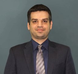 Fouladi Daniel F合作论文数Drug Applied Research Center, Tabriz University of Medical Sciences4
 Shahrokh Mehrpisheh合作论文数Ghazvin Univercity of Medical Sciences4
 Mohsen Motalebi合作论文数4hossein vahidtari合作论文数3
 Marzieh Nojomi合作论文数Dept Community Med, Iran Univ Med Sci3
 Mohammadreza Chehrassan合作论文数3
 Farzam Mokarami合作论文数3
 Payam Kabiri合作论文数Faculty of Health, Tehran University of Medical Sciences3Abouzar Azizi合作论文数2
 Arvin Eslami合作论文数2Ali Habibollahzadeh合作论文数2
 Seyed-Mohammad Fereshtehnejad合作论文数University of Ottawa2
 Ali Tabrizi合作论文数2Bouzari Behshad合作论文数2
 Safdari Farshad合作论文数Akhtar Orthopedic Hospital, Shahid Beheshti University of Medical Sciences2
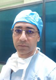 Heshmati Afshin Ahmadzadeh合作论文数Department of Orhthopedic Surgry Dr Bahonar University Hospital, Kerman Medical University2
 Behrooz A Akbarnia合作论文数Department of Orthopaedic Surgery, University of California, San Diego;Department of Orthopaedic Surgery, Rady Children's Hospital2Kamran Ali合作论文数1Mohammad Sadegh Saba合作论文数1Behrooz Givechian合作论文数1Shoeib Majdi合作论文数1Milad Haji Agha Bozorgi合作论文数1Navid Nabizadeh合作论文数1
 Hamed Tayyebi合作论文数1Mohamadsaleh Ganjavian合作论文数1
 Ali Andalib合作论文数1Mansour Karimi合作论文数1Mostafa Salehpour合作论文数1




