Muscle activity during sleep is a new area of interest in sleep research. No precise brain structures are known to be involved in sleep movement. The etiology of periodic movements during sleep is unknown. The present study was dedicated to evaluate involvement of basal ganglia in periodic movements of the legs during sleep (PMS) in Parkinson's diseased patients. Sleep was monitored in 3 patients suffering from Parkinson's disease and PMS (PMS/PD) and in 3 patients suffering from restless legs syndrome and PMS (PMS/non PD). Following treatment, the six patients were monitored again during sleep. It was found that only the PMS/PD group of patients had improved significantly with appropriate treatment. Improved motor function in PD patients is associated with decreased PMS, regardless of wether the patients are treated with dopaminergic or anticholinergic agents. This is consistent with our previous data. It may be suggested that the striopallidal system is involved in periodic sleep movements of Parkinson's diseased patients.
Growth hormone (hGH) secretion was measured during sleep in 10 healthy male subjects isolated from all time cues. HGH concentrations following sleep onset were compared between scheduled sleep episodes (entrainment) and self-selected sleep episodes (free-running). Peak sleep-related hGH values were decreased significantly during free-running. The duration of the first slow wave sleep (SWS) episode and the latency to the first REM sleep episode also decreased significantly during free-running. The latencies from sleep onset to SWS and to peak hGH secretion did not differ between entrainment and free-running. These results suggest that sleep-related hGH secretion begins 'on time' during free-running, but is terminated earlier. Thus, while sleep onset facilitates hGH release, the timing of other stages of sleep such as REM may alter the magnitude of sleep-related hGH secretion.
Une perfusion continue de melatonine diminue de facon significative les taux permatiques de LH chez des femmes menopausees. Cette experience plaide en faveur d'un effet antigonadotrope de la melatonine
Prepubertal children with major depressive disorder have shown increased growth hormone (GH) secretion during sleep while in a depressive episode. When restudied in a fully recovered state (for at least three months) and drug free (for at least one month), their increased GH secretory pattern during sleep had not changed. Illness-recovery correlations using area under the curve for GH secretion during sleep were highly significant, whereas paired comparisons showed no significant differences. In addition, children who had recovered from major depressive episodes secreted significantly more GH during sleep than did nondepressed neurotic and normal children. No significant differences in delta-sleep were found in the depressed group between ill and recovered states nor among those who had recovered from major depressive episodes or controls. It is concluded that increased GH secretion during sleep is independent of depressive episodes, remains unaltered after full recovery, and may be a true marker of trait for major depressive disorder in prepuberty.
It has been postulated that the sudden infant death syndrome (s.i.d.s.) may sometimes be due to abnormal maturation or injury to the brain stem centres that regulate respiration. This functional abnormality of the brain stem respiratory centres may result in the interruption of the automatic inspiratory/expiratory cycle by recurrent periods of apnoea. There is a subgroup of infants known as ‘near-miss’ for s.i.d.s., who survived a prolonged apnoeic episode during sleep which may have resulted in death. In a number of recent studies, the near-miss infant has been clearly identified as an infant at high risk for s.i.d.s. Clinical studies conducted by using polygraphic and behavioural monitoring of near-miss infants have revealed numerous apnoeic episodes during sleep. Consequently, the clinical necessity of monitoring these babies in a paediatric care unit has become well established both for diagnostic purposes and for subsequent outpatient care. In view of these findings, it has become increasingly more important to develop advanced sophisticated computer methods for the on-line detection and processing of apnoeas during in-hospital monitoring of infants. This paper describes a digital computer method of on-line apnoea processing for application during the in-hospital monitoring of infants. The method is based on the application of Walsh transformations to the expired CO2 signal measured in infants using a Beckman CO2 analyser.
In order to test the hypothesis that cortisol secretion is inhibited during sleep, six healthy young men (ages 18-24) were studied in a 4-day protocol. A baseline nocturnal sleep period was followed by one night's total sleep deprivation, then sleep at 180 degrees phase shift, and then return to a normal nocturnal sleep episode (SP-3). Plasma cortisol concentrations were measured every 20 min (obtained by an indwelling venous catheter), rectal temperature was measured every minute, and sleep was polygraphically defined. During the first 4 h of sustained sleep, cortisol secretion was decreased even when sleep occurred during a time when the subject was usually awake; conversely, it was elevated if awake at the usual daily time of sleeping. This was not the case for the last 4 h of sleep. Body temperature was also decreased but during each entire 7- to 8-h sleep period. Meals produced only a small brief rise of cortisol and produced no change in body temperature. Stage 4 sleep was increased during the 180 degrees inverted sleep episode and decreased during SP-3, REM sleep however was increased during SP-3. A reciprocal relationship was found between REM and stages 3 and 4 for the second, third, and fourth, and sixth h of sleep for SP-3. These results demonstrate the inhibitory effect of the behavioral complex of sleeping on cortisol secretion superimposed on its endogenous circadian and ultradian rhythm. These neurophysiological events may be used to entrain and time the period and phase of biological rhythms in relation to shift work, sleep deprivation, and transmeridian jet travel.
Obstructive sleep apnea syndrome was studied in 32 children, aged 2 to 14 years, in the sleep-wake disorders center at Montefiore Hospital and Medical Center during the years 1977 to 1980. All children underwent all-night polysomnograms; 17 of these children had surgery to relieve airway obstruction and seven had a repeat polysomnographic study 4 to 6 weeks following the surgery. There was a significant improvement in the number of obstructive apneas and in other apnea indices following surgery. There was no significant effect on the durations and the proportions of the various sleep stages, on sleep efficiency, or on the number of awakenings.
Under the organizational aegis of Project Sleep and the Association of Sleep Disorders Centers (ASDC), nearly 5,000 patient records from 11 sleep-wake disorders clinics were analyzed in a cooperative study. These cases represented the diagnostic experience of each of these centers over a two-year period. Each patient underwent polysomnographic study, and his or her condition was diagnosed according to the ASDC classification system, a new, standardized nosology of sleep disorders medicine. The most common major diagnostic category was "disorders of excessive sleepiness (hypersomnia)," 42%; this was followed by "disorders of initiating and maintaining sleep (insomnia)," 26%; "penile tumescence evaluations for impotency," 17%; "parasomnias," 3%; and "disorders of the sleep-wake schedule," 2%. If the impotency evaluations performed in the sleep clinics are removed from the total, leaving only the population that was studied because of sleep complaints, the proportions of the diagnostic categories are hypersomnia, 51%; insomnia, 31%; parasomnias, 15%; and sleep-wake schedule disturbances, 3%. The most prevalent diagnoses in the hypersomnia category were sleep apnea (43%) and narcolepsy (25%). Psychiatric disorders (35%) comprised the most frequent group of insomnia diagnoses, though a variety of other disorders were common. The applications of these results for the practicing physician are discussed. (JAMA1982;247:997-1003)
—Studies were carried out on a group of six young (ages 23–30) and six older (ages 53–70) normal men who lived under conditions of temporal, but not social isolation, from three to eight weeks. During entrained and non-entrained (free-running) conditions, comparative measurements were made of sleep-wake cycles, sleep stages and rectal temperature rhythms for these two age groups. Results demonstrated a reduction in the period and amplitude of the body temperature rhythms during free-running in the older group. Sleep efficiency, total sleep time, REM sleep latency, REM episode length, percent REM in the last 2 hours of sleep, the length and frequency of arousals during sleep, and the terminal wake latency were all age related and dependent on entrainment. The period of the sleep-wake cycle, terminal awakenings from REM and percent REM in the first 3 hours of sleep were not age related but were dependent on entrainment. Sleep stages as percents of total sleep time were found to be age related but independent of entrainment, while sleep latency, mid-REM to mid-REM cycle length and the ratio of sleep to total time were neither age related nor dependent on entrainment. In addition, individual chronobiological differences were prominent in the older group. Changes of period and of the phase relationship of sleep-wake and temperature rhythms occurred in several subjects during the non-entrained condition.
We studied the effect of flurazepam, a known suppressor of stages 3 and 4 of sleep, on nocturnal sleep patterns and on growth hormone release immediately following sleep onset in normal young adults. Polysomnography and sampling of growth hormone (every 20 min for 26 h) were performed before and after 2 weeks of nightly flurazepam (30 mg, 8 subjects) or placebo (8 subjects) administration. There were no significant changes in growth hormone release in spite of a significant decrease in stage 4 sleep (69%) and total waking time (46%) and an increase in total sleep time (23%) in the drug group. REM sleep was not changed. These results indicate that the normal GH release following sleep onset continues to occur despite stage 4 suppression by pharmacological means.
The acute and chronic effects of phenobarbital and phenobarbital withdrawal on sleep patterns and on plasma growth hormone (GH) and cortisol fluctuations occurring during sleep were studied. Before bed, five healthy men, aged 21 to 25, were given a placebo on three baseline nights, phenobarbital (100 mg p.o.) for nine nights, and a placebo on a final withdrawal night. Beginning on the third of three consecutive nights in the laboratory, all-night polygraphic sleep recordings and blood samples (obtained every 20 min through indwelling venous cannulae) were collected for the placebo, acute phenobarbital, chronic phenobarbital, and phenobarbital withdrawal conditions. Blood phenobarbital levels ranged between 5 to 9 micrograms/100 mL across all hours of the chronic drug night. At this low sedative dose, latency to sleep onset and stage 4 sleep were significantly reduced in the chronic drug condition, but REM sleep was not significantly reduced. No significant sleep change was observed on the withdrawal night. Both peak GH level and total integrated GH across the night were unaffected by the acute, chronic, and withdrawal conditions. The pattern of GH release appeared to be altered on the phenobarbital and phenobarbital withdrawal nights as compared with placebo. Nighttime plasma cortisol levels were not significantly altered by any experimental condition.
This chapter discusses the regularity in the patterns of unsynchronized circadian rhythms in man. When activity–rest and body temperature cycles become unsynchronized in man, large variations in sleep duration and timing occur. Both these cycles have become unsynchronized (NS) in every subject free-running longer than 2 months. The average period of activity–rest typically lengthened during the free-run whereas sleep duration varied predictably with body temperature at sleep onset, even in subjects with activity-rest cycles of 24 hrs. The rate of rapid eye movement (REM) sleep accumulation varied with the phase of body temperature, as did plasma cortisol and subjective assessments of alterness.
— Man is the only eukaryotic organism in which it has been reported that the circadian system cannot be entrained to a 24-h period by a simple light-dark (LD) cycle. In this paper, we reexamine the evidence for that claim and demonstrate that there were some fundamental flaws in the experimental design of the previous studies on which this conclusion was based. We report new studies in which we tested the efficacy of LD cycles in entraining the circadian rhythms of human subjects living in isolation from environmental time cues. We found that the cyclic alternation of light and dark, when applied to human subjects in a comparable way to experiments in other species, was an effective entraining agent. Our results and a critical review of the literature indicate that a LD cycle alone can be an effective environmental synchronizer of the human circadian timing system. Other factors, such as the knowledge of time of day, social contacts, the feeding schedule, and the imposed rest-activity schedule may contribute to stable entrainment, although their relative strengths as synchronizers have yet to be determined.
The developmental pattern of the in vitro rat liver melatonin degrading activity was studied. Livers from rats 3, 12, 16, 21, 30 and 90 days old were incubated with [3H] melatonin. A high pressure liquid chromatography (HPLC) technique was used to determine the appearance of melatonin metabolic product--6-hydroxymelatonin--in the liver extract. Catabolic activity belong very low at age 3 days, increased rapidly with age and reached maximum activity between the ages 21 to 30 days.
We describe a new syndrome called "delayed sleep phase insomnia." Thirty of 450 patients seen for a primary insomniac complaint had the following characteristics: (1) chronic inability to fall asleep at a desired clock time; (2) when not on a strict schedule, the patients have a normal sleep pattern and after a sleep of normal length awaken spontaneously and feel refreshed; and (3) a long history of unsuccessful attempts to treat the problem. These patients were younger than the general insomniac population and as a group did not have a specific psychiatric disorder. Six patients' histories are described in detail, including the successful nonpharmacological chronotherapy regimen (resetting the patients' biological clock by progressive phase delay). Delayed sleep phase insomnia is proposed to be a disorder of the circadian sleep-wake rhythm in which the "advance" portion of the phase response curve is small.
The meal patterns of men who were initiating meals for extended periods in the absence of time cues were examined. A significant, positive correlation was found between the sizes (in kcals) of mixed and varied meals and the lengths of postprandial intervals. This quantitative relationship between meal size and meal timing is similar to patterns reported for freely feeding rats. If postprandial correlations reflect a short term mechanism for energy regulation then when humans schedule their meals due to social or time considerations they may negate this contribution to the regulation of their energy intake.
The relationship between plasma levels of the tricyclic antidepressant desmethylimipramine (DMI) and plasma levels of melatonin-like immunoreactivity was studied in 32 endogenously depressed patients. An inverse correlation between plasma levels of DMI and plasma levels of melatonin-like immunoreactivity was found in the group of clinical responders to the chronic administration of the drug. The nonresponders had higher levels of melatonin-like immunoreactivity at comparable levels of DMI. This finding is consistent with the hypothesis that chronic high plasma levels of DMI may down-regulate the beta-adrenergic receptors in man. However, some other homeostatic mechanisms may be involved in the clinical response.
The acute and chronic effects of alcohol and alcohol withdrawal on sleep patterns and plasma GH and cortisol fluctuations occurring during sleep were studied. Five healthy men, aged 21-26 yr, consumed a placebo drink for 3 baseline nights, alcohol (0.8 g/kg) for 9 nights, and a placebo drink on final withdrawal night. All-night polygraphic sleep recordings and blood samples (every 20 min with a venous catheter) were collected for 1 placebo, 1 acute alcohol, 1 chronic alcohol (night 9), and 1 alcohol withdrawal night. Acute and chronic alcohol consumption reduced rapid eye movement sleep nonsignificantly during the first half of the night, whereas slow wave sleep (stages 3 and 4) was increased significantly after acute alcohol, returning to baseline values on the chronic alcohol night. On the withdrawal night, rapid eye movement sleep and slow wave sleep were not significantly different from placebo sleep. Alcohol significantly suppressed plasma GH values (70-75%) on acute and chronic nights. All measures of GH, including total integral for bedtime hours, mean hourly rate, and peak level, were similarly affected by alcohol. GH returned to placebo values on the withdrawal night. Measures of nighttime plasma cortisol were not significantly altered by alcohol or alcohol withdrawal at this dose level.
 L Hellman合作论文数30S Kapen合作论文数19Robert M Boyar合作论文数16
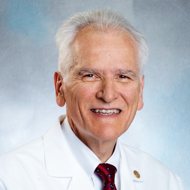 Charles A. Czeisler合作论文数Division of Sleep Medicine, Harvard Medical School;Division of Sleep and Circadian Disorders, Departments of Medicine and Neurology, Brigham and Women's Hospital14J. F. Sassin合作论文数12Cp Pollak合作论文数11J C Zimmerman合作论文数9M Perlow合作论文数9Jordan W Finkelstein合作论文数9Dk Fukushima合作论文数7J Sassin合作论文数6Martin Moore Ede合作论文数Circadian Technologies, Inc.6Peter K Mcgregor合作论文数6H ROFFWARG合作论文数6U Weinberg合作论文数5D Fukushima合作论文数4Es Tauber合作论文数4Jh Jacoby合作论文数4T. F. GALLAGHER合作论文数4S Laxminarayan合作论文数4
 Daniel Kripke合作论文数University of California, San Diego4C. Nogeire合作论文数4Jacob Kream合作论文数4Julien Mendlewicz合作论文数4R. S. Rosenfeld合作论文数4Paul Linkowski合作论文数4RM COLEMAN合作论文数3Ac Cornwell合作论文数3RE KRAVATH合作论文数3O Mills合作论文数3Spielman A J合作论文数3Howard P Roffwarg合作论文数3M. Greenstein合作论文数3Y Frank合作论文数3J Jacoby合作论文数2J. Ronda合作论文数2Lj Graziani合作论文数2Ej Sachar合作论文数2Mi Surks合作论文数2E Smith合作论文数2TA Roehrs合作论文数2ML MOLINE合作论文数2
 Eve Van Cauter合作论文数2K. YOSHIDA合作论文数2
 Leon H Leon Bradlow合作论文数David and Alice Jurist Institute for Research, Hackensack University Medical Center2Erlich S合作论文数2Weinberg U合作论文数2Andrew G Frantz合作论文数2A. Marmarou合作论文数2




