Soil fungi and oomycetes play a critical role in soil ecosystems, contributing to nutrient cycling, carbon sequestration, soil structure formation and plant health as symbionts and pathogens. Existing approaches to identify and quantify these including molecular methods and labour-intense manual observation provide limited spatial and temporal information.,In this study, we present a novel framework that integrates microfluidic chips with convolutional neural networks (CNNs) for fungal classification and quantification. Eight fungi and oomycete species were selected to represent both a broad range of morphologies but also a set of closely taxonomically related taxa with similar morphology for model validation. The YOLO11 nano object detection model was used for classification, and YOLO11 nano instance segmentation model was used for hyphae quantification.,The classification model achieved a mean 0.947 F1-score, successfully differentiating closely related taxa. The segmentation model reached a 0.852 Dice score, accurately detecting and quantifying hyphae. Both models performed well on multispecies images despite mainly being trained on single-species images supplemented with a subset of multispecies images. Performance varied amongst species and was mainly influenced by class availability and hyphal morphology, where thicker and less dense networks generally yielding higher performance.,This work presents a system for direct pore-scale visualization of mycelia and quantitative analysis of species composition, biomass, and mycelial morphology. The framework complements both molecular and traditional culturing methods and provides a foundation for image-based monitoring of fungi and oomycetes in soil environments.
Understanding metabolic processes of soil fungi is essential for elucidating their ecological roles in biogeochemical cycles and responses to emergent environmental stressors. Here, we demonstrate the potential of using stable isotope probing Raman (SIP-Raman) microspectroscopy in microfluidics technology-based soil chips to trace glucose metabolism rates and stress responses in laboratory grown filamentous soil fungus Psilocybe cf. subviscida. The time evolution of Raman spectral band intensities resulting from deuterated glucose uptake in the fungal hyphae allowed us to assess glucose metabolism rates. Under excess copper (Cu) stress, we observed suppression of both glucose metabolic activity and growth. In addition, reduced spectral signatures of intracellular cytochrome c further implied impaired mitochondrial function and potential onset of cell death. However, laser-induced radiation damage hampered repeated Raman measurements, including multispectral mapping, on individual hyphae, especially when exposed to the Cu stress. To overcome this, we employed stimulated Raman scattering (SRS) microscopy, which offers much higher sensitivity and mapping speeds, and therefore much lower radiation doses. This enabled localization of the uptaken glucose at the inner edges of the P. cf. subviscida hyphae and Cu-induced formation of putative vacuolar structures. While integration of this approach with soil chips requires future modifications to the chip design for increased optical transparency and ensured sterility, overall, our results demonstrate the potential of Raman-based microspectroscopy for spatially resolved, in situ analysis of fungal primary metabolism and stress physiology.
Soil aggregation is a dynamic process influenced by physical, chemical and biological factors; however, their individual and combined effect on the formation and turnover of aggregates is not well understood. The aim of this study was to examine incorporation of fresh litter inputs of different physicochemical properties including their carbon-to-nitrogen (C/N) ratio - maize (C/N =12) and straw (C/N =103) - into aggregates, de novo formed from mineral soil with or without the presence of microbiota. Using rare-earth element oxides, we labelled structures formed during a four-week incubation with a single litter type and traced their incorporation into newly formed aggregates after mixing them together and incubating for a subsequent seven-week period. To visualize them, we used synchrotron-based X-ray fluorescence microspectroscopy, which allowed us to demonstrate that presence of the plant-derived particulate organic matter was the key factor for the aggregate formation. Within the timescale of the experiment, neither microbial abundance nor the community composition had any significant effect. However, the relative increase in straw-associated soil in aggregates larger than 250 mu m provided support for our hypothesis regarding impact of carbon-rich organic matter on macroaggregation, likely via promotion of fungal growth and hyphal enmeshing. Phospholipid fatty acid analysis further confirmed relatively higher abundance of fungi in macroaggregates in straw-containing soil. All in all, our study provides insights into the initial stages of aggregate formation following litter additions and development of associated microbial community. The spatial analysis enabled by the X-ray fluorescence microspectroscopy enabled visualization of internal aggregate structures, shedding light on the processes involved, which is not possible with bulk analysis alone.
Nanoplastics are emerging environmental contaminants that increasingly threaten soil ecosystems, yet their effects on microbial behavior remain poorly understood. This is mainly due to the lack of experimental tools capable of directly observing microbial dynamics in situ under realistic soil-like conditions. Here, we present a proof-of-concept system that enables real-time, high-throughput monitoring of soil protists within microfluidic soil chips under nanoplastic exposure. Using microscopy video analysis integrated with a deep learning-based detection model and a transformer-based trajectory reconstruction algorithm, we quantitatively measured the movement of three morpho-/locomotion type groups-flagellates, ciliates, and amoebae-across a gradient of nanoplastic concentrations (0, 2, and 10 mg/L). Our results showed reduced movement velocities for flagellates and ciliates under high nanoplastic conditions with a 24%-30% reduction in speed, while no effect on amoebae was detected. The trajectory data also provides novel insights into how protists navigate soil-like structures. Beyond these specific findings, our approach establishes a transformative framework for observing microbial life directly within its microenvironment, comparable to how animal behavior is monitored in ecological studies. By bridging real-time imaging and artificial intelligence, this method offers a new angle to study protist-environment interactions without the need for culture extraction. It opens the door to rethinking how microbial ecology, soil contamination, and biotic responses to environmental stressors are investigated, advancing opportunities from static, population-level measurements to dynamic, behavioral-level understanding within realistic habitats.
Soil pore space, considered the most complex biomaterial that exists, generates a complex environment, that gives rise to a wide variety of properties, such as microbial diversity and carbon storage. Soils contain, at the same time, the largest carbon reservoir on earth and an immense amount of nutrient-limited microbial biomass. The reason why this carbon is not consumed by soil microbes is attributed to the complex nature of soil, which forms a labyrinth where carbon and microbes cannot be in direct contact. In the present study, by using microfluidics, we tested the effect of labyrinth-like structures of decreasing accessibility on the decomposing activity of soil microbial communities from a soil inoculum. The two parameters used to study the effect of microhabitat accessibility were either the turning angle in an array of channel-like pore structures or the fractal order in an array of maze-like pore structures. We found that in both cases, channels and mazes, decreasing accessibility produced a higher peptide substrate degradation. When we analyzed the degradation within the structures, we found that most of the activity is concentrated in the regions of intermediate accessibility. We think that the increased degradation activity in low accessibility mazes might be due to the reduced interactions within the microbial communities which leads to a reduction in competition. Lowered competition allows different communities with a wide range of metabolic strategies to cohabit in the structures, which resulted in a bulk increase of the peptide substrate degradation.IMPORTANCEThe role microbes have in the environment is highly influenced by the characteristics of their habitat. Here, we show that a complex habitat enhances the enzymatic activity of a soil microbial inoculum. This might occur due to a reduced competition in complex habitats, which allows a more diverse community to coexist and explore a wider variety of metabolic strategies. The different rates of enzymatic activity in different levels of complexity suggest emergent properties of microbial communities in complex microhabitats which could have important implication for microbial processes, such as soil carbon storage and nutrient cycling.
Nanoplastics are emerging contaminants that have a significant impact on soil microorganisms. To fully understand the effect of plastic contamination on soil ecosystems, it is necessary to advance techniques that can monitor nanoplastic-microbe interactions under realistic conditions. In this work, we investigated the effects of nanoplastic contamination on a community of soil protists monitored through microfluidic soil chips, and analysed changes in their behavior via microscopy videos and a deep learning approach. The presented method employs a deep learning-based detection model combined with a transformer-based matching model for video frame interpolation, enabling accurate reconstruction of protist movement trajectories and velocities within soil chips. The results revealed reduced movement velocities for the groups of flagellates and ciliates under high nanoplastic conditions, a 24-30% reduction at a marginal significance level, while amoebae were unaffected. Our trajectory data provides novel insights into how protists navigate soil-like structures. By facilitating comprehensive assessments of protist–environment interactions, it opens new avenues for understanding their ecological roles and the broader implications of hazardous contaminants in both soil and aquatic ecosystems at microbial community level without need for culture extraction. This proof-of-concept system enables continuous, high-throughput monitoring of soil protist behavior and can be readily adapted to investigate protist responses to diverse chemical and physical soil hazards.
The keystone species concept holds that certain members of an ecological community, despite their low abundance, exert disproportionately large effects on species diversity and composition. In microbial ecology, experimental validation of this concept has been limited because targeted removal of individual species remains technically challenging. Here, we developed a procedure to test the keystone species concept within a soil microbial food web by selectively suppressing a protist predator at the microscale via ultraviolet-induced phototoxicity in a microfluidic soil chip system. We targeted a hypotrich ciliate (subclass Hypotrichia), and combined microscopy with high-throughput amplicon sequencing of microbial taxonomic markers to assess, across multiple trophic levels, how its suppression affected microbial community abundance, diversity, and composition. Over the 20-day incubation, the chip system supported complex communities of bacteria, fungi, and protists. Following Hypotrichia suppression, two distinct ecological responses were observed: first, an increase in the relative abundance of flagellates, consistent with mesopredator release, accompanied by a significant rise in overall protist diversity; second, a convergence in protist community composition, indicative of biotic homogenization. Bacterial community abundance, richness, and composition remained unchanged, likely due to compensatory predation from a relative increase in bacterivorous flagellates. In contrast, fungal diversity decreased, presumably because the altered protist community favored facultative fungal consumers. Collectively, these findings provide direct experimental evidence that low-abundance microbial predators can function as keystone species, modulating predator community composition and diversity, and exerting cascading effects on lower trophic levels within microbial brown food webs.
Arctic wildfires disrupt biogeochemical cycles of carbon (C), nitrogen (N), and phosphorus (P), which challenges exposed tundra ecosystems. In this study, we investigated the legacy of variable fire intensity on soil microbial nutrient cycling in field experiments on Disko Island, West Greenland, three years post-fire. Despite finding no significant differences in gross N mineralization and consumption rates, high-intensity fire-treated soil microbes exhibited reduced degradation of a protein substrate in soil chips, suggesting altered microbial activity in organic N cycling pathways. These results highlight the return of some biogeochemical processes over time, and also reveal potential vulnerabilities in microbial communities and their functionality in legacy after high-intensity fire. As fire frequency in the Arctic is expected to increase due to climate change, long-term consequences for ecosystems may include shifts in microbial composition and nutrient cycling, and slow ecosystem recovery. The feedback could alter greenhouse gas emissions, accelerate permafrost thaw and cause ecosystem transformation. Understanding these processes is critical for predicting the wider ecological effects of more frequent and intense fires. Future research should focus on multi-temporal sampling and microbial dynamics to better capture fire-induced alterations and their cascading effects on Arctic ecosystems and global climate regulation.
Cracks in concrete compromise structural integrity by exposing steel reinforcement to corrosion agents, shortening its service life. Fungal-induced calcium carbonate (CaCO3) precipitation via urea hydrolysis offers a fast and robust self-healing mechanism to seal the cracks, extending the lifespan while reducing the carbon (C) footprint of concrete infrastructure. However, current studies rely on bulk-scale analytical methods, which lack the spatial resolution and chemical sensitivity to distinguish and map CaCO3 polymorphs at the nanoscale. This study combined scanning electron microscopy (SEM) and synchrotron-based scanning transmission X-ray microscopy (STXM) with near-edge X-ray absorption fine structure (NEXAFS) spectroscopy to characterize fungal CaCO3 polymorphs at the nanoscale. CaCO3 biominerals precipitated by three urease-positive fungi were sectioned into 75-200 nm thin layers. STXM data were collected from at least two spots per section, focusing on Ca (L-edge) and C (K-edge) chemical speciation and elemental quantitative mapping. Calcite, the thermodynamically most stable polymorph, was identified as the predominant mineral phase precipitated by all fungi species, while aragonite and non-CO3-Ca species (CaCl2 or Ca adsorbed onto extracellular polymeric substances (EPS)) occurred as minor components. In fungal species 2, we observed nanoscale heterogeneity in Ca phases across five analyzed spots, three dominated by calcite with minor contributions of other Ca species, while the others showed mixed CaCO3/non-CO3 phases, as confirmed by NEXAFS spectra. These findings suggest that biomineralization in the fungal micro and nanoenvironment is influenced by localized physicochemical and metabolic conditions that shape mineral phases. C NEXAFS spectra further supported the Ca data, showing C-specific spectral features in the calcite-rich regions across all samples. This underscores STXM's capability to resolve complexities and mechanisms of fungal CaCO3 formation (e.g., mineral phase composition, fungal organic-mineral interactions, and spatial heterogeneity). Overall, this study provides critical nanoscale insights into fungal CaCO3 precipitation, thus providing valuable guidance in optimizing fungal systems in self-healing concrete applications.
Micro- and nanoplastics have become very common pollutants of soil ecosystems, yet their impact on soil microorganisms remains poorly understood. We exposed a model soil bacterium (Pseudomonas putida) and a model soil fungus (Coprinopsis cinerea) to different concentrations of nanosized polystyrene beads in microfluidic soil chips. The transparent micromodels allowed us to perform direct investigation of the effect of beads on abundance of the microbes and on interactions of individual cells with the nanobeads. Growth of both the bacteria and the fungi was reduced by the exposure to nanoplastics, along with a reduction in bacterial enzymatic activity. Nanobeads were strongly attracted to fungal hyphae, causing a high concentration of beads along the first hyphae to enter a pore space, and thus freeing the surrounding from a large proportion of the beads. We also found plastic particle accumulation along fungal hyphae in setups with soil inocula. Chips with soil inocula also allowed us to investigate direct interactions of microbes with plastic particles, and particle aggregation under the influence of the microbe-affected soil solution over time. These studies contribute to our understanding of direct toxicity effects and interactions of nanoplastics and soil microbes.
Routine methods of concrete production contribute 9% to anthropogenic CO2 emissions and demands 2-3% energy, along with 9% water consumption. Despite these environmental costs, concrete structures frequently undergo deterioration due to unavoidable physical, chemical, and biochemical stressors, resulting in cracks that permit gas diffusion, water, and pollutants penetration, ultimately compromising its integrity and internal steel reinforcement. Microbially induced CaCO3 precipitation has emerged as a sustainable way of concrete protection and self-healing. However, the detailed mechanisms and formation of various CaCO3 polymorphs remain inadequately explored. In this ongoing study, samples were prepared by inoculating a growth medium, containing urea and nutrients, with different fungi under diverse growth conditions. High-resolution Scanning Transmission X-ray Microscopy (STXM) in the Ca 2p energy range (340−360 eV) were employed to investigate the fungal-induced formation and chemical speciation of CaCO3 at the cellular base or interface between hypha and the surrounding ions. To discriminate potential absorption saturation effects, only spectra (NEXAFS) extracted from thin regions (≈ 30 nm) of the entire sample thickness were considered for spectral analysis. Furthermore, SEM with EDS was used to reveal morphology and elemental distribution, and composition in studied sample thin sections. The preliminary results suggest that the samples spectra resembled those of pure calcite and aragonite, according to reference spectra. These are the most stable CaCO3 biomaterials. Notably, the intensity of weak peaks preceding each main resonance peak of the Ca L3 and Ca L2 edges were relatively smaller for aragonite-dominated spots than in calcite-dominated spots. As revealed by the spectral analysis, some fungi showed the ability to form CaCO3, predominantly in the form of either calcite or aragonite. Other fungal strains demonstrated a more heterogeneous precipitation behavior by forming both phases, albeit in distinct nano spots within the same sample. Furthermore, a few fungal species exhibited the ability to precipitate other crystalline Ca minerals, most likely CaPO4, as shown by SEM/EDS analyses. In conclusion, the results of this ongoing investigation provided not only valuable insight on distinctive fungal behaviors in the biomineralization process, but also revealed spatial nanoscale heterogeneity in CaCO3 speciation under the same fungal conditions.
Soil protists are increasingly recognized as key players in organic matter turnover, yet their role as direct decomposers (i.e., saprotrophs) remains underexplored compared to that of bacteria and fungi. Here, we synthesize ecological, physiological, and genomic evidence to highlight the potential of protists to actively decompose organic matter and influence soil carbon cycling. We distinguish two saprotrophic strategies within protists—lysotrophic (extracellular) and phagotrophic (intracellular)—with the latter being unique to protists among microbial decomposers. By directly ingesting particulate or dissolved organic matter, phagotrophic saprotrophic protists may bypass constraints associated with extracellular decomposition, potentially providing an advantage in breaking down recalcitrant substrates. In contrast, lysotrophic saprotrophy in protists involves the secretion of enzymes, similar to bacterial and fungal decomposers. We propose that integrating protist saprotrophy into conceptual and quantitative models of soil organic matter decomposition could address critical knowledge gaps. This integration involves employing functional genomics and functional ecology methodologies to determine, in vitro, the capacity of protists to function as saprotrophs, elucidate the genetic pathways underpinning saprotrophic activities, and assess, in situ, their direct contributions to organic matter decomposition processes. Ultimately, a clearer view of the organic matter decomposition capacities of soil protists will refine our understanding of microbially driven carbon fluxes.
The way microbes interact in nature can vary widely depending on the spatial characteristics they are located in. This aspect of the microbial environment can determine whether processes such as organic matter turnover, community dynamics, or microbial speciation, among others, occur and their impact on soil functions. Investigating how the geometry of microhabitats influences microbes has been traditionally challenging due to methodological limitations. A major challenge in soil microbial ecology is to reveal the mechanisms that allow a wide diversity of microorganisms to co-exist. This study is directed towards answering the question of how spatial complexity affects bacterial competition, and how this can lead to organic matter turnover. Using microfluidic chips that mimic the inner soil pore physical geometry, and fluorescence microscopy, we followed the effect of an increasing complexity in the growth and substrate degradation of two soil bacterial strains. The parameters used to define complexity were two: the turning angle and order of pore channels, and the fractal order of pore mazes. When we tested the effect of an increasing in turning angle sharpness on microbial growth, we found that in sharper angles, both species coexisted, but only until certain sharpness where both populations decreased. We also found that substrate degradation was highest in the same sharp angles that permitted the coexistence of both strains. Our next series of experiments, testing the effect of maze fractal complexity showed that both strains could coexist and degrade the most substrate in complex mazes that had dead ends as opposed to mazes that were highly connected. Our results demonstrate the relevance of microhabitat complexity in bacterial competition and substrate degradation, showing that complex habitats allow bacterial strains to coexist and perform functions with higher efficiency than in less complex ones.
Established ecological theory has focused on unitary organisms, and thus its concepts have matured into a form that often hinders rather than facilitates the ecological study of modular organisms. Here, we use the example of filamentous fungi to develop concepts that enable integration of non-unitary (modular) organisms into the established community ecology theory, with particular focus on its spatial aspects. In doing so, we provide a link between fungal community ecology and modern coexistence theory (MCT). We first show how community processes and predictions made by MCT can be used to define meaningful scales in fungal ecology. This leads to the novel concept of the unit of community interactions (UCI), a promising conceptual tool for applying MCT to communities of modular organisms with indeterminate clonal growth and hierarchical individuality. We outline plausible coexistence mechanisms structuring fungal communities, and show at what spatial scales and in what habitats they are most likely to act. We end by describing challenges and opportunities for empirical and theoretical research in fungal competitive coexistence.
Net rhizodeposition corresponds to the portion of living root carbon (C) that remains in the soil after microbial processing and partial decomposition. Although it is assumed that this C input exerts an important role in the formation of soil organic matter (SOM), its contribution to distinct SOM pools is still not fully understood. In this study, we aimed to (i) quantify the retention of net rhizodeposition C in the different SOM fractions and in reactive Al and Fe mineral phases and (ii) investigate how rhizodeposition drives the spatial distribution of microbial communities in the rhizosphere. To track the transfer of net rhizodeposition into the soil, we used artificially labeled eucalypt (Eucalyptus spp.) seedlings under a 13C-CO2 atmosphere (multiple-pulse labeling). Combining physical SOM fractionation and the chemical extraction of aluminum (Al) and iron (Fe) reactive phases, we studied the distribution of net rhizodeposition into different soil fractions. We also assessed the 13C incorporation into microbial phospholipid fatty acids (PLFAs) at different distances from the roots. Our results show that 76 % of the net rhizodeposition 13C was retained within the mineral-associated organic matter (MAOM) fraction. About 28 % of net rhizodeposition 13C within the MAOM fraction was retained within the Al and Fe reactive phases, indicating that this is a sizeable mechanism for the retention of net rhizodeposition in soil. Rhizodeposition increased the abundance of microbial PLFAs exclusively in the soil close to the roots (0–4 mm), with prominent incorporation of net rhizodeposition 13C into fungal biomarkers. Overall, our findings underscore the importance of mineral associations for the retention of net rhizodeposition in the soil. We also highlight the role of fungi in transferring the root-derived C beyond the root vicinity and promoting the formation of occluded SOM.
Arbuscular mycorrhizal fungi (AMF) are considered ecosystem engineers, but the interactions of their mycelium with their immediate surroundings are largely unknown. In this study, we used microfluidic chips, simulating artificial soil structures, to study foraging strategies and habitat modification of Rhizophagus irregularis symbiotically associated to carrot roots. AMF hyphae foraged over long distances in nutrient-void spaces, preferred straight over tortuous passages, anastomosed and showed strong inducement of branching when encountering obstacles. We measured bi-directional transport of cellular content inside active hyphae and documented stra-tegic allocation of biomass within the mycelium via cytoplasm retraction from inefficient paths. R. irregularis modified pore-spaces in the chips by clogging pores with irregularly shaped spores. We suggest that studying AMF hyphal behaviour in spatial settings can explain phenomena reported at bulk scale such as AMF modification of water retention in soils. The use of microfluidic soil chips in AMF research opens up novel opportunities to study their ecophysiology and interactions with both biotic and abiotic factors.
Soil microbes play an important role in governing global processes such as carbon cycling, but it is challenging to study them embedded in their natural environment and at the single cell level due to the opaque nature of the soil. Nonetheless, progress has been achieved in recent years towards visualizing microbial activities and organo-mineral interaction at the pore scale, especially thanks to the development of microfluidic ‘soil chips’ creating transparent soil model habitats. Image-based analyses come with new challenges as manual counting of bacteria in thousands of digital images taken from the soil chips is excessively time-consuming, while simple thresholding cannot be applied due to the background of soil minerals and debris. Here, we adopt the well-developed deep learning algorithm Mask-RCNN to quantitatively analyze the bacterial communities in soil samples from different locations in the world. This work demonstrates analysis of bacterial abundance from three contrasting locations (Greenland, Sweden and Kenya) using deep learning in microfluidic soil chips in order to characterize population and community dynamics. We additionally quantified cell- and colony morphology including cell size, shape and the cell aggregation level via calculation of the distance to the nearest neighbor. This approach allows for the first time an automated visual investigation of soil bacterial communities, and a crude biodiversity measure based on phenotypic cell morphology, which could become a valuable complement to molecular studies.
<p>Empirical soil models reproducing soil characteristics can help to reduce the inherent complexity of soils in experiments. Microengineered or microfluidic soil chips can simulate the soil pore space at microscale in a transparent material that enables direct visual investigation of soil- and soil microbial processes including monitoring of single cells and their interactions in communities. Through the chips it is possible to control and closely monitor microhabitat conditions including oxygen levels and pH, and to single out factors such as spatial relations, pore space structure or resource patch size. They can be designed either close to realistic conditions such as based on &#181;CT measurements, or using simple geometrical patterns that can be frequently replicated and modified within the chip design. They can thus be tailored to fit scenarios of spatially explicit soil computer models and used for iterative in-silico &#8211; in-situ experiments. We found amongst others that the geometric shape of a pore space and its connectivity influences bacterial and fungal growth, their interactions and enzymatic activity. We can measure those factors spatially resolved at cellular scale.&#160;&#160;We want to initiate a discussion for future collaborations between soil chip experimentalists and computer modelers.</p>
Understanding microbes in nature requires consideration of their microenvironment. Here, we present a protocol for quantifying biomass and nutrient degradation of bacterial and fungal cultures (Pseudomonas putida and Coprinopsis cinerea, respectively) in microfluidics. We describe steps for mask design and fabrication, master printing, polydimethylsiloxane chip fabrication, and chip inoculation and imaging using fluorescence microscopy. We include procedures for image analysis, plotting, and statistics. For complete details on the use and execution of this protocol, please refer to Arellano-Caicedo et al. (2023).1
<p>Arctic ecosystems are experiencing a strong and fast warming in the realms of climate change, and understanding the involved processes are important to predict impacts and feedbacks on their C cycling. Winter warming leads to frequent and reoccurring snow melts and as a consequence exposed bare ground. This leads to accelerated freeze-thaw cycles, since the snow cover that was insulating the soil below to temperature variations around a few degrees minus now can be exposed to much harsher freezes. We experimentally exposed soil crusts from Greenland to freezing-thawing cycles of different intensities and frequencies and measured the abundance of the three soil microbial groups bacteria, fungi and protists with help of microfluidic soil chips. The soil chips are brought into tight contact with the soil sample, and the microbial community colonizes their transparent pore spaces which enable us to image-based analysis of microbial abundance and interactions. We found that increased freezing frequency (daily versus bi-weekly) strikingly reduced bacterial populations, stronger than increased freezing intensity (-5&#176;C vs -18&#176;C). We also exposed the soil chips to live-freezing under the microscope to analyze direct effects of the approaching ice front on the microbial community. At intermediate freezing temperatures, dead-end pockets in the pore space remained liquid-filled and could act as refugia for the organisms. Fast approaching ice fronts caught fleeing organisms and in some cases led to detrimental outcomes, especially for protists. Disturbances in the trophic network differently affecting predators and pray may thus also contribute to changes in the bacterial carbon cycling.</p> <p>&#160;</p>
 Pelle D. Ohlsson合作论文数Department of Micro and Nanotechnology, Technical University of Denmark29
 Pucetaite Milda合作论文数†Department of General Physics and Spectroscopy, Vilnius University13Hanbang Zou合作论文数12
 Pål Axel Olsson合作论文数Department of Biology, Faculty of Science, Lund University12
 Carlos Arellano合作论文数11
 Aleklett Kristin合作论文数Biology, University of British Columbia Okanagan11
 Håkan Wallander合作论文数Department of Biology, Lund University9
 Matthias C. Rillig合作论文数Department of Biology, Chemistry, Pharmacy, Freie Universitat Berlin;Institute of Biology, Freie Universitat Berlin;Berlin-Brandenburg Institute of Advanced Biodiversity Research8
 Martin Bengtsson合作论文数7
 Per Persson合作论文数Centre for Environmental and Climate Science, Lund University7Paola Micaela Mafla Endara合作论文数6Jan Pallon合作论文数6
 Jason P. Beech合作论文数5
 Tancredi Caruso合作论文数School of Biology and Environmental Science, University College Dublin4
 Iver Jakobsen合作论文数Department of Plant and Environmental Sciences, University of Copenhage3
 Josef Kohler合作论文数Department of Soil and Water Conservation, CSIC-Centro de Edafología y Biología Aplicada del Segura3
 Jan Jansa合作论文数3
 Carsten Müller合作论文数Institute of Ecology, Faculty VI - Planning Building Environment, Technische Universität München3
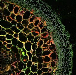 Francoise Maillard合作论文数Inserm U953, université Pierre-et-Marie-Curie3Anders Tunlid合作论文数Lunds Universitet2
 Mohsen Barin合作论文数Faculty of Agriculture, Urmia University2
 Alix Vidal合作论文数UMR Milieux environnementaux, transferts et interactions dans les hydrosystèmes et les sols (METIS), UMR 7619, Sorbonne Université UPMC-CNRS-EPHE, 4 place Jussieu, F-75252 Paris, France2
 Stavros Veresoglou合作论文数School of Ecology, Sun Yat-sen University2
 Hempel Stefan合作论文数Inst Biol, Free Univ Berlin2
 Erik Verbruggen合作论文数Department of Biology, University of Antwerp2
 Johannes Lehmann合作论文数School of Integrative Plant Science, College of Agriculture and Life Sciences, Cornell University2
 Peter G. Kennedy合作论文数Department of Biology, Lewis and Clark College2Hafedh Nasr合作论文数2Martijn Van Praagh合作论文数2
 Andreas Prinzing合作论文数State Museum of Natural History;Department of Zoology;Department of Zoology, State Museum of Natural History2Martin Obst合作论文数2
 Enrique Lara合作论文数WSL Wetlands Research Group, Swiss Federal Research Institute2
 Jens Dauber合作论文数2
 Ylva Lekberg合作论文数Department of Biology, University of Copenhagen2
 Katrin Böhning-Gaese合作论文数Helmholtz-Centre for Environmental Research;Leipzig University2Nasera Hammouti合作论文数2Louise C. Andresen合作论文数2
 Toby Kiers合作论文数Faculty of Science, Ecology & Evolution, Vrije Universiteit Amsterdam2Daniel L. Evans合作论文数1Hanwen Chen合作论文数1Manfred Forstreuter合作论文数1Luis F. J. Almeida合作论文数1Ivan F. Souza合作论文数1Laura S. Schnee合作论文数1Fatemeh Hashemnejad合作论文数1
 Paola Micaela Mafla-Endara合作论文数1
 Hanwen Kang合作论文数1Merlin Schäfer合作论文数1Camilla Simongini合作论文数1




