Neurovascular complications of carotid artery stenting (CAS) include intracranial embolism and hemorrhage, carotid dissection, in-stent thrombosis and vasospasm. Management of these crises requires knowledge of the intracranial vascular anatomy and collateral circulation, correct identification of the complication and its clinical implications, and a response plan that is appropriate to the complication and to the skill set of the operator. In this chapter the authors discuss techniques for the avoidance and management of CAS procedural complications.
OBJECTNeuroendoscopic approaches to lesions of the central nervous system and spine are limited by the loss of stereoscopic vision and high-fidelity image quality inherent in the operating microscope. Image-guided endoscopy (IGE) and image-guided surgery (IGS) have the potential to overcome these limitations. The goal of this study was to evaluate IGE for its potential applications in neurosurgery.METHODSTo determine the feasibility of IGE, a rigid endoscope was tracked using an IGS system that provided navigational data for the endoscope tip and trajectory as well as a computer-generated, three-dimensional, virtual representation of the image provided by the endoscope. The IGE procedure was successfully completed in 14 patients (nine with pituitary adenomas, one with a temporal cavernous malformation, and four with unruptured aneurysms). No complications could be attributed to the procedure. Compared with direct microscopy performed using anatomical landmarks, registration of the endoscope, and virtual image were highly accurate.CONCLUSIONSThis procedure offers many potential advantages for central nervous system and spinal endoscopy. Advances in IGE may enable its application to regions outside the central nervous system as well.
OBJECT Cranial nerve injuries, particularly motor nerve injuries, following carotid endarterectomy (CEA) can be disabling and therefore patients should be given reliable information about the risks of sustaining such injuries. The reported frequency of cranial nerve injury in the published literature ranges from 3 to 23%, and there have been few series in which patients were routinely examined before and after surgery by a neurologist. METHODS The authors investigated the risk of cranial nerve injuries in patients who underwent CEA in the European Carotid Surgery Trial (ECST), the largest series of patients undergoing CEA in which neurological assessment was performed before and after surgery. Cranial nerve injury was assessed and recorded in every patient and persisting deficits were identified on follow-up examination at 4 months and 1 year after randomization. Risk factors for cranial nerve injury were examined by performing univariate and multivariate analyses. There were 88 motor cranial nerve injuries among the 1739 patients undergoing CEA (5.1% of patients; 95% confidence interval [CI] 4.1-6.2). In 23 patients, the deficit had resolved by hospital discharge, leaving 3.7% of patients (95% CI 2.9-4.7) with a residual cranial nerve injury: 27 hypoglossal, 17 marginal mandibular, 17 recurrent laryngeal, one accessory nerve, and three Homer syndrome. In only nine patients (0.5%; 95% CI 0.24-0.98) the deficit was still present at the 4-month follow-up examination; however, none of the persisting deficits resolved during the subsequent follow up. Only duration of operation longer than 2 hours was independently associated with an increased risk of cranial nerve injury (hazard ratio 1.56, p < 0.0001). CONCLUSIONS The risk of motor cranial nerve injury persisting beyond hospital discharge after CEA is approximately 4%. The vast majority of neurological deficits resolve over the next few months, however, and permanent deficits are rare. Nevertheless, the risk of cranial nerve injury should be communicated to patients before they undergo surgery.
This text provides insights into current trends and controversial issues regarding state-of-the-art techniques in neurosurgery, interventional neuroradiology, and endovascular surgery. The authors confront such questions as: Are unruptured aneurysms managed most effectively by neurosurgical clipping or endovascular coiling? Is surgery or endovascular coiling the best treatment for ruptured cerebral aneurysms of the anterior or posterior cerebral circulation? In managing arteriovenous malformations and fistulas, should clinicians use neurointerventional techniques or surgery, or a combination of both? Highlights: More than 240 excellent illustrations, figures, and radiological images reviewing key concepts Detailed coverage of treatments for ischemic diseases, including extracranial and intracranial atherosclerosis Discussion of the organizational issues of integrating subspecialties and training subspecialists This text also offers you the opportunity to earn valuable CME credits online. Neurosurgeons, neuroradiologists, neurologists, vascular surgeons, and specialists will find the book's balanced coverage of critical current issues in neurosurgery an indispensable aid in making informed decisions. (A co-publication of Thieme and the American Association of Neurological Surgeons)
 Marc R. Mayberg合作论文数Department of Neurological Surgery, School of Medicine, University of Washington3
 Colin T. McDonald合作论文数1Stanley Kim合作论文数1Richard J. Parkinson合作论文数1Mitchell F. Berman合作论文数1
 Sean Lavine合作论文数1L Nelson Hopkins合作论文数1
 Michael DiLuna合作论文数Department of Neurosurgery, Yale School of Medicine, Yale University1Amar Arun Paul合作论文数1Nesbit Gary M合作论文数1Shownkeen Harish合作论文数1
 Peter Rothwell合作论文数Medical Sciences Division, Nuffield Department of Clinical Neurosciences, University of Oxford1
 David F. Kallmes合作论文数Department of Radiology, Mayo Clinic;Comprehensive Cancer Center, Mayo Clinic1
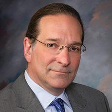 Lee R Guterman合作论文数Catholic Health1
 John C. Flickinger合作论文数Department of Radiation Oncology, School of Medicine, University of Pittsburgh1
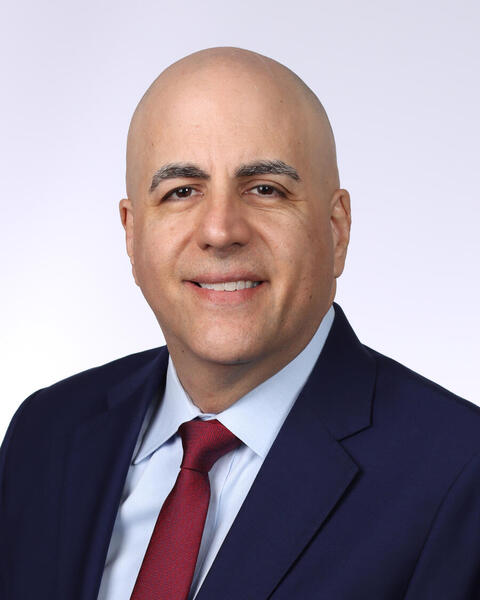 Bernard R. Bendok合作论文数Comprehensive Cancer Center, Mayo Clinic;Department of Radiology, Mayo Clinic1
 Richard a Bond合作论文数Stroke Prevention Research Unit, University Department of Clinical Neurology1Nerissa U. Ko合作论文数1
 Douglas Kondziolka合作论文数Center for Advanced Radiosurgery, NYU Langone Health;Department of Neurosurgery, Grossman School of Medicine, New York University1Alan S. Boulos合作论文数1
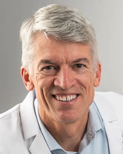 Clay Johnston合作论文数Harbor Health;Soley Therapeutics1
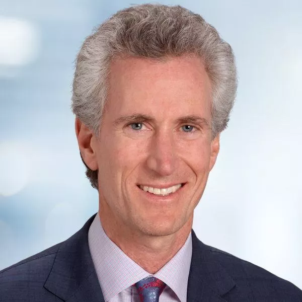 Michael T. Lawton合作论文数Department of Neurosurgery, Barrow Neurological Institute1
 Philip M. Meyers合作论文数College of Physicians & Surgeons, Columbia University1Eric Lapresto合作论文数Dept. Neurosurgery, Cleveland Clinic1Thomas J. Masaryk合作论文数1Daryl R. Gress合作论文数1
 L. Dade Lunsford合作论文数Department of Neurological Surgery, School of Medicine, University of Pittsburgh;Center for Image-Guided Neurosurgery, University of Pittsburgh Medical Center1Christopher C. Getch合作论文数1Stanley L. Barnwell合作论文数1
 Christopher S. Ogilvy合作论文数Beth Israel Deaconess Medical Center;Harvard Medical School1Thomas C. Origitano合作论文数1
 Howard A. Riina合作论文数Department of Neurology, Grossman School of Medicine, New York University;Department of Radiology, Grossman School of Medicine, New York University;Langone Health Center for Stroke and Neurovascular Diseases, New York University1
 Randall Higashida合作论文数Department of Radiology & Biomedical Imaging, University of California, San Francisco;School of Medicine, University of California, San Francisco;Division of Interventional Neurovascular Radiology, UCSF Medical Center1Brian E. Snell合作论文数1
 Jacques Dion合作论文数Department of Radiology and Imaging Sciences, Emory University School of Medicine;Terumo Neuro1Christopher M Putman合作论文数1Charles Warlow合作论文数1Robert J. Wienecke合作论文数1




