Direct electrical stimulation (DES) during awake brain surgery offers a unique opportunity to explore the neural basis of language. Although evidence from existing studies has shown increased involvement of the non-dominant (right) hemisphere in second language processing (particularly in late bilinguals) evidence from DES studies remains limited. We report the case of a 36-year-old right-handed late bilingual female (L1: Bulgarian; L2: Greek), who underwent awake craniotomy to treat an arteriovenous malformation beneath the right middle frontal gyrus (rMFG). Preoperative assessments revealed no clinically significant cognitive or linguistic deficits. During intraoperative mapping, DES on rMFG elicited anomias and phonemic paraphasias in L2, but not in L1, a finding consistent with reports in the literature regarding the role of rMFG in the episodic memory and the mental lexicon. Although coming from a single case, our findings provide direct evidence for the role of the rMFG in supporting second language processing in late bilinguals.
Abstract:Low-grade gliomas (LGGs) may affect language functions and health-related quality of life (HRQoL), particularly when located in brain areas essential for language. The present study investigates linguistic performance and HRQoL in patients with prefrontal cortex (PFC) and motor-sensory cortex LGGs. Data from 15 patients who underwent a standardized preoperative assessment, including linguistic and HRQoL measures, were analyzed and compared with healthy controls and published norms. Self-reported physical and communication functioning were relatively preserved in both tumor groups, whereas psychosocial well-being was notably reduced, particularly in the PFC group. Linguistic performance in both groups was generally lower than that of controls, with the PFC group showing selective vulnerabilities in semantic processing, most consistently in the time-limited semantic odd-image out task. Correlations within the PFC group indicated links between semantic processing, naming, and perceived communication functioning. These findings reveal subtle yet functionally and clinically meaningful difficulties that may not be evident in routine examinations, suggesting a need to assess both language and quality of life, including time-limited measures, in patients with LGGs.
The vestibular system plays a fundamental role in body orientation, posture control, and spatial and body motion perception, as well as in gaze and eye movements. We aimed to review the current knowledge regarding the location of the cortical and subcortical areas, implicated in the processing of vestibular stimuli. The search was performed in PubMed and Scopus. We focused on studies reporting on vestibular manifestations after electrical cortical stimulation. A total of 16 studies were finally included. Two main types of vestibular responses were elicited, including vertigo and perception of body movement. The latter could be either rotatory or translational. Electrical stimulation of the temporal structures elicited mainly vertigo, while stimulation of the parietal lobe was associated with perceptions of body movement. Stimulation of the occipital lobe produced vertigo with visual manifestations. There was evidence that the vestibular responses became more robust with increasing current intensity. Low-frequency stimulation proved to be more effective than high-frequency in eliciting vestibular responses. Numerous non-vestibular responses were recorded after stimulation of the vestibular cortex, including somatosensory, viscero-sensory, and emotional manifestations. Newer imaging modalities such as functional MRI (fMRI), Positron Emission Tomography (PET), SPECT, and near infra-red spectroscopy (NIRS) can provide useful information regarding localization of the vestibular cortex.
OBJECTIVE:EPI DWI is a routinely used sequence in brain imaging but it has limitations when it comes to SNR and artifact reduction. PROPELLER DWI has the benefit of improving image quality compared to EPI DWI. The aim of this study is to compare the EPI DWI sequence in brain MR imaging with the PROPELLER DWI sequence. The objective is to identify which sequence is more beneficial in brain imaging by evaluating image quality and the depiction of pathologies.MATERIALS AND METHODS:A total of 101 patients (55 females and 46 males, mean age 56 years) underwent brain MRI examination on a 1.5 T scanner. EPI DWI and PROPELLER DWI sequences were acquired in every exam and were reviewed by 2 radiologists. The images were evaluated by performing a quantitative analysis based on Relative Contrast and a qualitative analysis (overall image quality, conspicuousness of lesions, artifact reduction, etc.).RESULTS:In both the qualitative and quantitative analysis PROPELLER DWI achieved better results than EPI DWI. PROPELLER DWI showed statistical significance in the overall image quality (P < 0.001), the elimination of susceptibility (P < 0.001) and flow pulsation artifacts (P < 0.001), as well as in the contrast between CSF with white (P < 0.001) and grey matter (P < 0.001). Also, PROPELLER DWI presented better delineation of pathologies like ischemic strokes, metastasis, tumors and vasogenic edemas than conventional EPI DWI.CONCLUSION:PROPELLER DWI was the preferred sequence during the image evaluation. Compared to EPI DWI, PROPELLER DWI managed to reduce susceptibility and flow pulsation whilst achieving higher image quality and lesion delineation and earlier depiction of ischemic strokes than the conventional EPI DWI. PROPELLER DWI may be incorporated in brain MR imaging replacing EPI DWI.
Introduction: Obstructive sleep apnea hypopnea syndrome (OSAHS) is associated with many risk factors, but its pathophysiology is still not fully understood. Iron overload in brain regions isa hallmark of many neurodegenerative processes Objectives: The aim of this study is to evaluate iron deposition in the brain of the patients with OSAHS. Methods: All participants underwent a multi-recording sleep study and a brain magnetic resonance imaging (MRI) including: pre- and post-contrast T1-weighted, T2-weighted, FLAIR, and SWI at 3T Results: The study involved 26 patients with moderate to severe OSAHS [mean age 57.54±13.176 years (61.5% male)] and 34 healthy volunteers [mean age 57.59±10.79 years (58.8% men)]. MRIs showed that iron deposits in the brainstem were present in 65.4% of patients and 11.8% of the control group respectively (p<0.001), in the globus pallidus in 73.1% of patients and 11.8% of the control group (p<0.001), in the cerebellum in 46.2% of the patients and 8.8% of the control group (p<0.001) and in the temporal lobe in 30.8% of the patients andnone of the healthy volunteers (p<0.001). Furthermore, none of the patients and healthy controls had iron depositionsin the frontal, parietal and occipital lobe. OSAHS patients had a significantly higher degree of total brain iron deposition compared to the control group (p<0.001). Conclusions: Our study revealed a statistically significant association of OSAHS with increased iron deposition in the brain. The correlation of OSAHS with increased iron deposition in the brainstem needs further research as it plays an important role in breathing control.
BACKGROUND:White matter hyperintensities (WMHs) may be observed on Magnetic Resonance Imaging (MRI) in patients with Parkinson disease with or without vascular risk factors. Whether WMHs may influence motor and non-motor aspects of Parkinson disease is a subject of debate. The aim of this study is to evaluate the impact of WMH severity on various aspects of Parkinson disease in combination to the estimation of the impact of cerebrovascular risk factors. MATERIALS AND METHODS:We included a cohort of patients with Parkinson's disease who underwent MRI examination. The Fazekas visual rating scale was used to assess the severity and location of WMHs, and patient clinical characteristics were correlated with MRI data. RESULTS:All vascular risk factors were associated with higher Fazekas score in both periventricular and deep white matter. Periventricular white matter hyperintensities (PWMHs) and deep white matter hyperintensities (DWMHs) were associated with lower scores in the ACE-R cognitive assessment scale (p < 0.001). Furthermore, PWMHs and DWMHs severity was associated with higher UPDRS motor score (p < 0.001), while the Postural Instability Gait Difficulty (PIGD) phenotype was correlated with higher burden of WMHs. CONCLUSIONS:Comorbid WMHs may contribute to multi-dimension dysfunction in patients with Parkinson disease and consequently the management of vascular risk factors may be crucial to maintain motor and non-motor functions in PD.
BACKGROUND:Aggressive resection without compromising the patient's neurological status remains a significant challenge in treating intracranial gliomas. Our current study aims to evaluate the efficacy and safety of extra-operative stimulation and mapping via implanted subdural electrodes with or without depth (EOCSM), offering an alternative approach when awake mapping is contraindicated.METHODS:Fifty-one patients undergoing EOCSM for glioma resection in our institution formed the sample study of our current retrospective study. We assessed the effectiveness and safety of our approach by measuring the extent of resection and recording the periprocedural complications, respectively.RESULTS:The mean age of our participants was 58 years (±9.4 years). The lesion was usually located on the left side (80.4%) and affected the frontal lobe (51.0%). EOCSM was successful in 94.1% of patients. The stimulation and electrode implantation procedures lasted for a median of 2.0 h and 75 h, respectively. Stimulation-induced seizures and CSF leakage occurred in 13.7% and 5.9% of our cases. The mean extent of resection was 91.6%, whereas transient dysphasia occurred in 21.6% and transient hemiparesis in 5.9% of our patients, respectively.CONCLUSIONS:Extraoperative stimulation and mapping constitute a valid alternative mapping option in glioma patients who cannot undergo an awake craniotomy.
The differential diagnosis of bilateral lower extremity weakness is broad. We present a very rare case of a 48-year old male patient, with walking difficulties due to Bing-Neel syndrome. On clinical examination, there was a significant loss of muscle power in all his lower extremities key-muscle groups. The lumbar spine magnetic resonance imaging (MRI) showed only mild degenerative changes, whereas the MRI of the head indicated a diffuse meningeal thickening at the right temporal region, characterized by significant enhancement after contrast administration. Serum protein electrophoresis detected an IgM-kappa monoclonal protein. The patient received intrathecal chemotherapy with methotrexate and cytarabine, and was started on oral ibrutinib 420mg daily. In conclusion, a past medical history of Waldenstrom macroglobulinemia in conjunction with neurological manifestations should alert the treating physician for Bing-Neel syndrome. A complete diagnostic imaging and serologic protocol helps in setting the final diagnosis. Steroids are part of the treatment, but should be given after the diagnosis is set. Neurosurgical intervention is indicated for histologic confirmation in the case of diagnostic uncertainty.
AbstractCerebellar metastases represent the most common lesion requiring surgical treatment in the daily neurosurgical praxis. We report a rare case with Lhermitte–Duclos disease presenting to the neurosurgical outpatient department and describe our diagnostic and treatment challenges. A 57-year-old man presented with transient cerebellar manifestations, including walking difficulties, dizziness, and a persistent occipital headache. The magnetic resonance imaging demonstrated a high-intensity lesion in the right cerebellar hemisphere on T2-weighted images. We initially considered surgery to establish a histopathological diagnosis. However, the magnetic resonance spectroscopy was compatible with a dysplastic gangliocytoma of the cerebellum. Therefore, we preferred a more conservative approach. Posterior fossa contrast-enhancing lesions are much more than metastases. The role of advanced magnetic resonance in avoiding unnecessary surgical interventions is priceless.
Awake brain surgery allows for maximal tumor resection, while minimizing postoperative deficits, even when the tumor is located within eloquent brain regions. In the current study, we present the case of a patient who underwent awake craniotomy to remove a space-occupying lesion located at the left (dominant) temporal lobe. During subcortical mapping, electrical stimulation of the inferior longitudinal fasciculus caused severe errors (paralexias) on a semantic odd-word out task but not on other tasks that use different input routes and processes. The cognitive neuropsychological model for single word processing assisted us to associate a specific structure (inferior longitudinal fascicle [ILF]) with a specific cognitive process (i.e., access to the semantic system). Our results highlight the importance of subcortical fascicles in reading and agree with previous studies regarding the critical role of ILF in reading comprehension.
Purpose of this study was to evaluate the effect of a robust magnetic resonance reconstruction pipeline equipped with a deep convolutional neural network on the overall image quality, in terms of Gibbs artifact reduction, and SNR improvement. Sixteen (16) healthy volunteers enrolled in this study and were imaged at 3T. Representative images of each image series that were reconstructed through the pipeline that leverages a deep learning (DL) algorithm were retrospectively benchmarked against corresponding images reconstructed through a conventional pipeline. DL-reconstructed images showed significant SNR improvements compared to the corresponding conventionally reconstructed images. In addition to that, Gibbs artifacts were effectively eliminated, when the raw data were reconstructed through the DL pipeline. Gibbs artifact reduction was qualitatively assessed by two experienced medical physicists and two experienced radiologists. DL-based reconstruction can lead to an SNR surplus which can be further invested into either higher spatial resolution and thinner slices, or into shorter scan times.
Meningioma is one of the most frequent primary central nervous system tumors. While magnetic resonance imaging (MRI), is the standard radiologic technique for provisional diagnosis and surveillance of meningioma, it nevertheless lacks theprima faciecapacity in determining meningioma biological aggressiveness, growth, and recurrence potential. An increasing body of evidence highlights the potential of machine learning and radiomics in improving the consistency and productivity and in providing novel diagnostic, treatment, and prognostic modalities in neuroncology imaging. The aim of the present article is to review the evolution and progress of approaches utilizing machine learning in meningioma MRI-based sementation, diagnosis, grading, and prognosis. We provide a historical perspective on original research on meningioma spanning over two decades and highlight recent studies indicating the feasibility of pertinent approaches, including deep learning in addressing several clinically challenging aspects. We indicate the limitations of previous research designs and resources and propose future directions by highlighting areas of research that remain largely unexplored. Level of Evidence 5 Technical Efficacy Stage 2
AIM: To report the clinical characteristics and diagnostic procedures used in patients with spasm of the near reflex (SNR), in order to present common investigation strategies and diagnostic pitfalls. METHODS: Retrospective case series of twenty-two patients, mainly children, with SNR or accommodation spasm (AS). AS was diagnosed on the basis of blurred vision and a difference of >= 2 dioptres between manifest and cycloplegic retinoscopy. If esotropia and miosis were present, the patients were diagnosed with SNR. All patients underwent visual acuity testing, orthoptic evaluation, assessment of refraction before and after cycloplegia, and dilated fundoscopy. Additional diagnostic investigations, such as neuroimaging, lumbar puncture (LP), electrophysiology and blood tests, were also recorded. Screen use among children was assessed in hours per day. RESULTS: There were 19 female and 3 male patients (age range 7-33y, median=10y). Seventeen patients had AS and 5 patients had SNR, with episodic blurry vision and headaches being the most common symptoms. Brain neuroimaging was performed in six patients (27%), although only one had a history of brain trauma. Two of those patients underwent visual evoked potentials and three also underwent LP and received intravenous steroid therapy. The majority of patients (90%) reported prolonged daily screen time (>2h/d), and in 55% of cases there were concurrent social problems or psychological triggers. Treatment consisted of careful explanation of the condition, atropine 1% eye drops and full cycloplegic correction by means of bifocal glasses. CONCLUSION: The diagnosis of SNR and AS may be challenging, because symptoms are usually intermittent and nonspecific, and a large number of patients are often subjected to redundant and potentially time-consuming examinations and treatment, that may exaggerate the underlying psychological disorder. Hence, detailed clinical testing and assessment of psychosocial profile is necessary, in order to avoid unnecessary investigations. Neuroimaging should be performed only in selected cases. Finally, due to prolonged screen use SNR and AS may become more frequent in the future.
Purpose: To study the agreement between digital subtraction angiography (DSA) and computed tomographic angiography (CTA) measurements on the aneurysmal neck and sac of ruptured intracranial aneurysms (IAs). Material and Methods: Through a retrospective agreement analysis of all consecutive patients who reached our Tertiary Hospital with aneurysmal subarachnoid haemorrhage, we measured the intra-class correlation, Lin’s concordance correlation and Bland-Altman analysis estimates on the maximal neck and sac diameters. We included patients who underwent both CTA and DSA in the period between 2012 and 2018. All CTA examinations were acquired using one of two CT scanners: a Toshiba Aquilion 16 CT scanner and a multi-detector Philips Ingenuity 128 CT scanner. Results: Thirty-two patients (mean age of 55 years) and an equal number of IAs fulfilled our eligibility criteria. Most IAs (87.5%) were located at the anterior circulation. Based on CTA measurements, the inter-observer agreement of the CTA was “weak” regarding the neck diameter, and ranged from “strong” (2D-CTA) to “very-strong” (3D-CTA) regarding the sac measurements. Based on DSA readings, the 2D-CTA was more precise than 3D-CTA regarding the neck (overestimated by 0.91 mm and 0.94 mm, respectively) and the sac diameters (overestimated by 0.49 and 0.51 mm, respectively). Accordingly, the mean normalised smallest detectable differences (MNSDD) of 2D-CTA were 0.93 mm and 0.41 mm for the neck and sac, respectively. Likewise, the MNSDD regarding 3D-CTA were 0.93 mm and 0.36 mm for the neck and sac, respectively. Conclusions: CTA is an imaging modality, which seems to describe the size of a ruptured IA sac accurately, but fails to delineate the morphology of the aneurysmal neck. CTA seems to overestimate the aneurysm neck, and thus, it is probably inferior to DSA in the decision-making process for the management of ruptured IAs of the anterior circulation.
Purpose The Thrombolysis in Ischemic Stroke Patients (TRISP) collaboration was a concerted effort initiated in 2010 with the purpose to address relevant research questions about the effectiveness and safety of intravenous thrombolysis (IVT). The collaboration also aims to prospectively collect data on patients undergoing endovascular treatment (EVT) and hence the name of the collaboration was changed from TRISP to EVA-TRISP. The methodology of the former TRISP registry for patients treated with IVT has already been published. This paper focuses on describing the EVT part of the registry. Participants All centres committed to collecting predefined variables on consecutive patients prospectively. We aim for accuracy and completeness of the data and to adapt local databases to investigate novel research questions. Herein, we introduce the methodology of a recently constructed academic investigator-initiated open collaboration EVT registry built as an extension of an existing IVT registry in patients with acute ischaemic stroke (AIS). Findings to date Currently, the EVA-TRISP network includes 20 stroke centres with considerable expertise in EVT and maintenance of high-quality hospital-based registries. Following several successful randomised controlled trials (RCTs), many important clinical questions remain unanswered in the (EVT) field and some of them will unlikely be investigated in future RCTs. Prospective registries with high-quality data on EVT-treated patients may help answering some of these unanswered issues, especially on safety and efficacy of EVT in specific patient subgroups. Future plans This collaborative effort aims at addressing clinically important questions on safety and efficacy of EVT in conditions not covered by RCTs. The TRISP registry generated substantial novel data supporting stroke physicians in their daily decision making considering IVT candidate patients. While providing observational data on EVT in daily clinical practice, our future findings may likewise be hypothesis generating for future research as well as for quality improvement (on EVT). The collaboration welcomes participation of further centres willing to fulfill the commitment and the outlined requirements.
Background: The efficacy of laser interstitial thermal therapy (LITT) in mesial temporal lobe epilepsy (MTLE) has not been clearly established yet. Objective: We conducted a meta-analysis to estimate the efficacy of LITT for TLE (Q1). We also examined the effect of the patient's age (Q2), the total ablation volume (TAV) (Q3), the strength of the MRI unit (Q4), the type of the utilized stereotactic platform (Q5), and the follow up period (Q6) on the patient's outcome. Methods: Fixed- and random-effects model meta-analysis was conducted to assess the proportion estimate for each parameter individually. Kaplan-Meier survival-analysis was performed on the available individual patient time-to-first seizure data. Results: Sixteen studies with 575 patients fulfilled our eligibility criteria. The efficacy of LITT was 0.547 (95%CI: 0.506-0.588). Our statistical analysis had robust results after stratification according to the study population (Q2; p = 0.3418), and the type of the utilized stereotactic platform (Q5; p = 0.286), whereas the role of the TAV (Q3; p = 0.058) and strength of the magnetic field (Q4; p = 0.062) in seizure control remained unclear. The median seizure-free period (Q6) was 0.643 (0.569-0.726) and 0.467 (0.385-0.566) for the one- and the two-year follow up. Conclusions: LITT seems to offer a viable alternative to resective surgery, with a moderate efficacy and enduring results. Higher ablation volumes may be associated with improved seizure control, although our current study provided no statistically significant data. More high-quality studies are required to highlight the role of LITT in epilepsy surgery, particularly in the pediatric population.
Obstructive sleep apnea hypopnea syndrome (OSAHS) is associated with increased risk of cerebrovascular disease. The aim of the present study was to investigate the association between the presence of the small vessel disease (SVD) of the brain in patients with OSAHS. The study included 24 patients with moderate to severe OSAHS and 34 healthy volunteers. All the subjects underwent magnetic resonance imaging (MRI) of the brain, in order to sought periventricular white matter (PVWM), deep white matter (DWM) and brainstem SVD. Among patients with OSAHS, 79.1% had SVD (grade 1–3, Fazekas score) in DWM and 91.7% in PVWM while 22.4% had brainstem—white matter hyperintensities (B-WMH). Patients with OSAHS had a much higher degree of SVD in the DWM and PVWM compared to the control group (p < 0.001). The multivariate analysis showed an independent significant association of OSAHS with SVD (DWM and PVWM) (p = 0.033, OR 95% CI: 8.66 (1.19–63.08) and: p = 0.002, OR 95% CI: 104.98 (5.15–2141)). The same analysis showed a moderate association of OSAHS with B-WMH (p = 0.050, OR 15.07 (0.97–234.65)). Our study demonstrated an independent significant association of OSAHS with SVD and a moderate association of OSAHS with B-WMH.
 Kostas N. Fountas合作论文数Faculty of Medicine, School of Health Sciences, University of Thessaly110
 Ioannis Tsougos合作论文数Department of Medical Physics, University Hospital of Larissa45
 Ioannis V. Fezoulidis合作论文数Department of Radiology, University Hospital of Larissa37
 Joe S. Robinson合作论文数31
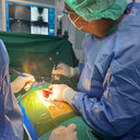 Alexandros G Brotis合作论文数University Hospital of Larissa, University of Thessaly18
 Panagiotis Georgoulias合作论文数16Machinis Theofilos G合作论文数15
 Dardiotis Efthimios合作论文数Department of Neurology, Faculty of Medicine, University of Thessaly;Multi-disciplinary Multiple Sclerosis Centre, Department of Neurology, General University Hospital of Larissa15Kw Johnston合作论文数14Hf Smisson合作论文数14Konstantinos Fountas合作论文数14
 Varvara Valotassiou合作论文数Department of Nuclear Medicine, University Hospital of Larissa14
 Hadjigeorgiou Georgios合作论文数Medical School, University of Cyprus;Department of Neurology, Faculty of Medicine, University of Thessaly;School of Health Sciences, University of Thessaly;European Restless Legs Syndrome Study Group14Kousi Evanthia合作论文数Department of Medical Physics, University of Thessaly13
 Patricia Svolos合作论文数Medical Physics Department, University Hospital of Larissa13Kyriaki Theodorou合作论文数13
 Efstathios Gotsis合作论文数Encephalos MRI Unit Athens Greece12Ch Feltes合作论文数11
 Chara K Tzavara合作论文数Dept Nucl Med, Univ Hosp Larissa11
 Kiki Theodorou合作论文数Physics Department;Medical School;Patras University;Medical School, Patras University11
 Konstantinos N. Paterakis合作论文数11J. Papatriantafyllou合作论文数10
 Constantin Kappas合作论文数Medical School, University of Patras10Joe Sam Robinson合作论文数10
 Tsolaki Evangelia合作论文数Medical School, University of Thessaly9
 Nicolas Sifakis合作论文数Institute for Space Applications and Remote Sensing;National Observatory of Athens;Institute for Space Applications and Remote Sensing, National Observatory of Athens9G. Chadjigeorgiou合作论文数8
 Lavdas Eleftherios合作论文数Department of Medical Radiological Technologists, Technological Education Institute of Athens8Vassilios G. Dimopoulos合作论文数8D. Psimadas合作论文数7Siasios Ioannis合作论文数7
 Paschalis Thanasis合作论文数7Joseph R. Smith合作论文数7Vg Dimopoulos合作论文数6Smisson Hugh F合作论文数6Anastasia Tasiou合作论文数6
 Katerina Vassiou合作论文数Department of Radiology, University Hospital of Larissa6Kim W. Johnston合作论文数6
 Gregory P. Lee合作论文数Medical College of Georgia6I Karampelas合作论文数5J. Tsougos合作论文数5
 Alexandros Vamvakas合作论文数5P. Toulas合作论文数5
 Georgios Koukoulis合作论文数Dept Gen Surg, Gen Hosp Larissa5Nikolakakos Leonidas G合作论文数5
 Marianna Vlychou合作论文数Department of Radiology, University Hospital of Larissa5Arthur A. Grigorian合作论文数5
 Georgia Xiromerisiou合作论文数5Maria Dardioti合作论文数5




