Introduction Socio-economic status (SES) influences paediatric visual health by affecting eye care access, refractive error prevalence and spectacle provision. This study examines the link between household income and paediatric vision outcomes in Orange County, California, USA using data from the University of California, Irvine EyeMobile Pediatric Clinic (2021–2024).Methods Vision screening was conducted on 15 842 children (ages 3–10). Subsequent comprehensive eye examinations were performed on 3350 children who failed initial screening. Socio-economic stratification was based on zip-code-level median household income (MHI), categorised as below-average or above-average for Orange County. A retrospective cross-sectional analysis of examination data compared visual acuity, refractive error and spectacle provision rates between the two SES groups.Results Children from below-average MHI areas had higher screening failure rates than those from above-average MHI areas (24.6% vs 18.5%) and a higher odds of screening failure (OR 1.44, 95% CI 1.33 to 1.57). They also demonstrated a higher need for spectacles (70.3% vs 64.8%), with an increased odds of requiring spectacles (OR 1.28, 95% CI 1.05 to 1.56). The prevalence of astigmatism was higher among children from areas with below-average MHI (75.3% vs 66.1%), corresponding to a significantly higher odds of astigmatism (OR 1.56, 95% CI 1.36 to 1.79). Best-corrected distance visual acuity improved with the provision of spectacles in both income groups; however, children from lower-income communities were more likely to present with uncorrected visual impairment during screening, consistent with the higher screening failure rates observed.Conclusion Children from lower-income communities demonstrated higher rates of uncorrected visual impairment and a greater need for vision care services, while children from higher-income communities also faced substantial unmet vision needs. A mobile school-based vision screening programme, such as the EyeMobile, could significantly benefit both groups by promoting equitable access to vision care.
Purpose:The University of California, Irvine (UCI) Eye Mobile provides vision screenings and comprehensive eye exams for children, with referrals to pediatric ophthalmologists as needed. This study evaluates the indications for these referrals, follow-up rates, patient-reported outcomes, and appointment wait times. Methods:We conducted a retrospective chart review of de-identified records from UCI Eye Mobile patients referred to pediatric ophthalmologists. Additionally, telephone interviews were held with parents of referred children. Results:Between the academic years of 2018-19 and 2022-23, 3619 children received eye exams through the UCI Eye Mobile. Of these, 75 (2.1%; 95% CI 1.7-2.6%) were referred to pediatric ophthalmologists. The primary referral reasons included strabismus (32.0%), optic nerve or posterior segment evaluation (26.7%), and amblyopia (14.7%). Among surveyed families, 95.5% (95% CI 84.9-98.7%) secured a follow-up appointment. Outcomes for children who were evaluated by a specialist included new glasses or patching therapy (78.1%), routine follow-up (9.4%), and surgery (9.4%). Among children with available post-referral outcomes, 93.5% (95% CI 79.3-98.2%) showed caregiver-reported improvement. Follow-up and improvement outcomes were based on caregiver telephone survey responses, obtained from 44 of 75 referred families (58.7%). The median wait time for a pediatric ophthalmologist was 8.0 weeks (IQR 6.0-13.0 weeks). Conclusions:The UCI Eye Mobile plays a crucial role in early detection and referral for pediatric ocular conditions requiring ophthalmologic care. However, delays in care due to prolonged wait times and limited healthcare access persist, particularly in underserved populations. Addressing these barriers is critical to improving timely access to pediatric ophthalmologic services.
Membrane frizzled-related protein (MFRP), present in the retinal pigment epithelium (RPE), is an integral membrane protein essential for ocular development and the normal physiology of the retina. Mutations in MFRP are associated with autosomal recessive nonsyndromic nanophthalmos, leading to severe hyperopia and early-onset retinitis pigmentosa. While several preclinical gene-augmentation and gene-editing trials hold promise for future therapies aimed at stopping degeneration and restoring retinal function, the molecular mechanisms involved in MFRP biology are still not well understood. Here, we studied the biochemical properties of MFRP and the molecular consequences of its loss of function in the retinal degeneration 6 (rd6) mouse model. Using transcriptomic and lipidomic approaches, we observed that accumulation of docosahexaenoic acid (DHA) constitutes a primary defect in the MFRP-deficient RPE. In biochemical assays, we showed that MFRP undergoes extensive glycosylation, and it preferentially binds lipids of several classes, including phosphatidylserine and phosphatidylinositol-4-phosphate; as well as binding to several transmembrane proteins, notably adiponectin receptor 1 (ADIPOR1) and inward rectifier potassium channel 13 (KCNJ13). Moreover, MFRP determines the subcellular localization of ADIPOR1 and KCNJ13 in the RPE in vivo. This feature is altered by MFRP deficiency and can be restored by gene-therapy approaches. Overall, our observations suggest that MFRP constitutes an important interaction hub within the apical membrane of RPE cells, coordinating protein trafficking and subcellular localization within the RPE, and lipid homeostasis within the entire retina.
Purpose: With the growing popularity of badminton worldwide, the incidence of badminton-related ocular injuries is expected to rise. The high velocity of shuttlecocks renders ocular traumas particularly devastating, especially with the possibility of permanent vision loss. This study investigated the mechanism behind ocular complications through simulation analyses of mechanical stresses and pressures upon shuttlecock impact. Design: Computational simulation study. Participants: None. Methods: A 3-dimensional human eye model was reconstructed based on the physiological and biomechanical properties of various ocular tissues. Finite element analysis simulations involved a frontal collision with a shuttlecock projectile at 128.7 km/hour (80 mph). Intraocular pressure (IOP) changes and tissue stress were mapped and quantified in the following ocular structures: the limbus, ciliary body, zonular fibers, ora serrata, retina, and optic nerve head. Main Outcome Measures: Intraocular pressure and tissue stress. Results: Upon shuttlecock impact, compressive force was transferred to the anterior pole of the cornea, propagating posteriorly to the optic nerve head. Deflection of forces anteriorly contributed to refractory oscillations of compressive and tensile stress of ocular tissue. Initial impact led to a momentary (<1 ms) spike in IOP 5.66 MPa (42.5 x 10(3) mmHg) that radially distributed for a very brief instance (<1 ms) of pressure at the trabecular meshwork of the iridocorneal angle of 1.25 MPa (9.4 x 10(3) mmHg). The lens had a maximal posterior displacement of 1.5 mm with peak zonular fiber tensile strain of 52%. The limbus, ciliary body, and ora serrata had a peak tensile stress of 5.16 MPa, 1.90 MPa, and 0.62 MPa, respectively. Compressive force from the sclera concentrated at the optic nerve head for a peak stress of 5.97 MPa while peak pressure from vitreous humor was 7.99 MPa. Conclusions: Shuttlecock impact led to a very brief, substantial rise in pressure and stress significant for tissue damage and subsequent complications, such as secondary glaucoma, angle recession, lens subluxation, hyphema, or retinal dialysis. Our findings offer valuable mechanistic insights into how ocular structures are affected by shuttlecock projectile impact to inform clinical assessments and treatment strategies, while highlighting the importance of protective eyewear in racket sports.
PURPOSE. Homeostatic plasticity is crucial for maintaining stable neural activity by adjusting strength and intrinsic properties of synapses. This mechanism is vital for normal nervous system function and plays a role in various neurological conditions, including retinal degenerations. Sensitive night vision has been shown in P23H/Gnat2-/- retinitis pigmentosa (RP) mice, which lack cone phototransduction and rely solely on rods, even after losing more than half of their rod photoreceptors. While homeostatic plasticity has been proposed as a potential explanation, the underlying molecular mechanisms remain unclear. The aim of this study was to investigate the molecular basis of this phenomenon. METHODS. Single cell RNA-sequencing (scRNA-seq) and bulk retina proteomics were used to investigate the transcriptomic and proteomic changes of the degenerating retinas in 1-month-old P23H/Gnat2-/- RP and Gnat2-/-control mice. Immunohistochemistry was used to analyze the expression of synaptic SNARE complex and vesicle proteins, SNAP25 and SYT1, in the outer plexiform layer, the site of rod axon terminals. RESULTS. This study shows a significant upregulation of genes encoding synaptic SNARE complex and vesicle proteins (Snap25, Stxbp1, and Syt1) in P23H mouse rods. Bulk retina proteomics analysis shows trends toward upregulation of the corresponding proteins as well as upregulation of many matrix-associated and trans-synaptic-complex proteins. Immunohistochemistry shows persistent SYT1 and SNAP25 expression in the outer plexiform layer of P23H/Gnat2-/- mice despite significant rod death. CONCLUSIONS. Rod degeneration induces molecular changes in the P23H/Gnat2-/- mouse rods that suggest synaptic plasticity and strengthening of rod-rod bipolar cell synaptic transmission in early RP.
Background:Despite the common association between bilateral retinal hemorrhage and shaken baby syndrome (SBS), unilateral retinal hemorrhage does not necessarily exclude this diagnosis. This study used computational simulations to elucidate the biomechanical phenomena within the eye under asymmetrical shaking forces. Methods:Finite element analysis (FEA) incorporating the vitreous, vitreoretinal interface, retinal layers, and retinal vessels was performed under asymmetrical shaking conditions. To assess the stress-strain response at the preretinal, intraretinal, and subretinal locations, we divided the retinal mesh into three equally spaced layers with an element height of 0.083 mm. The remaining space within the retina was filled with the vitreous humor and attached to it via the main retinal vessels extracted from a standard fundus image. The resulting changes in shear stress and intraocular pressure (IOP) were quantified. Results:The FEA model demonstrated that increasing the rotational radius from 10 cm to 14 cm or 17 cm led to a significant increase in shear stress and IOP across the vitreoretinal interface and within the retinal layers. Specifically, shear stress in the preretinal layer increased by 70.2% (8.0 kPa vs. 4.7 kPa), in the intraretinal layer by 20.0% (5.4 kPa vs. 4.5 kPa), and in the subretinal layer by 6.1% (3.5 kPa vs. 3.3 kPa). Simultaneously, IOP in the central region increased by 157.5% (39.4 mmHg vs. 15.3 mmHg) and in the posterior region by 162.3% (41.7 mmHg vs. 15.9 mmHg) when the rotational radius was increased to 17 cm from 10 cm. Increasing the rotational radius to 17 cm led to more pronounced changes in peak IOPs, with the central region showing a change of 39.4 mmHg and the posterior region a change of 41.7 mmHg. These results indicate a direct correlation between the rotational radius and the magnitude of IOP changes in the vitreous. Conclusions:These findings highlight the critical impact of rotational radius on the biomechanical forces exerted within the eye during asymmetrical shaking events, leading to variations in shear stress and IOP that could contribute to unilateral retinal hemorrhage in SBS. These insights reveal the complexity of diagnosing SBS and emphasize the need for careful consideration of the biomechanical evidence in patients presenting with asymmetrical or unilateral retinal hemorrhage.
Cellular retinaldehyde-binding protein (CRALBP) supports production of 11-cis-retinaldehyde and its delivery to photoreceptors. It is found in the retinal pigment epithelium (RPE) and Müller glia (MG), but the relative functional importance of these two cellular pools is debated. Here, we report RPE- and MG-specific CRALBP knockout (KO) mice and examine their photoreceptor and visual cycle function. Bulk visual chromophore regeneration in RPE-KO mice is 15-fold slower than in controls, accounting for their delayed rod dark adaptation and protection against retinal phototoxicity, whereas MG-KO mice have normal bulk visual chromophore regeneration and retinal light damage susceptibility. Cone pigment regeneration is significantly impaired in RPE-KO mice but mildly affected in MG-KO mice, disclosing an unexpectedly strong reliance of cone photoreceptors on the RPE-based visual cycle. These data reveal a dominant role for RPE-CRALBP in supporting rod and cone function and highlight the importance of RPE cell targeting for CRALBP gene therapies.
AbstractInherited retinopathies are devastating diseases that in most cases lack treatment options. Disease-modifying therapies that mitigate pathophysiology regardless of the underlying genetic lesion are desirable due to the diversity of mutations found in such diseases. We tested a systems pharmacology-based strategy that suppresses intracellular cAMP and Ca2+ activity via G protein-coupled receptor (GPCR) modulation using tamsulosin, metoprolol, and bromocriptine coadministration. The treatment improves cone photoreceptor function and slows degeneration in Pde6βrd10 and RhoP23H/WT retinitis pigmentosa mice. Cone degeneration is modestly mitigated after a 7-month-long drug infusion in PDE6A-/- dogs. The treatment also improves rod pathway function in an Rpe65-/- mouse model of Leber congenital amaurosis but does not protect from cone degeneration. RNA-sequencing analyses indicate improved metabolic function in drug-treated Rpe65-/- and rd10 mice. Our data show that catecholaminergic GPCR drug combinations that modify second messenger levels via multiple receptor actions provide a potential disease-modifying therapy against retinal degeneration.
Rhodopsin, the prototypical class-A G-protein coupled receptor, is a highly sensitive receptor for light that enables phototransduction in rod photoreceptors. Rhodopsin plays not only a sensory role but also a structural role as a major component of the rod outer segment disc, comprising over 90% of the protein content of the disc membrane. Mutations in RHO which lead to structural or functional abnormalities, including the autosomal recessive E150K mutation, result in rod dysfunction and death. Therefore, correction of deleterious rhodopsin mutations could rescue inherited retinal degeneration, as demonstrated for other visual genes such as RPE65 and PDE6B. In this study, we describe a CRISPR/Cas9 adenine base editing strategy to correct the E150K mutation and demonstrate precise in vivo editing in a Rho -E150K mouse model of autosomal recessive retinitis pigmentosa (RP). Using ultraviolet-visible spectroscopy, mass spectrometry, and the G-protein activation assay, we characterized wild-type rhodopsin and rhodopsin variants containing bystander base edits. Subretinal injection of dual-adeno-associated viruses delivering our base editing strategy yielded up to 44% Rho correction in homozygous Rho -E150K mice. Injection at postnatal day 15, but not later time points, restored rhodopsin expression, partially rescued retinal function, and partially preserved retinal structure. These findings demonstrate that in vivo base editing can restore the function of mutated structural and functional proteins in animal models of disease, including rhodopsin-associated RP and suggest that the timing of gene-editing is a crucial determinant of successful treatment outcomes for degenerative genetic diseases.
Delivering ribonucleoproteins (RNPs) for in vivo genome editing is safer than using viruses encoding for Cas9 and its respective guide RNA. However, transient RNP activity does not typically lead to optimal editing outcomes. Here we show that the efficiency of delivering RNPs can be enhanced by cell-penetrating peptides (covalently fused to the protein or as excipients) and that lipid nanoparticles (LNPs) encapsulating RNPs can be optimized for enhanced RNP stability, delivery efficiency and editing potency. Specifically, after screening for suitable ionizable cationic lipids and by optimizing the concentration of the synthetic lipid DMG-PEG 2000, we show that the encapsulation, via microfluidic mixing, of adenine base editor and prime editor RNPs within LNPs using the ionizable lipid SM102 can result in in vivo editing-efficiency enhancements larger than 300-fold (with respect to the delivery of the naked RNP) without detectable off-target edits. We believe that chemically defined LNP formulations optimized for RNP-encapsulation stability and delivery efficiency will lead to safer genome editing. The safety and efficacy of ribonucleoproteins for genome editing can be enhanced by formulations of lipid nanoparticles optimized for enhanced stability, delivery efficiency and editing potency of the protein complexes.
MicroRNAs (miRs) are short, evolutionarily conserved non-coding RNAs that canonically downregulate expression of target genes. The miR family composed of miR-204 and miR-211 is among the most highly expressed in the retinal pigment epithelium (RPE) in both mouse and human, and also retains high sequence identity. To assess the role of this miR family in the developed mouse eye, we generated two floxed conditional knockout mouse lines crossed to the RPE65-ERT2-Cre driver mouse line to perform an RPE-specific conditional knockout of this miR family in adult mice. After Cre-mediated deletion, we observed retinal structural changes by optical coherence tomography; dysfunction and loss of photoreceptors by retinal imaging; and retinal inflammation marked by subretinal infiltration of immune cells by imaging and immunostaining. Single-cell RNA sequencing of diseased RPE and retinas showed potential miR-regulated target genes, as well as changes in non-coding RNAs in the RPE, rod photoreceptors, and Müller glia. This work thus highlights the role of miR-204 and miR-211 in maintaining RPE function and how the loss of miRs in the RPE exerts effects on the neural retina, leading to inflammation and retinal degeneration.
Rhodopsin (Rho) and cone opsins are essential for detection of light. They respond via photoisomerization, converting their Schiff-base-adducted 11-cis-retinylidene chromophores to the all-trans configuration, eliciting conformational changes to activate opsin signaling. Subsequent Schiff-base hydrolysis releases all-trans-retinal, initiating two important cycles that maintain continuous vision – the Rho photocycle and visual cycle pathway. Schiff-base hydrolysis has been thoroughly studied with photoactivated rhodopsin but not with cone opsins. Using established methodology, we directly measured the formation of Schiff-base between retinal chromophores with mammalian visual and non-visual opsins of the eye. Next, we determined the rate of light-induced chromophore hydrolysis. We found that retinal hydrolysis from photoactivated cone opsins was markedly faster than from photoactivated rhodopsin. Bovine retinal G protein-coupled receptor RGR (bRGR) displayed rapid hydrolysis of its 11-cis-retinylidene photoproduct to quickly supply 11-cis-retinal, and re-bind all-trans-retinal. Hydrolysis within bRGR in native retinal pigment epithelium (RPE) microsomal membranes was > 6-times faster than that of bRGR purified in detergent micelles. N-terminal-targeted antibodies significantly slowed bRGR hydrolysis, while C-terminal antibodies had no effect. Our study highlights the much faster photocycle of cone opsins relative to Rho and the crucial role of RGR in chromophore recycling in daylight. By contrast, in our experimental conditions, bovine peropsin (bRRH) did not form pigment in the presence of all-trans-retinal, nor with any mono-cis retinal isomers, leaving uncertain the role of this opsin as a light sensor.
Purpose: To analyze referral rates, patient demographics, referral indications, and the impact of socioeconomic factors on ocular health from the University of California Irvine (UCI) Eye Mobile for Children, particularly during the coronavirus disease 2019 (COVID-19) pandemic. Methods: A retrospective chart review was performed on de-identified records of children examined on the UCI Eye Mobile. GraphPad Prism 10.0.0 and Python software were used for statistical analyses. Results: In the academic years from 2018 to 2022, 3,619 children received comprehensive eye examinations on the UCI Eye Mobile. Among them, 76 were referred to a pediatric ophthalmologist. The majority of these children were Hispanic (72.6%, 54 of 74), followed by Asian (10.9%, 8 of 74). A significant proportion (82.9%, 63 of 76) attended school districts with median incomes below that of Orange County. Statistically significant differences were found in age ( P = .001; pre-COVID: 3.98 +/- 1.08 years vs COVID: 5.75 +/- 2.92 years) and gender ( P = .023; pre-COVID female: 31 of 41 vs COVID female: 15 of 32) between the pre-COVID and COVID years. Additionally, there were significant differences in the proportion of children with hyperopia with astigmatism between the pre-COVID and COVID years ( P = .044; pre-COVID: 23 of 40 vs COVID: 12 of 35). The most common indications for ophthalmologist referrals were for strabismus evaluation/treatment (28.9%, 22 of 76), followed by abnormal cup-to-disc ratio (21.1%, 16 of 76). Conclusions: The study highlights the pivotal role of the UCI Eye Mobile for children in identifying ocular conditions needing referrals to subspecialty care. The majority of children needing these referrals attended schools in lower economic communities. Additionally, the COVID-19 pandemic appears to have influenced the demographic and clinical characteristics.
In daylight, demand for visual chromophore (11-cis-retinal) exceeds supply by the classical visual cycle. This shortfall is compensated, in part, by the retinal G-protein-coupled receptor (RGR) photoisomerase, which is expressed in both the retinal pigment epithelium (RPE) and in Müller cells. The relative contributions of these two cellular pools of RGR to the maintenance of photoreceptor light responses are not known. Here, we use a cell-specific gene reactivation approach to elucidate the kinetics of RGR-mediated recovery of photoreceptor responses following light exposure. Electroretinographic measurements in mice with RGR expression limited to either cell type reveal that the RPE and a specialized subset of Müller glia contribute both to scotopic and photopic function. We demonstrate that 11-cis-retinal formed through photoisomerization is rapidly hydrolyzed, consistent with its role in a rapid visual pigment regeneration process. Our study shows that RGR provides a pan-retinal sink for all-trans-retinal released under sustained light conditions and supports rapid chromophore regeneration through the photic visual cycle.
Chronic, progressive retinal diseases, such as age-related macular degeneration (AMD), diabetic retinopathy, and retinitis pigmentosa, arise from genetic and environmental perturbations of cellular and tissue homeostasis. These disruptions accumulate with repeated exposures to stress over time, leading to progressive visual impairment and, in many cases, legal blindness. Despite decades of research, therapeutic options for the millions of patients suffering from these disorders remain severely limited, especially for treating earlier stages of pathogenesis when the opportunity to preserve the retinal structure and visual function is greatest. To address this urgent, unmet medical need, we employed a systems pharmacology platform for therapeutic development. Through integrative single-cell transcriptomics, proteomics, and phosphoproteomics, we identified universal molecular mechanisms across distinct models of age-related and inherited retinal degenerations, characterized by impaired physiological resilience to stress. Here, we report that selective, targeted pharmacological inhibition of cyclic nucleotide phosphodiesterases (PDEs), which serve as critical regulatory nodes that modulate intracellular second messenger signaling pathways, stabilized the transcriptome, proteome, and phosphoproteome through downstream activation of protective mechanisms coupled with synergistic inhibition of degenerative processes. This therapeutic intervention enhanced resilience to acute and chronic forms of stress in the degenerating retina, thus preserving tissue structure and function across various models of age-related and inherited retinal disease. Taken together, these findings exemplify a systems pharmacology approach to drug discovery and development, revealing a new class of therapeutics with potential clinical utility in the treatment or prevention of the most common causes of blindness.
As public and private institutions recognize the role of space exploration as a catalyst for economic growth, various areas of innovation are expected to emerge as drivers of the space economy. These include space transportation, in-space manufacturing, bioproduction, in-space agriculture, nuclear launch, and propulsion systems, as well as satellite services and their maintenance. However, the current nature of space as an open-access resource and global commons presents a systemic risk for exuberant competition for space goods and services, which may result in a “tragedy of the commons” dilemma. In the race among countries to capture the value of space exploration, NASA, American research universities, and private companies can avoid any coordination failures by collaborating in a public–private research and development partnership (PPRDP) structure. We present such a structure founded upon the principles of polycentric autonomous governance, which incorporate a decentralized autonomous organization framework and specialized research clusters. By advancing an alignment of incentives among the specified participatory members, PPRDPs can play a pivotal role in stimulating open-source research by creating positive knowledge spillover effects and agglomeration externalities as well as embracing the nonlinear decomposition paradigm that may blur the distinction between basic and applied research.
Genome-editing technologies have ushered in a new era in gene therapy, providing novel therapeutic strategies for a wide range of diseases, including both genetic and nongenetic ocular diseases. These technologies offer new hope for patients suffering from previously untreatable conditions. The unique anatomical and physiological features of the eye, including its immune-privileged status, size, and compartmentalized structure, provide an optimal environment for the application of these cutting-edge technologies. Moreover, the development of various delivery methods has facilitated the efficient and targeted administration of genome engineering tools designed to correct specific ocular tissues. Additionally, advancements in noninvasive ocular imaging techniques and electroretinography have enabled real-time monitoring of therapeutic efficacy and safety. Herein, we discuss the discovery and development of genome-editing technologies, their application to ocular diseases from the anterior segment to the posterior segment, current limitations encountered in translating these technologies into clinical practice, and ongoing research endeavors aimed at overcoming these challenges.
 Krzysztof Palczewski合作论文数Department of Chemistry, School of Physical Sciences, University of California, Irvine;School of Medicine, University of California, Irvine32
 Susie Suh合作论文数Department of Dermatology, University of California at Irvine22
 Nolan J Brown合作论文数16Philip D Kiser合作论文数15Zhiqian Dong合作论文数14
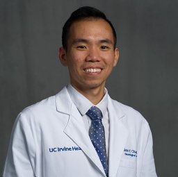 Alvin Y Chan合作论文数Dept Neurol Surg, Univ Calif Irvine13Michael Y. Oh合作论文数11
 Brian V Lien合作论文数Department of Neurological Surgery, University of California11
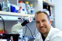 Henri Leinonen合作论文数A.I. Virtanen Institute, University of Eastern Finland11Samuel W Du合作论文数9
 Avindra Nath合作论文数Section of Infections of the Nervous System, Division of Neuroimmunology & Neurovirology, National Institute of Neurological Diseases and Stroke, National Institutes of Health9
 David R. Liu合作论文数Department of Chemistry and Chemical Biology, Faculty of Arts and Sciences, Harvard University9
 Shane Shahrestani合作论文数Keck School of Medicine, University of Southern California8
 Ronald Sahyouni合作论文数Department of Neurosurgery, University of California San Diego8
 Seth Blackshaw合作论文数Department of Otolaryngology-Head and Neck Surgery, School of Medicine, Johns Hopkins University8
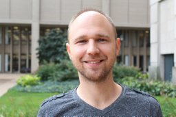 Lewandowski Dominik合作论文数8Tongguang Wang合作论文数Neuropharmacology Section, Laboratory of Pharmacology and Chemistry, NCI, National Institute of Environmental Health Sciences, National Institutes of Health Sciences, Mail Drop F1-01, P.O. Box 12233, Research Triangle Park, NC 27709, USA8Chen Yi Yang合作论文数7
 David Salom合作论文数7
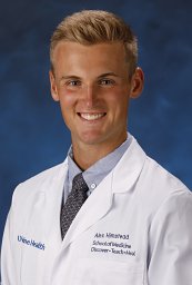 Alexander Himstead合作论文数Department of Neurosurgery, University of California Irvine7Grazyna Palczewska合作论文数Polgenix Inc7
 Jianye Zhang合作论文数School of Medicine, Case Western Reserve University7
 Aleksander Tworak合作论文数Institute of Bioorganic Chemistry, Polish Academy of Sciences7
 Andrzej Tomasz Foik合作论文数Nencki Institute of Experimental Biology, Warsaw, Poland.7
 Gregory Newby合作论文数McKusick-Nathans Department of Genetic Medicine, School of Medicine, Johns Hopkins University;Institute for Nanobiotechnology, Johns Hopkins University;Department of Biomedical Engineering, Johns Hopkins University7Hong John D合作论文数7
 Marcin Tabaka合作论文数Institute of Physical Chemistry, Polish Academy of Sciences7
 Frank P K Hsu合作论文数6Donny W. Suh合作论文数6
 Dorota Skowronska-Krawczyk合作论文数Department of Biochemistry, University of Geneva6
 Kourosh Shahraki合作论文数5Seth C Ransom合作论文数5
 Aditya Raguram合作论文数The Raguram Lab, Whitehead Institute5
 Thanh V. Hoang合作论文数Solomon H. Snyder Department of Neuroscience, Johns Hopkins University School of Medicine, Baltimore, MD 21205, USA.5
 Katelynn Tran合作论文数5
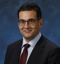 Vladimir Jivkov Kefalov合作论文数Dept Ophthalmol & Visual Sci, Washington Univ5Sander C合作论文数Department of Physics, University of Notre Dame5Fangyuan Gao合作论文数East China University of Science and Technology, East China University of Science and Technology5
 Roman Smidak合作论文数Institute of Molecular Biology, Slovak Academy of Sciences5Antônio F M Pinto合作论文数4Kiarash Golshani合作论文数4
 David E Einstein合作论文数Res Serv, VA Long Beach Healthcare Syst4
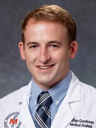 Julian Gendreau合作论文数Johns Hopkins Whiting Sch Engn4
 Samagya Banskota合作论文数Nvelop Therapeutics;College of Engineering, Boston University;Biological Design Center, Boston University4Sandra Gattas合作论文数4
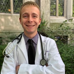 Nathan A Shlobin合作论文数Department of Neurological Surgery, Northwestern University Feinberg School of Medicine4
 Vera Ong合作论文数4Alexander V. Kolesnikov合作论文数4
 Tafreshi Ali R合作论文数4




