To evaluate clinical and cardiac magnetic resonance (CMR) short-term follow-up (FU) in patients with vaccine-associated myocarditis, pericarditis or myo-pericarditis (VAMP) following COVID-19 vaccination. We retrospectively analyzed 44 patients (2 women, mean age: 31.7 ± 15.1 years) with clinical and CMR manifestations of VAMP, recruited from 13 large tertiary national centers. Inclusion criteria were troponin raise, interval between the last vaccination dose and onset of symptoms < 25 days and symptoms-to-CMR < 20 days. 29/44 patients underwent a short-term FU-CMR with a median time of 3.3 months. Ventricular volumes and CMR findings of cardiac injury were collected in all exams. Mean interval between the last vaccination dose and the onset of symptoms was 6.2 ± 5.6 days. 30/44 patients received a vaccination with Comirnaty, 12/44 with Spikevax, 1/44 with Vaxzevria and 1/44 with Janssen (18 after the first dose of vaccine, 20 after the second and 6 after the "booster" dose). Chest pain was the most frequent symptom (41/44), followed by fever (29/44), myalgia (17/44), dyspnea (13/44) and palpitations (11/44). At baseline, left ventricular ejection fraction (LV-EF) was reduced in 7 patients; wall motion abnormalities have been detected in 10. Myocardial edema was found in 35 (79.5%) and LGE in 40 (90.9%) patients. Clinical FU revealed symptoms persistence in 8/44 patients. At FU-CMR, LV-EF was reduced only in 2 patients, myocardial edema was present in 8/29 patients and LGE in 26/29. VAMPs appear to have a mild clinical presentation, with self-limiting course and resolution of CMR signs of active inflammation at short-term follow-up in most of the cases.
Abstract Funding Acknowledgements Type of funding sources: None. Introduction Data supporting the role of stress test myocardial perfusion imaging (MPI) before non-cardiac surgery (NCS) are sparse and out of date. In particular, there is no specific evidence on the use of stress cardiac magnetic resonance (sCMR) in this setting. The aim of the present study was to evaluate the impact MPI stress test with sCMR and single-photon emission computed tomography myocardial perfusion imaging (SPECT-MPI) on surgery-related cardiac events. Method In this single-centre retrospective study, we included all patients with CAD or at least two cardiovascular risk factors undergoing intermediate-to-high-risk non-cardiac surgery. After a pre-operative cardiological assessment, including EKG and echocardiography, the indication to perform a pre-operative MPI-stress-test relied on the clinical judgement of the cardiologist. Cardiac events occurring within 30 days from surgery were evaluated. A composite primary endpoint included myocardial infarction, unstable angina, cardiac death, cardiogenic shock and pulmonary oedema. The secondary endpoint was the rate of acute coronary syndromes. Results A total of 1590 patients were included, of whom 669 underwent an MPI stress test strategy (287 sCMR, 382 SPECT-MPI). The rate of 30-day cardiac events was lower in the stress test group vs. the non-stress test group (1.2% vs 3.4%; p 0,006; figure 1). Adopting a stress test strategy showed a significant reduction of composite endpoint (OR: 0.334, IC: 0.155 – 0.766, p 0.009) and acute coronary syndrome (OR: 0.414, IC: 0.174 – 0.984, p 0.046) at multivariable analysis. Stress CMR was non-inferior to SPECT in predicting cardiac events and showed a greater accuracy to predict coronary artery revascularizations (AUC for sCMR: 0.95 with a percentage of myocardial ischemia cut-point of 5.5%; figure 2). Conclusions A stress-test strategy is associated with a lower incidence of cardiac events in high-risk patients candidate to intermediate-to-high-risk non-cardiac surgery. The rate of cardiac complications is similarly predicted by sCMR and SPECT-MPI, although sCMR is more accurate in predicting coronary artery revascularizations.
As of September 18th, 2021, global casualties due to COVID-19 infections approach 200 million, several COVID-19 vaccines have been authorized to prevent COVID-19 infection and help mitigate the spread of the virus. Despite the vast majority having safely received vaccination against SARS-COV-2, the rare complications following COVID-19 vaccination have often been life-threatening or fatal. The mechanisms underlying (multi) organ complications are associated with COVID-19, either through direct viral damage or from host immune response (i.e., cytokine storm). The purpose of this manuscript is to review the role of imaging in identifying and elucidating multiorgan complications following SARS-COV-2 vaccination—making clear that, in any case, they represent a minute fraction of those in the general population who have been vaccinated. The authors are both staunch supporters of COVID-19 vaccination and vaccinated themselves as well.
To assess clinical and cardiac magnetic resonance (CMR) imaging features of patients with peri-myocarditis following Coronavirus Disease 2019 (COVID-19) vaccination. We retrospectively collected a case series of 27 patients who underwent CMR in the clinical suspect of heart inflammation following COVID-19 vaccination, from 16 large tertiary centers. Our patient’s cohort was relatively young (36.6 ± 16.8 years), predominately included males (n = 25/27) with few comorbidities and covered a catchment area of approximately 8 million vaccinated patients. CMR revealed typical mid-subepicardial non-ischemic late gadolinium enhancement (LGE) in 23 cases and matched positively with CMR T2 criteria of myocarditis. In 7 cases, typical hallmarks of acute pericarditis were present. Short-term follow-up (median = 20 days) from presentation was uneventful for 25/27 patients and unavailable in two cases. While establishing a causal relationship between peri-myocardial inflammation and vaccine administration can be challenging, our clinical experience suggests that CMR should be performed for diagnosis confirmation and to drive clinical decision-making and follow-up. • Acute onset of dyspnea, palpitations, or acute and persisting chest pain after COVID-19 vaccination should raise the suspicion of possible myocarditis or pericarditis, and patients should seek immediate medical attention and treatment to help recovery and avoid complications. • In case of elevated troponin levels and/or relevant ECG changes, cardiac magnetic resonance should be considered as the best non-invasive diagnostic option to confirm the diagnosis of myocarditis or pericarditis and to drive clinical decision-making and follow-up.
Abstract Aims Mitral annulus disjunction (MAD) has been associated with sudden cardiac death in selected patients with arrhythmic presentation, while its clinical significance in unselected cohorts remains unknown. Our purpose was to assess the prevalence and clinical significance of MAD in consecutive patients referred to cardiovascular-magnetic-resonance (CMR). Methods and results Our population included 103 consecutive patients undergoing CMR at our Institution, between August and September 2021. MAD was defined as a ≥ 1 mm atrial displacement of the mitral leaflet hinge point in standard long-axis cine images during end-systole. MAD analysis was performed in 97 patients (feasibility = 94%) and resulted positive in 49 (51%). MAD—patients were more often males (75% vs. 57%; P = 0.045) and affected by ischaemic (35% vs. 12%, P = 0.01) and non-ischaemic cardiomyopathy (38% vs. 16%, P = 0.026) compared to MAD+ patients. No significant differences were found in terms of age, history of ventricular arrhythmias, bi-ventricular and bi-atrial volumes, bi-ventricular ejection fraction, native T1 and T2 mapping values, extracellular volume, and prevalence of late gadolinium enhancement (P > 0.05 for all) between MAD + vs. MAD—patients. MAD extent was higher in patients with mitral valve prolapse (MVP; n = 7), (3.5 ± 1.5 mm in MVP+ vs. 2.0 ± 1.0 mm in MVP– patients; P = 0.004). No significant differences were conversely found in MAD extent between patients with and without ventricular arrhythmias (2.5 ± 1.1 mm vs. 2.3 ± 1.1 mm; P = 0.815). Conclusions Our findings suggest a high prevalence of MAD in unselected cohorts of patients, with no clinical significance. Prospective studies are needed to further elucidate the interplay between MAD and malignant ventricular arrhythmias in unselected cohorts of patients.
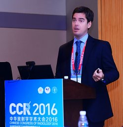 Marco Francone合作论文数Policlinico Umberto I, Università degli Studi di Roma “La Sapienza”,5
 Lorenzo Monti合作论文数4
 Luca Saba合作论文数3
 Mantini Cesare合作论文数3
 Antonio Esposito合作论文数2
 Giovanni Donato Aquaro合作论文数2Paolo Di Renzi合作论文数2
 Riccardo Cau合作论文数Department of Radiology, Azienda Ospedaliero Universitaria2Riccardo Mantovani合作论文数IRCCS, Humanitas Clin & Res Ctr2Fabio Fazzari合作论文数2
 Marco Gatti合作论文数Div Radiol, Univ Turin2
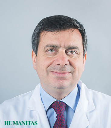 Gianluigi Condorelli合作论文数Department of Cardiovascular Diseases, Humanitas Research Hospital;Post-Graduate School of Cardiology, Humanitas University2
 Stefanini Giulio G合作论文数Dept Cardiol, Univ Hosp Bern2
 Fabrizio Ricci合作论文数Institute of Cardiology, G. d'Annunzio University2
 Andrea Barison合作论文数2
 Stefano Figliozzi合作论文数Institute of Cardiology, Università Cattolica del Sacro Cuore2Liguori Carlo合作论文数2Luigi Lovato合作论文数2Cristina Chimenti合作论文数2
 Riccardo Faletti合作论文数2
 Francesco Secchi合作论文数IRCCS Policlinico San Donato, Università degli Studi di Milano2
 Nicola Galea合作论文数Department of Radiological, Onchological and Anatomopathological Sciences, Sapienza University of Rome2
 Alessandra Volpe合作论文数1Marzia Olivieri合作论文数1Kamil Stankowski合作论文数1John Roubil合作论文数1Sara Bombace合作论文数1Davide Vignale合作论文数Radiol Unit, IRCCS San Raffaele Sci Inst1
 Mauro Gitto合作论文数1Mirko Curzi合作论文数1
 Mauro Chiarito合作论文数Dept Biomed Sci, Human Univ1Antonio Cirò合作论文数1
 Ludovica Lofino合作论文数Department of Biomedical Sciences, Humanitas University1
 Federica Catapano合作论文数1
 Caterina Beatrice Monti合作论文数Department of Biomedical Sciences for Health, Università degli Studi di Milano1
 Giulia Cundari合作论文数1G Cerrone合作论文数1
 Tevfik Ismail合作论文数Faculty of Life Sciences & Medicine, King’s College London;School of Biomedical Engineering & Imaging Sciences, King’s College London;Department of Cardiovascular Imaging, King’s College London1
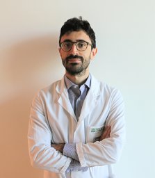 Francesco Cannata合作论文数Cardio Center, Humanitas Research Hospital;Department of Biomedical Sciences, Humanitas University;Humanitas Research Hospital, Humanitas University1
 Anna Palmisano合作论文数1
 Hua Alina合作论文数Department of Medicine, Imperial College London1Donghi Valeria合作论文数1
 Refky Nicola合作论文数Department of Radiology, University of Rochester Medical Center1Niro Fabio合作论文数1Carlo Catalano合作论文数1
 Michele Porcu合作论文数1Mahammedi Abdelkader合作论文数Russell H. Morgan Department of Radiology and Radiological Science, the Johns Hopkins Medical Institutions1
 Jasjit Suri合作论文数AtheroPoint1Daniela Fanni合作论文数University of Trieste Eye Clinic1




