Xanthine oxidoreductase is a metalloenzyme that catalyzes the final steps in purine metabolism by converting hypoxanthine to xanthine and then uric acid. Allopurinol, an analog of hypoxanthine, is widely used as an antigout drug, as xanthine oxidoreductase-mediated metabolism of allopurinol to oxypurinol leads to oxypurinol rotation in the enzyme active site and reduction of the molybdenum Mo(VI) active center to Mo(IV), inhibiting subsequent urate production. However, when oxypurinol is administered directly to a mouse model of hyperuricemia, it yields a weaker urate-lowering effect than allopurinol. To better understand its mechanism of inhibition and inform patient dosing strategies, we performed kinetic and structural analyses of the inhibitory activity of oxypurinol. Our results demonstrated that oxypurinol was less effective than allopurinol both in vivo and in vitro. We show that upon reoxidation to Mo(VI), oxypurinol binding is greatly weakened, and reduction by xanthine, hypoxanthine, or allopurinol is required for reformation of the inhibitor-enzyme complex. In addition, we show oxypurinol only weakly inhibits the conversion of hypoxanthine to xanthine and is therefore unlikely to affect the feedback inhibition of de novo purine synthesis. Furthermore, we observed weak allosteric inhibition of purine nucleoside phosphorylase by oxypurinol which has potentially adverse effects for patients. Considering these results, we propose the single-dose method currently used to treat hyperuricemia can result in unnecessarily high levels of allopurinol. While the short half-life of allopurinol in blood suggests that oxypurinol is responsible for enzyme inhibition, we anticipate multiple, smaller doses of allopurinol would reduce the total allopurinol patient load.
l‐2‐Haloacid dehalogenases, industrially and environmentally important enzymes that catalyse cleavage of the carbon‐halogen bond in S‐2‐halocarboxylic acids, were known to hydrolyse chlorinated, brominated and iodinated substrates but no activity towards fluorinated compounds had been reported. A screen for novel dehalogenase activities revealed four l‐2‐haloacid dehalogenases capable of defluorination. We now report crystal structures for two of these enzymes, Bpro0530 and Rha0230, as well as for the related proteins PA0810 and RSc1362, which hydrolyse chloroacetate but not fluoroacetate, all at ∼2.2 Å resolution. Overall structure and active sites of these enzymes are highly similar. In molecular dynamics (MD) calculations, only the defluorinating enzymes sample more compact conformations, which in turn allow more effective interactions with the small fluorine atom. Structural constraints, based on X‐ray structures and MD calculations, correctly predict the defluorination activity of the homologous enzyme ST2570.
Protein tyrosine phosphatases constitute a family of cytosolic and receptor-like signal transducing enzymes that catalyze the hydrolysis of phospho-tyrosine residues of phosphorylated proteins. PTP1B, encoded by PTPN1, is a key negative regulator of insulin and leptin receptor signaling, linking it to two widespread diseases: type 2 diabetes mellitus and obesity. Here, we present crystal structures of the PTP1B apo-enzyme and a complex with a newly identified allosteric inhibitor, 2-(2,5-dimethyl-pyrrol-1-yl)5-hydroxy-benzoic acid, designated as P00058. The inhibitor binding site is located about 18A away from the active center. However, the inhibitor causes significant re-arrangements in the active center of enzyme: residues 45-50 of catalytic Tyr-loop are shifted at their Ca-atom positions by 2.6 to 5.8 A. We have identified an event of allosteric signal transfer from the inhibitor to the catalytic area using molecular dynamic simulation. Analyzing change of complex structure along the fluctuation trajectory we have found the large Ca-atom shifts in external strand, residues 25-40, which occur at the same time with the shifts in adjacent catalytic p-Tyr-loop. Coming of the signal to this loop arises due to dynamic fluctuation of protein structure at about 4.0 nanoseconds after the inhibitor takes up its space.
For the two proteins myoglobin and fluoroacetate dehalogenase, we present a systematic comparison of crystallographic diffraction data collected by serial femtosecond (SFX) and serial synchrotron crystallography (SSX). To maximize comparability, we used the same batch of micron-sized crystals, the same sample delivery device, and the same data analysis software. Overall figures of merit indicate that the data of both radiation sources are of equivalent quality. For both proteins, reasonable data statistics can be obtained with approximately 5000 room-temperature diffraction images irrespective of the radiation source. The direct comparability of SSX and SFX data indicates that the quality of diffraction data obtained from these samples is linked to the properties of the crystals rather than to the radiation source. Therefore, for other systems with similar properties, time-resolved experiments can be conducted at the radiation source that best matches the desired time resolution.
ORGANIC CHEMISTRY Schulz, Eike (Max Planck Institute for Structure and Dynamics of Matter (MPSD), Hamburg, GER); Mehrabi, Pedram (Max Planck Institute for Structure and Dynamics of Matter (MPSD), Hamburg, GER); Dsouza, Raison (Max Planck Institute for Structure and Dynamics of Matter (MPSD), Hamburg, GER); Müller-Werkmeister, Henrike M. (University of Potsdam, Potsdam, GER); Tellkamp, Friedjof (Max Planck Institute for Structure and Dynamics of Matter (MPSD), Hamburg, GER); Miller, R. J. Dwayne (Max Planck Institute for the Structure and Dynamics of Matter, Hamburg, GER); Pai, Emil F. (University of Toronto, Toronto, CAN)
Cellular stress response has dual aspects; cell-protective or lethal. Mitochondria have their unique organellar response termed "mitochondrial unfolded protein response (UPRmt)" induced by damaged mitochondrial (mt) matrix proteins. While recent discoveries have successfully targeted BCL2, a regulator of mt integrity in acute myeloid leukemia (AML), the significance of UPRmt is unknown. We hypothesized that priming UPRmt towards cell death would be a novel therapeutic strategy for AML. UPRmt is generally induced by dysregulation of mt protein pools. Therefore, to test if UPRmt signaling is also operational in AML cells, we selected classical or putative UPRmt inducers; the mt translation inhibitors tetracycline and tigecycline, the mt protein transport inhibitor MitoBlock6, and the mtDNA damaging agent ethidium bromide. In OCI-AML3 and HL60 cells, these agents indeed induced the transcription factor ATF5, which was reported as a central inducer of UPRmt, and its targets (e.g., LonP, HSPA9), triggering apoptosis in AML cells. In addition, we here report imipridones (ONC201 and ONC212), the activators of mt protein degradation, as novel UPRmt inducers. We recently reported that imipridones non-covalently bind the mt protease ClpP and allosterically activate it. They induced prominent apoptosis in primary AML progenitor and leukemia initiating cells (LICs) in vitro and in vivo, but not in normal bone marrow cells, following "mitochondrial proteolysis" with reduction of selective mt matrix proteins (e.g., SDHB, NDUFA12) and resultant inhibition of oxidative phosphorylation (Oxphos) (Ishizawa, Zarabi et al, Cancer Cell 2019). We then postulated that dysregulation of mt protein pools by mitochondrial proteolysis can also induce UPRmt. Indeed, our gene expression profiles of ONC201-treated Z138 and Jeko-1 cells were highly enriched for previously published UPRmt gene signatures, and UPRmt effectors were induced also in AML cells. Of potentially high clinical significance is the finding of synergistic anti-leukemia effects of imipridones when combined with the selective BCL2 inhibitor venetoclax, in vitro and in vivo (Ishizawa et al. Science Signaling 2016, and Nii et al. Blood 2019). However, its underlying molecular mechanism is unclear. Since BCL2 is reported to be induced by UPRmt, we hypothesized that BCL2 is critical for the ClpP-mediated UPRmt to have the cell protective effects, contrary to lethal effects, as dual aspects of stress response. We utilized the tetracycline-inducible system of an activated mutant (Y118A) form of ClpP in OCI-AML3 cells, and demonstrated that venetoclax treatment sensitizes OCI-AML3 cells to genetic activation of ClpP towards apoptosis. Furthermore, other UPRmt inducers (tetracycline, tigecycline, and MitoBlock6) in combination with venetoclax also synergistically induced apoptosis in AML cells, suggesting that BCL2 inhibition and UPRmt induction generally exerts synergistic anti-leukemia effects. We next focused on the enhanced effect observed for the combination of imipridones with venetoclax as compared to other UPRmt inducers, searching for other targets that could further enhance the synergy. We then hypothesized that the synergism between ClpP activation and BCL2 inhibition involves SDHB, a respiratory chain complex II subunit degraded by activated ClpP but not targeted by any of other UPRmt inducers. Consistently, SDHB knockdown sensitized OCI-AML3 cells to venetoclax-induced apoptosis, indicating that SDHB reduction and UPRmt by ClpP activation concomitantly enhance the cell lethality by BCL2 inhibition. Collectively, UPRmt is a new potential therapeutic target for AML, which significantly enhances the cell death effects of BCL2 inhibition on AML cells. In particular, ClpP activation induces UPRmt and, concomitantly, downregulates SDHB, thus targeting the respiratory chain complex II, which results in improved synergistic leukemia cell apoptosis when combined with BCL2 inhibition. Oxphos is also a hallmark of drug resistant AML stem cells, which supports the notion that Oxphos inhibition by this combination targets LICs. Based on promising preclinical anti-tumor efficacy, ONC201 as a single agent is being evaluated in early phase clinical trials, showing clinical responses in AML and midline gliomas. A clinical trial testing the combinatorial strategy of targeting ClpP and Bcl-2 is under development. Disclosures Borthakur: Novartis: Research Funding; NKarta: Consultancy; Eisai: Research Funding; Oncoceutics: Research Funding; BioLine Rx: Consultancy, Membership on an entity's Board of Directors or advisory committees, Research Funding; Cyclacel: Research Funding; Strategia Therapeutics: Research Funding; Eli Lilly and Co.: Research Funding; Arvinas: Research Funding; Merck: Research Funding; AstraZeneca: Research Funding; PTC Therapeutics: Consultancy; Agensys: Research Funding; Argenx: Membership on an entity's Board of Directors or advisory committees; FTC Therapeutics: Membership on an entity's Board of Directors or advisory committees; GSK: Research Funding; Incyte: Research Funding; Janssen: Research Funding; AbbVie: Research Funding; BMS: Research Funding; Oncoceutics, Inc.: Research Funding; Bayer Healthcare AG: Research Funding; BioTheryX: Membership on an entity's Board of Directors or advisory committees; Tetralogic Pharmaceuticals: Research Funding; Cantargia AB: Research Funding; Polaris: Research Funding; Xbiotech USA: Research Funding. Stogniew:Oncoceutics, Inc.: Employment. Oster:Oncoceutics, Inc.: Employment. Kantarjian:BMS: Research Funding; AbbVie: Honoraria, Research Funding; Ariad: Research Funding; Amgen: Honoraria, Research Funding; Jazz Pharma: Research Funding; Pfizer: Honoraria, Research Funding; Cyclacel: Research Funding; Immunogen: Research Funding; Agios: Honoraria, Research Funding; Actinium: Honoraria, Membership on an entity's Board of Directors or advisory committees; Novartis: Research Funding; Takeda: Honoraria; Astex: Research Funding; Daiichi-Sankyo: Research Funding. Schimmer:Novartis Pharmaceuticals: Consultancy; Medivir Pharmaceuticals: Research Funding; Jazz Pharmaceuticals: Consultancy; Otsuka Pharmaceuticals: Consultancy. Andreeff:Eutropics: Equity Ownership; Daiichi Sankyo, Inc.: Consultancy, Patents & Royalties: Patents licensed, royalty bearing, Research Funding; Jazz Pharmaceuticals: Consultancy; Celgene: Consultancy; Aptose: Equity Ownership; Reata: Equity Ownership; 6 Dimensions Capital: Consultancy; AstaZeneca: Consultancy; Amgen: Consultancy; Breast Cancer Research Foundation: Research Funding; CPRIT: Research Funding; NIH/NCI: Research Funding; Center for Drug Research & Development: Membership on an entity's Board of Directors or advisory committees; Cancer UK: Membership on an entity's Board of Directors or advisory committees; NCI-CTEP: Membership on an entity's Board of Directors or advisory committees; German Research Council: Membership on an entity's Board of Directors or advisory committees; Leukemia Lymphoma Society: Membership on an entity's Board of Directors or advisory committees; NCI-RDCRN (Rare Disease Cliln Network): Membership on an entity's Board of Directors or advisory committees; CLL Foundation: Membership on an entity's Board of Directors or advisory committees; BiolineRx: Membership on an entity's Board of Directors or advisory committees; Oncolyze: Equity Ownership; Oncoceutics: Equity Ownership; Senti Bio: Equity Ownership, Membership on an entity's Board of Directors or advisory committees. Ishizawa:Daiichi Sankyo: Patents & Royalties: Joint submission with Daiichi Sankyo for a PTC patent titled "Predictive Gene Signature in Acute Myeloid Leukemia for Therapy with the MDM2 Inhibitor DS-3032b," United States, 62/245667, 10/23/2015, Filed.
Bacterial ClpP is a highly conserved, cylindrical, self-compartmentalizing serine protease required for maintaining cellular proteostasis. Small molecule acyldepsipeptides (ADEPs) and activators of self-compartmentalized proteases 1 (ACP1s) cause dysregulation and activation of ClpP, leading to bacterial cell death, highlighting their potential use as novel antibiotics. Structural changes in Neisseria meningitidis and Escherichia coli ClpP upon binding to novel ACP1 and ADEP analogs were probed by X-ray crystallography, methyl-TROSY NMR, and small angle X-ray scattering. ACP1 and ADEP induce distinct conformational changes in the ClpP structure. However, reorganization of electrostatic interaction networks at the ClpP entrance pores is necessary and sufficient for activation. Further activation is achieved by formation of ordered N-terminal axial loops and reduction in the structural heterogeneity of the ClpP cylinder. Activating mutations recapitulate the structural effects of small molecule activator binding. Our data, together with previous findings, provide a structural basis for a unified mechanism of compound-based ClpP activation.
A comprehensive understanding of protein function demands correlating structure and dynamic changes. Using time-resolved serial synchrotron crystallography, we visualized half-of-the-sites reactivity and correlated molecular-breathing motions in the enzyme fluoroacetate dehalogenase. Eighteen time points from 30 milliseconds to 30 seconds cover four turnover cycles of the irreversible reaction. They reveal sequential substrate binding, covalent-intermediate formation, setup of a hydrolytic water molecule, and product release. Small structural changes of the protein mold and variations in the number and placement of water molecules accompany the various chemical steps of catalysis. Triggered by enzyme-ligand interactions, these repetitive changes in the protein framework's dynamics and entropy constitute crucial components of the catalytic machinery.
Trypanothione reductase (TR) plays a key role in the unique redox metabolism of trypanosomatids, the causative agents of human African trypanosomiasis (HAT), Chagas' disease, and leishmaniases. Introduction of a new, lean propargylic vector to a known class of TR inhibitors resulted in the strongest reported competitive inhibitor of Trypanosoma (T.) brucei TR, with an inhibition constant K-i of 73 nm, which is fully selective against human glutathione reductase (hGR). The best ligands exhibited in vitro IC50 values (half-maximal inhibitory concentration) against the HAT pathogen, T. brucei rhodesiense, in the mid-nanomolar range, reaching down to 50 nm. X-Ray co-crystal structures confirmed the binding mode of the ligands and revealed the presence of a HEPES buffer molecule in the large active site. Extension of the propargylic vector, guided by structure-based design, to replace the HEPES buffer molecule should give inhibitors with low nanomolar K-i and IC50 values for in vivo studies.
 Lakshmi Kotra合作论文数Leslie Dan Faculty of Pharmacy, University of Toronto;Center for Molecular Design and Preformulations, University Health Network;FSD Pharma Inc.;Lucid Psycheceuticals Inc.44
 N.Y. Chirgadze合作论文数27
 Angelica M. Bello合作论文数Toronto General Research Institute, University Health Network27V. Romanov合作论文数25B.T. Eger合作论文数25Pedram Mehrabi合作论文数Department for Atomically Resolved Dynamics, Max-Planck-Institute for Structure and Dynamics of Matter, Luruper Chaussee 149, 22761 Hamburg, Germany25R. Lam合作论文数21
 Wolfgang Kabsch合作论文数Max Planck Institute for Medical Research19Bryson Steve合作论文数18
 Alfred Wittinghofer合作论文数Max Planck Institute of Molecular Physiology18Vivian Saridakis合作论文数Department of Biology, York University17
 Roger S. Goody合作论文数Max Planck Institute of Molecular Physiology16K.P. Battaile合作论文数15E.C. Schulz合作论文数15Ewa Poduch合作论文数14
 Aled Edwards合作论文数Department of Medical Biophysics, Temerty Faculty of Medicine, University of Toronto;Structural Genomics Consortium;M4K Pharma;McGill University14Masahiro Fujihashi合作论文数14Matthew S. Kimber合作论文数Department of Molecular and Cellular Biology, University of Guelph14
 Takeshi Nishino合作论文数The University of Tokyo13
 Ilme Schlichting合作论文数Department of Biosciences, Heidelberg University;Department of Physics and Astronomy, Heidelberg University;Department of Biomolecular Mechanisms, Max Planck Institute for Medical Research13Ning Wu合作论文数12K. Lam合作论文数11
 Dinesh Christendat合作论文数Department of Cell & Systems Biology, Faculty of Arts and Science, University of Toronto11Chirgadze Nickolay Y合作论文数11
 Jean-Philippe Julien合作论文数Department of Biochemistry, Temerty Faculty of Medicine, University of Toronto11Lianhu Wei合作论文数Toronto General Research Institute, University Health Network11Bryan T. Eger合作论文数11
 Jacob John合作论文数Department of Biosystems and Agricultural Engineering, University of Minnesota11W. Qiu合作论文数10
 Dwayne Miller合作论文数Department of Chemistry, Faculty of Arts & Science, University of Toronto10Ute Krengel合作论文数10V.S. Stoll合作论文数9
 Vladimir Romanov合作论文数9Battaile Kevin P合作论文数9Ken Okamoto合作论文数9
 Régis Pomès合作论文数Department of Biochemistry, Temerty Faculty of Medicine, University of Toronto9
 Braden Sweeting合作论文数9I. Beletskaya合作论文数8J. Vodsedalek合作论文数8A. Dharamsi合作论文数8Ondrej Halgas合作论文数8R. Luise Krauth-Siegel合作论文数8M. Soloveychik合作论文数7F. Diederich合作论文数7
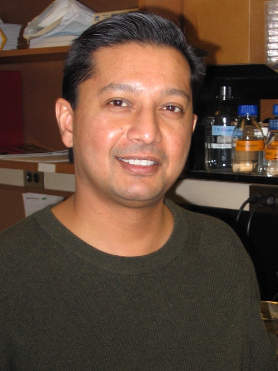 Avi Chakrabartty合作论文数Department of Biochemistry, Temerty Faculty of Medicine, University of Toronto7Payandeh Jian合作论文数7
 Ian Crandall合作论文数Department of Laboratory Medicine and Pathobiology, Temerty Faculty of Medicine, University of Toronto7François Diederich合作论文数7M. Fujihashi合作论文数6




