Variants in the LRRK2 and GBA1 genes are among the most common risk factors associated with Parkinson's disease (PD). Both patients carrying PD-associated variants in GBA1, encoding lysosomal enzyme glucocerebrosidase (GCase), and a subset of non-carrier patients have been shown to have reduced GCase enzymatic activity, suggesting that reduced GCase activity may be a feature of both genetic and a subset of sporadic PD. However, the effect of PD-associated variants in LRRK2, encoding a serine/threonine kinase, on GCase activity remains controversial, with conflicting results in various tissues and cell types. Moreover, rare patients carrying both GBA1 and LRRK2 risk alleles seem to have a more benign disease course than carriers of GBA1 variants alone, suggesting a complex interplay between these two genes in PD. Here, we evaluate the effect of LRRK2 kinase activity on GCase activity in human induced pluripotent stem cell (iPSC)-derived microglia (iMGs), a PD-relevant brain cell type expressing high levels of LRRK2. Using CRISPR editing, isogenic control iPSC lines were generated to match PD patient-derived iPSC lines harbouring the LRRK2 p.G2019S, p.M1646T, or p.N551K-p.R1398H protective haplotype variants. Whereas iMGs harbouring the p.M1646T variant, and the protective haplotype, respectively increased and decreased phosphorylation of canonical LRRK2 substrate, Rab10, GCase protein levels and activity were not altered in any of the LRRK2 variant lines. Additionally, whereas pharmacological inhibition of LRRK2 kinase activity had no impact on GCase activity in iMGs under basal conditions, it attenuated the increase in GCase activity elicited in response to interferon γ (IFNγ) treatment. Moreover, GCase activity induced by IFNγ was reduced in PD risk LRRK2 p.M1646T iMGs and increased in p.N551K-p.R1398H protective haplotype iMGs compared to their isogenic corrected controls, congruent with their respective effects on LRRK2 kinase activity and PD risk. Thus, our data suggest a role for LRRK2 kinase activity in regulation of GCase activity in response to neuroinflammation.
There are around 100 genes or copy-number variations used in genetic testing for autism spectrum disorder (ASD)1,2. The established genes are protein coding, and the associated phenotypes usually extend beyond sociobehavioural traits seen in autism, including cognitive/medical complexities and attention deficit hyperactivity disorder (ADHD)3,4. We examined whole-genome sequencing data in cases of ASD (9,349) and controls (8,332) and identify 27 male individuals with ASD with X-chromosome microdeletions that implicate the long non-coding RNA PTCHD1-AS as an ASD-susceptibility gene (odds ratio = 2.56, P = 0.01). Two Ptchd1-as-knockout mouse models, which were created by disrupting/deleting the evolutionarily conserved exon 3, show ASD-like features in male mice, including increased repetitive behaviours and impaired social behaviour and communication without cognitive comorbidities or ADHD-like behaviours. Hippocampus-dependent synaptic function, complex learning and locomotor activity are unaffected in knockout mice. Native nuclear-enriched mouse Ptchd1-as showed sustained expression from postnatal day 7 onwards in the dorsal striatum, a predominantly GABAergic brain region that is implicated in ASD5. Multi-omics analysis revealed transcriptomic alterations in striatal oligodendrocytes, astrocytes and neurons impacting myelination and synaptic plasticity. Disrupting Ptchd1-as led to reductions in conventional protein kinase C (cPKC) isoforms, altered SRC and GSK-3α/β phosphorylation and enhanced striatal synaptic plasticity (long-term potentiation and long-term depression). Together, these findings implicate striatal molecular and circuit-level dysregulation through PTCHD1-AS in ASD aetiology.
Biomedical research has been transformed by the discovery of human induced pluripotent stem cells (iPSCs), providing unlimited access to patient-specific human cell types for disease modeling and mechanistic studies [...]
The intercellular transmission of α-synuclein contributes to Parkinson's disease pathology. Yet, the mechanisms of α-synuclein spread are not fully understood. Here we used live-cell microscopy to examine the impact of Parkinson's disease associated lipid alterations on α-synuclein release. We discovered that increased glucosylceramides as a consequence of reduced β-glucocerebrosidase activity induce ectosome shedding from primary neurons and from dopaminergic neurons derived from induced pluripotent stem cells of a patient with Parkinson's disease harbouring mutations in GBA1 (N370S, L444P and W378G) and LRRK2 (G2019S and R1441H) compared with their isogenic control. We show that elevated glucosylceramide and the pharmacological inhibition of β-glucocerebrosidase similarly increase vesicle release and uptake by other neurons in mouse brains. Finally, we show that ectosomes are loaded with α-synuclein and lead to the transmission of α-synuclein pathology to neighbouring neurons. These data reveal ectosomes as a major route for α-synuclein transmission in Parkinson's disease.
A significant challenge in ALS research is the heterogeneity of the disease. Even mutations within the same gene can lead to different disease prognosis. For instance, in silico protein modeling predicts distinct properties for distinct SOD1 mutations. With this in mind, in this study, we generated and characterized 3D iPSC-derived MN spheroids carrying homozygous knock-in SOD1 mutations (D90A and G93A), as well as a double mutation (D90A/G93A), to evaluate potential synergistic effects. An isogenic control line with the same genetic background was used for phenotypic comparisons with the knock-in variants. Mutant SOD1 MN spheroids exhibited multiple ALS-related phenotypes including altered SOD1 expression, reduced cell viability, downregulation of neurofilament (NF) subunit expression, hypoactivity, and altered burst activity. Our results highlight the advantages of using 3D MN spheroids as a disease model and stress the importance of considering phenotype variability at the genetic level in ALS. ### Competing Interest Statement The authors have declared no competing interest.
There are ~100 genes or copy number variants (CNVs) used in genetic testing for Autism Spectrum Disorder (ASD, or autism). These genes are protein-coding, and the associated phenotypes often extend beyond socio-behavioral traits seen in autism including cognitive/medical complexities, epilepsy, and ADHD. Here, we characterize 27 males with ASD through whole genome sequencing (WGS), delineating X-chromosome microdeletions that implicate the long non-coding RNA (lncRNA) PTCHD1-AS as an ASD-susceptibility gene (OR=2.56, p=0.01). Two Ptchd1-as knockout (KO) murine models, created by removing the evolutionarily conserved exon-3, show ASD-like features in males, increasing repetitive behaviors and impairing typical social behavior and communication without overt cognitive comorbidities or ADHD-like behaviors. Hippocampus-dependent synaptic function, complex learning, and locomotor activity are unaffected in KO mice. Native nuclear-enriched mouse Ptchd1-as showed sustained expression from post-natal day-7 onward in the dorsal striatum, a predominantly GABAergic brain region implicated in ASD. Multi-omics revealed transcriptomic alterations in striatal oligodendrocyte, astrocyte and neurons impacting myelination and plasticity pathways. Disrupting Ptchd1-as led to reductions in conventional Protein Kinase-C, altered Src and GSK3α/β phosphorylation, and an enhancement of synaptic plasticity (long-term potentiation and long-term depression). Together, these findings implicate striatal molecular and circuit level dysregulation via Ptchd1-as in ASD etiology.
Loss-of-function mutations in the PINK1 and PRKN genes are the most common cause of early-onset Parkinson disease (PD). The encoded enzymatic pair selectively identifies, labels, and targets damaged mitochondria for degradation via the macroautophagy/autophagy-lysosome system (mitophagy). This pathway is cytoprotective and efforts to activate mitophagy are pursued as therapeutic avenues to combat PD and other neurodegenerative disorders. When mitochondria are damaged, the ubiquitin kinase PINK1 accumulates and recruits PRKN from the cytosol to activate the E3 ubiquitin ligase from its auto-inhibited conformation. We have previously designed several mutations that effectively derepress the structure of PRKN and activate its enzymatic functions in vitro. However, it remained unclear how these PRKN-activating mutations would perform endogenously in cultured neurons or in vivo in the brain. Here, we gene-edited neural progenitor cells and induced pluripotent stem cells to express PRKN-activating mutations in dopaminergic cultures. All tested PRKN-activating mutations indeed enhanced the enzymatic activity of PRKN in the absence of exogenous stress, but their hyperactivity was linked to their own PINK1-dependent degradation. Strikingly, in vivo in a mouse model expressing an equivalent activating mutation, we find the same relationship between PRKN enzymatic activity and protein stability. We conclude that PRKN degradation is the consequence of its structural derepression and enzymatic activation, thus resulting only in a temporary gain of activity. Our findings imply that pharmacological activation of endogenous PRKN will lead to increased turnover and suggest that additional considerations might be necessary to achieve sustained E3 ubiquitin ligase activity for disease treatment.Abbreviations: BSA: bovine serum album, CCCP: carbonyl cyanide 3-chlorophenylhydrazone; ECL: electrochemiluminescence; EGF: epidermal growth factor; ELISA: enzyme-linked immunosorbent assay; FGF: fibroblast growth factor; iPSC: induced pluripotent stem cell; KI: knock-in; KO: knockout; MAP2: microtubule associated protein 2; MFN2: mitofusin 2; MSD: Meso Scale Discovery; mt-Keima: mitochondrial targeted Keima; NPC: neural progenitor cell; PD: Parkinson disease; PDH: pyruvate dehydrogenase; p-S65-PRKN: Serine 65 phosphorylated PRKN; p-S65-Ub: Serine 65 phosphorylated ubiquitin; REP: repressor element of PRKN; TH: tyrosine hydroxylase; TX: Triton X-100, Ub: ubiquitin; UBL: ubiquitin-like; WT: wild-type.
Background & AimParkinson's disease (PD) is a neurodegenerative disorder with over fifty genes linked to its development. Only a few of those genes have been well characterized, partially due to the lack of research-grade antibodies, highlighting the need for efficient tools to study them. In this project, we use a split luciferase-based reporter to tag PD-linked genes, which is composed of the small subunit HiBiT and the larger LgBiT. When both subunits are present, they bind and emit luminescence, allowing ultrasensitive detection of proteins in a minimally invasive and quantitative manner that is applicable in both end points assays or real-time monitoring in live cells. In gene therapy regulatory context, HiBiT fusions in N-terminal can be very useful for the detection of truncated proteins, with potential residual and/or harmful activity, which might have escaped nonsense-mediated decay (NMD) following indel/frameshift-induced CRISPR knockouts that rely on premature termination codons.Methods, Results & ConclusionA template plasmid containing LgBiT expressed by a doxycycline-inducible promoter (iLgBiT) was designed to be inserted at the CLYBL safe harbor site into human iPSCs using CRISPR. Digital PCR (ddPCR) was used to screen edited cells, and a clone we named M2E3 was found to be monoclonal and heterozygous for the expression of iLgBiT. We showed a >40 fold increase in LgBiT transcript levels and a >10 fold increase in LgBiT protein expression measured in a luminescence assay. We validated full functionality and integrity of iLgBiT in M2E3 iPSCs and derived dopaminergic neurons. Then, the HiBiT subunit was successfully fused to several PD associated genes, namely SNCA, LRRK2, PRKN, VPS35, VPS13C, PINK1, GPNMB1 and GBA, in M2E3 iPSCs using CRISPR. We confirmed that M2E3-GBA-HiBiT iPSCs and neurons both show increased luminescence upon dox treatments. Furthermore, the therapeutic Ambroxol, known to increase lysosomal glucocerebrosidase (GCase; encoded by GBA) activity, was able to significantly upregulate GCase levels, detected by both luminescence assay and Western blot, in our M2E3-GBA-HiBiT dopaminergic neurons. This system will help with screening new therapeutics for PD. Antibody reproducibility issues and PD protein integrity will also be addressed as part of regulation of candidate gene therapies. Finally, this tool will serve as a regulation surveillance system to detect the expression of harmful truncated peptides, otherwise undetectable, that may have escaped NMD in knockouts.
With emerging genetic association studies, new genes and pathways are revealed as causative factors in the development of Parkinson’s disease (PD). However, many of these PD genes are poorly characterized in terms of their function, subcellular localization, and interaction with other components in cellular pathways. This represents a major obstacle towards a better understanding of the molecular causes of PD, with deeper molecular studies often hindered by a lack of high-quality, validated antibodies for detecting the corresponding proteins of interest. In this study, we leveraged the nanoluciferase-derived LgBiT-HiBiT system by generating a cohort of tagged PD genes in both induced pluripotent stem cells (iPSCs) and iPSC-derived neuronal cells. To promote luminescence signals within cells, a master iPSC line was generated, in which LgBiT expression is under the control of a doxycycline-inducible promoter. LgBiT could bind to HiBiT when present either alone or when tagged onto different PD-associated proteins encoded by the genes GBA1, GPNMB, LRRK2, PINK1, PRKN, SNCA, VPS13C, and VPS35. Several HiBiT-tagged proteins could already generate luminescence in iPSCs in response to the doxycycline induction of LgBiT, with the enzyme glucosylceramidase beta 1 (GCase), encoded by GBA1, being one such example. Moreover, the GCase chaperone ambroxol elicited an increase in the luminescence signal in HiBiT-tagged GBA1 cells, correlating with an increase in the levels of GCase in dopaminergic cells. Taken together, we have developed and validated a Doxycycline-inducible luminescence system to serve as a sensitive assay for the quantification, localization, and activity of HiBiT-tagged PD-associated proteins with reliable sensitivity and efficiency.
Recent years have witnessed unprecedented progress in therapeutic gene editing, revolutionizing the approach to treating genetic disorders. In this comprehensive review, we discuss the progression of milestones leading to the emergence of the clustered regularly interspaced short palindromic repeats (CRISPR)-based technology as a powerful tool for precise and targeted modifications of the human genome. CRISPR-Cas9 nuclease, base editing, and prime editing have taken center stage, demonstrating remarkable precision and efficacy in targeted ex vivo and in vivo genomic modifications. Enhanced delivery systems, including viral vectors and nanoparticles, have further improved the efficiency and safety of therapeutic gene editing, advancing their clinical translatability. The exploration of CRISPR-Cas systems beyond the commonly used Cas9, such as the development of Cas12 and Cas13 variants, has expanded the repertoire of gene editing tools, enabling more intricate modifications and therapeutic interventions. Outstandingly, prime editing represents a significant leap forward, given its unparalleled versatility and minimization of off-target effects. These innovations have paved the way for therapeutic gene editing in a multitude of previously incurable genetic disorders, ranging from monogenic diseases to complex polygenic conditions. This review highlights the latest innovative studies in the field, emphasizing breakthrough technologies in preclinical and clinical trials, and their applications in the realm of precision medicine. However, challenges such as off-target effects and ethical considerations remain, necessitating continued research to refine safety profiles and ethical frameworks.
Parkinson’s disease (PD) is a neurodegenerative disorder that results in the loss of dopaminergic neurons in the substantia nigra pars compacta. Despite advances in understanding PD, there is a critical need for novel therapeutics that can slow or halt its progression. Induced pluripotent stem cell (iPSC)-derived dopaminergic neurons have been used to model PD but measuring differences between PD and control cells in a robust, reproducible, and scalable manner remains a challenge. In this study, we developed a binary classifier convolutional neural network (CNN) to accurately classify microscopy images of PD models and matched control cells. We acquired images of iPSC-derived neural precursor cells (NPCs) and dopaminergic (DANs) and trained multiple CNN models comparing control cells to genetic and chemical models of PD. Our CNN accurately predicted whether control NPC cells were treated with the PD-inducing pesticide rotenone with 97.60% accuracy. We also compared control to a genetic model of PD (deletion of the Parkin gene) and found a predictive accuracy of 86.77% and 95.47% for NPC and DAN CNNs, respectively. Our cells were stained for nuclei, mitochondria, and plasma membrane, and we compared the contribution of each to the CNN’s accuracy. Using all three features together produced the best accuracy, but nuclear staining alone produced a highly predictive CNN. Our study demonstrates the power of deep learning and computer vision for analyzing complex PD-related phenotypes in DANs and suggests that these tools hold promise for identifying new targets for therapy and improving our understanding of PD.
Health Canada regulates gene therapy products and many cell therapy products as biological drugs under the Canadian Food and Drugs Act and its attendant regulations. Cellular products that meet certain criteria, including minimal manipulation and homologous use, may be subjected to a standards-based approach under the Safety of Human Cells, Tissues and Organs for Transplantation Regulations. The manufacture and clinical testing of cell and gene therapy products (CGTP) presents many challenges beyond those for protein biologics. Cells cannot be subjected to pathogen removal or inactivation procedures and must frequently be administered shortly after final formulation. Viral vector design and manufacturing control are critically important to overall product quality and linked to safety and efficacy in patients through concerns such as replication competence, vector integration, and vector shedding. In addition, for many CGTP, the value of nonclinical studies is largely limited to providing proof of concept, and the first meaningful data relating to appropriate dosing, safety parameters, and validity of surrogate or true determinants of efficacy must come from carefully designed clinical trials in patients. Addressing these numerous challenges requires application of various risk mitigation strategies and meeting regulatory expectations specifically adapted to the product types. Regulatory cooperation and harmonization at an international level are essential for progress in the development and commercialization of these products. However, particularly in the area of cell therapy, new regulatory paradigms may be needed to harness the benefits of clinical progress in situations where the resources and motivation to pursue a typical drug product approval pathway may be lacking. This chapter is dedicated to provide an overview of Health Canada regulatory oversight of CGTP.
Fragile X syndrome (FXS) is caused by a repression of the FMR1 gene that codes the Fragile X mental retardation protein (FMRP), an RNA binding protein involved in processes that are crucial for proper brain development. To better understand the consequences of the absence of FMRP, we analyzed gene expression profiles and activities of cortical neural progenitor cells (NPCs) and neurons obtained from FXS patients’ induced pluripotent stem cells (IPSCs) and IPSC-derived cells from FMR1 knock-out engineered using CRISPR-CAS9 technology. Multielectrode array recordings revealed in FMR1 KO and FXS patient cells, decreased mean firing rates; activities blocked by tetrodotoxin application. Increased expression of presynaptic mRNA and transcription factors involved in the forebrain specification and decreased levels of mRNA coding AMPA and NMDA subunits were observed using RNA sequencing on FMR1 KO neurons and validated using quantitative PCR in both models. Intriguingly, 40% of the differentially expressed genes were commonly deregulated between NPCs and differentiating neurons with significant enrichments in FMRP targets and autism-related genes found amongst downregulated genes. Our findings suggest that the absence of FMRP affects transcriptional profiles since the NPC stage, and leads to impaired activity and neuronal differentiation over time, which illustrates the critical role of FMRP protein in neuronal development.
Background The X-linked PTCHD1 locus is strongly associated with autism spectrum disorder (ASD). Males who carry chromosome microdeletions of PTCHD1 antisense long non-coding RNA ( PTCHD1-AS )/DEAD-box helicase 53 ( DDX53 ) have ASD, or a sub-clinical form called Broader Autism Phenotype. If the deletion extends beyond PTCHD1-AS / DDX53 to the next gene, PTCHD1 , which is protein-coding, the individuals typically have ASD and intellectual disability (ID). Three male siblings with a 90 kb deletion that affects only PTCHD1-AS (and not including DDX53 ) have ASD. We performed a functional analysis of DDX53 to examine its role in NGN2 neurons. Methods We used the clustered regularly interspaced short palindromic repeats (CRISPR) gene editing strategy to knock out DDX53 protein by inserting 3 termination codons (3TCs) into two different induced pluripotent stem cell (iPSC) lines. DDX53 CRISPR-edited iPSCs were differentiated into cortical excitatory neurons by Neurogenin 2 (NGN-2) directed differentiation. The functional differences of DDX53 -3TC neurons compared to isogenic control neurons with molecular and electrophysiological approaches were assessed. Results Isogenic iPSC-derived control neurons exhibited low levels of DDX53 transcripts. Transcriptional analysis revealed the generation of excitatory cortical neurons and DDX53 protein was not detected in iPSC-derived control neurons by western blot. Control lines and DDX53 -3TC neurons were active in the multi-electrode array, but no overt electrophysiological phenotype in either isogenic line was observed. Conclusion DDX53 -3TC mutation does not alter NGN2 neuronal function in these experiments, suggesting that synaptic deficits causing ASD are unlikely in this cell type.
There are hundreds of risk genes associated with autism spectrum disorder (ASD), but signaling networks at the protein level remain unexplored. We use neuron-specific proximity-labeling proteomics (BioID2) to identify protein-protein interaction (PPI) networks for 41 ASD risk genes. Neuron-specific PPI networks, including synaptic transmission proteins, are disrupted by de novo missense variants. The PPI network map reveals convergent pathways, including mitochondrial/metabolic processes, Wnt signaling, and MAPK signaling. CRISPR knockout displays an association between mitochondrial activity and ASD risk genes. The PPI network shows an enrichment of 112 additional ASD risk genes and differentially expressed genes from postmortem ASD patients. Clustering of risk genes based on PPI networks identifies gene groups corresponding to clinical behavior score severity. Our data report that cell type-specific PPI networks can identify individual and convergent ASD signaling networks, provide a method to assess patient variants, and highlight biological insight into disease mechanisms and sub-cohorts of ASD.
The GBA gene encodes the lysosomal enzyme glucocerebrosidase (GCase), responsible for the hydrolysis of glucocerebroside to glucose and ceramide. Heterozygous GBA mutations have been associated with the development of Parkinson's disease (PD) and dementia with Lewy bodies (DLB). We generated two induced pluripotent stem cell (iPSC) lines from PD patients carrying heterozygous GBA W378G or N370S mutations and subsequently produced isogenic control lines using CRISPR/Cas9 genome editing. The patient-derived iPSCs and isogenic control lines maintained full pluripotency, normal karyotypes, and differentiation capacity. All iPSC lines could be differentiated into dopaminergic neurons, thus providing valuable tools for studying PD pathogenesis.
Amyotrophic lateral sclerosis (ALS) represents a complex neurodegenerative disorder with significant genetic heterogeneity. To date, both the genetic etiology and the underlying molecular mechanisms driving this disease remain poorly understood, although in recent years several studies have highlighted a number of genetic mutations causative for ALS. With these mutations pointing to potential pathways that may be affected within individuals with ALS, having the ability to generate human neurons and other disease relevant cells containing these mutations becomes even more critical if new therapies are to emerge. Recent developments with the advent of induced pluripotent stem cells (iPSCs) and clustered regularly interspaced short palindromic repeats (CRISPR) gene editing fields gave us the tools to introduce or correct a specific mutation at any site within the genome of an iPSC, and thus model the specific contribution of risk mutations. In this study we describe a rapid and efficient way to either introduce a mutation into a control line, or to correct an allele-specific mutation, generating an isogenic control line from patient-derived iPSCs with a given mutation. The mutations introduced were the G94A (also known as G93A) mutation into SOD1 or H517Q into FUS, and the mutation corrected was a patient iPSC line with I114T mutation in SOD1. A combination of small molecules and growth factors were used to guide a stepwise differentiation of the edited cells into motor neurons in order to demonstrate that disease-relevant cells could be generated for downstream applications. Through a combination of iPSCs and CRISPR editing, the cells generated here will provide fundamental insights into the molecular mechanisms underlying neuron degeneration in ALS.
SNCA, the first gene associated with Parkinson's disease, encodes the α-synuclein protein, the predominant component within pathological inclusions termed Lewy bodies. The presence of Lewy bodies is one of the classical hallmarks found in the brain of patients with Parkinson's disease, and Lewy bodies have also been observed in patients with other synucleinopathies. However, the study of α-synuclein pathology in cells has relied largely on two-dimensional culture models, which typically lack the cellular diversity and complex spatial environment found in the brain. Here, to address this gap, we use three-dimensional midbrain organoids, differentiated from human-induced pluripotent stem cells derived from patients carrying a triplication of the SNCA gene and from CRISPR/Cas9 corrected isogenic control iPSCs. These human midbrain organoids recapitulate key features of α-synuclein pathology observed in the brains of patients with synucleinopathies. In particular, we find that SNCA triplication human midbrain organoids express elevated levels of α-synuclein and exhibit an age-dependent increase in α-synuclein aggregation, manifested by the presence of both oligomeric and phosphorylated forms of α-synuclein. These phosphorylated α-synuclein aggregates were found in both neurons and glial cells and their time-dependent accumulation correlated with a selective reduction in dopaminergic neuron numbers. Thus, human midbrain organoids from patients carrying SNCA gene multiplication can reliably model key pathological features of Parkinson's disease and provide a powerful system to study the pathogenesis of synucleinopathies.
 Guy Sauvageau合作论文数Institut de Recherche en Immunologie et en Cancérologie, Universite de Montreal;RejuvenRx13
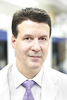 Stephen Scherer合作论文数Department of Molecular Genetics, Temerty Faculty of Medicine, University of Toronto;The Centre for Applied Genomics, The Hospital for Sick Children;Institute of Medical Science, University of Toronto;The McLaughlin Centre, Faculty of Medicine, University of Toronto13
 Thomas Durcan合作论文数Department of Neurology and Neurosurgery, Faculty of Medicine and Health Sciences, McGill University10
 Jennifer L Howe合作论文数9
 James Ellis合作论文数Department of Molecular Genetics, Temerty Faculty of Medicine, University of Toronto9Pellecchia Giovanna合作论文数9Carol X-Q Chen合作论文数8Bhooma Thiruvahindrapuram合作论文数8Zhipeng You合作论文数8
 Ryan K.C. Yuen合作论文数Department of Molecular Genetics, Temerty Faculty of Medicine, University of Toronto8
 Michael Nicouleau合作论文数8Narges Abdian合作论文数7
 Sonia Cellot合作论文数Department of Pathology, McGill University/Department of Pediatrics, Université de Montréal7
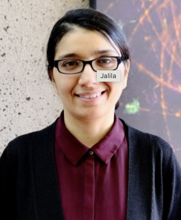 Jalila Chagraoui合作论文数7
 Susan Walker合作论文数Dept of Maternal Fetal Medicine, University of Melbourne;Division of Medicine, Dentistry and Health Sciences, University of Melbourne;Obstetrics and Gynaecology Royal Women's Hospital7
 Edward A Fon合作论文数Department of Neurology and Neurosurgery, Faculty of Medicine and Health Sciences, McGill University;Montreal Neurological Institute6
 P. Joel Ross合作论文数Program in Neurosciences and Mental Health, Hospital for Sick Children, Toronto, ON, Canada M5G 1X8;Program in Neurosciences and Mental Health, Hospital for Sick Children, Toronto, ON, Canada M5G 1X8;6Hoang Ny合作论文数6
 Christian Marshall合作论文数Division of Genome Diagnostics, Department of Pediatric Laboratory Medicine, The Hospital for Sick Children;Department of Laboratory Medicine & Pathobiology, Temerty Faculty of Medicine, University of Toronto;SickKids Genome Clinic6
 Mehdi Zarrei合作论文数Royal Botanic Gardens Kew6Lia D'Abate合作论文数5Alina Piekna合作论文数5
 Mohammed Uddin合作论文数5
 Evdokia Anagnostou合作论文数Institute of Medical Science, Temerty Faculty of Medicine, University of Toronto;Department of Paediatrics, University of Toronto;Bloorview Research Institute;Holland Bloorview Kids Rehabilitation Hospital5
 Kristin Hope合作论文数Department of Medical Biophysics, Temerty Faculty of Medicine, University of Toronto5
 Deivid De Carvalho Rodrigues合作论文数Instituto de Biofísica Carlos Chagas Filho, Universidade Federal do Rio de Janeiro5Brian T. Wilhelm合作论文数5
 Peter Szatmari合作论文数Department of Psychiatry, Temerty Faculty of Medicine, University of Toronto;Centre for Addiction and Mental Health5
 Nadine Mayotte合作论文数5Christina Chrysler合作论文数4
 Martin Sauvageau合作论文数4
 Muhammad Faheem Ph.D合作论文数4Zhuozhi Wang合作论文数4
 Gilles Maussion合作论文数4Ann Thompson合作论文数4
 Brendan J. Frey合作论文数Department of Electrical and Computer Engineering, University of Toronto;Best Department of Medical Research, University of Toronto;Department of Computer Science, University of Toronto4
 Daniele Merico合作论文数BioBox Analytics;Tahoe Therapeutics;The Hospital for Sick Children4Peter Pasceri合作论文数4
 Worrawat Engchuan合作论文数School of Information Technology, King Mongkut's University of Technology Thonburi4Stephen B. Ting合作论文数Rotary Bone Marrow Research Laboratories, c/o Royal Melbourne Hospital Post Office4Roumiana Alexandrova合作论文数3Tadeo Thompson合作论文数3Frédéric Barabé合作论文数3
 Alana Iaboni合作论文数Holland Bloorview Kids Rehabil Hosp, Bloorview Res Inst, Autism Res Ctr, Toronto, ON, Canada3
 Tara Ann Paton合作论文数3
 Jacob A.S. Vorstman合作论文数Department of Psychiatry, Temerty Faculty of Medicine, University of Toronto3Karun Singh合作论文数Department of Laboratory Medicine and Pathobiology, Temerty Faculty of Medicine, University of Toronto3
 Mayada Elsabbagh合作论文数Division of Neuroscience, Department of Neurology and Neurosurgery, Faculty of Medicine and Health Sciences, McGill University3Carole Shum合作论文数3




