Energy requirements in the human body are met by fuel mobilization stored in the form of fat, protein, and carbohydrate. Fat depots and glycogen storage in muscle and liver represent the largest contributors that sustain essential organic functions and mobility. Protein reserves are intended primarily for structural, biochemical, and physiological purposes and are, thus, spared as substrate for oxidation, except under conditions of prolonged starvation. Extracellular free fatty acid, triglycerides, and glucose account for only a minor fraction of the total energy storage, but they are essential sources of energy to organs and tissues in times of need. Therefore, maintenance of appropriate circulating levels of these substrates, particularly glucose, is an important component of fuel homeostasis. In this review, we focus on those mechanisms responsible for maintaining adequate blood glucose concentrations, with an emphasis on the role of the kidney. Renal adaptations to changes in the metabolic milieu, both in healthy and diabetic conditions, will be examined. We will provide a comprehensive description of the pharmacology and therapeutic actions of the sodium-glucose cotransporter 2 inhibitor class of drugs, which target the kidney and inhibit the reabsorption of glucose. The renal and cardiovascular benefits of the sodium-glucose cotransporter 2 inhibitor will be reviewed.
Introduction and Objective: Continuous glucose monitoring (CGM) yields information on glycemic variability (GV) which is linked to cardiovascular disease (CVD) in diabetes. However, CGM indicators have been less well studied in prediabetes. Methods: 49 participants underwent a 2-hour oral glucose tolerance test (OGTT) and euglycemic insulin clamp and were subsequently divided into four groups based on their OGTT glucose profile: Normal Glucose Tolerance (NGT, n=6), Isolated Impaired Fasting Glucose (IFG, n=9), Isolated Impaired Glucose Tolerance (IGT, n=4), and Combined IFG and IGT (CGI, n=30). For 5 days, subjects wore a CGM device. Matsuda insulin sensitivity index and Beta-cell function (BCF) (ΔI/ΔG and ΔI/ΔG x Matsuda Index=Disposition Index) were calculated from OGTT. Results: From CGM, mean glucose was similar in prediabetes and NGT (98±16 vs 98±8 mg/dL). Peak glucose, standard deviation (SD), coefficient of variation (CV), and time above 140 mg/dL (TAR) were all significantly higher in prediabetes than NGT (151 vs 126 mg/dL; 16 vs 10 mg/dL; 0.16 vs 0.10; and 12 vs 0.2% respectively, all p<0.05). Glucose Infusion Rate during the insulin clamp and Disposition Index from OGTT progressively declined in a stepwise fashion from NGT to IFG to IGT to CGI (p<0.01). In the CGI subgroup, the mean CGM glucose correlated negatively with Disposition Index (r= -0.381, p<0.05). Conclusion: Glycemic variability indices can identify individuals with prediabetes even when mean glucose levels with CGM are normal. Higher CV may provide a metric that these individuals are at higher risk of progression to diabetes. The present findings may inform the design of future studies examining this relationship. Disclosure P. Mahapol: None. A. Merovci: None. S.V. Rosembarque: None. A.A. Hansis-Diarte: None. J.M. Adams: None. E. Cersosimo: None. M. Abdul-Ghani: None. R. Belfort DeAguiar: None. R.A. DeFronzo: Advisory Panel; Ended; AstraZeneca. Research Support; Current; AstraZeneca. Advisory Panel; Current; Novo Nordisk. Research Support; Current; Eli Lilly and Company. Advisory Panel; Current; Corcept Therapeutics. Speaker's Bureau; Current; Corcept Therapeutics. Consultant; Current; Alnylam Pharmaceuticals, Inc. Advisory Panel; Current; Regeneron Pharmaceuticals Inc., Aardvark. A.O. Chavez-Velazquez: Advisory Panel; Ended; Crinetics Pharmaceuticals, Inc. Funding AZ ISSDAPA0002NIH R01 DK024092-37
Introduction and Objective: Antidiabetic agents differ in their mechanisms of action and can exert distinct long-term effects on β-cell function (BCF). Hyperglycemic clamp studies (HC) enable detailed assessment of first- and second-phase insulin secretion (ISecr), yet comparative longitudinal data across drug classes among individuals with PreDM remains limited. Methods: Sixty PreDM subjects were studied and treated with either metformin (MET n=15), pioglitazone (PIO n=15), saxagliptin (SAXA n=15), or dapagliflozin (DAPA n=15) for 2 years. All groups were matched for age and BMI. HC (+125 mg/dL) were performed before and after treatment. ISecr was calculated as the incremental AUC of insulin from 0-15 min (first phase) and during the 15-120 min (second phase) of the HC. Insulin sensitivity was estimated as glucose infusion rate during the steady state (GIR 90-120) of the HC. Results: As a whole (n=60), FPG decreased from 107 to 104 mg/dL (p < 0.05), with no significant change in HbA1c. First-phase ISecr increased from baseline to follow-up (949 vs 1174 µU/ml, p<0.001), with no change in second-phase ISecr, while GIR 90-120 improved (from 6.1 to 7.2 mg/kg.min, p<0.05). In treatment-specific analyses, MET, SAXA and PIO increased first-phase ISecr at 2 years (p < 0.05). GIR 90-120 improved with MET (p<0.05), DAPA (p < 0.05) and PIO (p = 0.05). Second-phase ISecr and disposition index (ISecr x GIR) remained unchanged across all four individual treatment groups. MET led to a net weight loss (p<0.005), whereas PIO led to weight gain (p<0.001). Conclusion: Treatment with antidiabetic agents for two years improved first phase ISecr and maintained second phase ISecr in individuals with PreDM, which carries a ~10% annual risk of progression to type 2 diabetes. Intervening early with pharmacological treatment can improve beta-cell function and modify the natural course toward progression to diabetes. Disclosure H. Zaitoon: None. A. Merovci: None. P. Mahapol: None. A.A. Hansis-Diarte: None. E. Cersosimo: None. J.M. Adams: None. C.L. Puckett: None. M. Abdul-Ghani: None. R. Belfort DeAguiar: None. R.A. DeFronzo: Advisory Panel; Ended; AstraZeneca. Research Support; Current; AstraZeneca. Advisory Panel; Current; Novo Nordisk. Research Support; Current; Eli Lilly and Company. Advisory Panel; Current; Corcept Therapeutics. Speaker's Bureau; Current; Corcept Therapeutics. Consultant; Current; Alnylam Pharmaceuticals, Inc. Advisory Panel; Current; Regeneron Pharmaceuticals Inc., Aardvark. A.O. Chavez-Velazquez: Advisory Panel; Ended; Crinetics Pharmaceuticals, Inc. Funding Astra Zeneca (ISSDAPA0002), National Institute of Health (R01 DK024092-37)
Introduction Sodium-glucose cotransporter (SGLT) inhibitors have shown substantial benefit in reducing cardiovascular and kidney events across diverse clinical populations, but the underlying physiological mechanisms remain unclear. However, existing mechanistic studies on renal and cardiovascular haemodynamics show variability in design, have limited statistical power and yield inconsistent outcomes, thus limiting the ability to draw generalisable conclusions. To address this gap, we conducted a systematic review and proposed the first meta-analysis to aggregate individual participant-level data from mechanistic studies to identify consistent physiological patterns and enhance understanding of the therapeutic effects of SGLT inhibition.Methods and analysis Gold-standard measured glomerular filtration rate (mGFR) was selected as the primary outcome for this systematic review, which aimed to identify all completed mechanistic studies investigating the effects of SGLT inhibition. Electronic databases including Ovid MEDLINE; Ovid Embase; Cochrane Database of Systematic Reviews; and Cochrane Central Register of Controlled Trials were searched using a detailed search strategy. In total, 24 studies (n=1296) were identified. This systematic review was reported according to the Preferred Reporting Items for Systematic Reviews and Meta-Analyses (PRISMA) guidelines. Key variables including demographics, medical history, concomitant medications, vital signs, mGFR, renal haemodynamics, urine and plasma biochemistry, tubular sodium handling, echocardiography, cardiac output monitoring, arterial stiffness and fluid volume will be extracted. A one-stage individual participant data meta-analysis under a Bayesian framework will be conducted, using hierarchical models to simultaneously analyse data from all eligible studies. The risk of bias due to missing results will be assessed. Sensitivity analyses and subgroup evaluations will be incorporated to explore sources of heterogeneity and assess robustness of findings.Ethics and dissemination Ethics approval was obtained from University Health Network, Toronto, Canada. Findings from the Mechanisms of SGLT Inhibitor Action and Physiological Mediators (MOSAIC) meta-analysis will be published in peer-reviewed journals and results will be disseminated at scientific conferences.PROSPERO registration number CRD420251001413.
OBJECTIVE:To examine the effects of sodium-glucose cotransporter 2 inhibitors (SGLT2is) alone or with glucagon-like peptide 1 receptor agonists (GLP-1RAs) on β-cell function (BCF) in type 2 diabetes. The hypothesis was that an SGLT2i combined with a GLP-1RA provides superior improvement in BCF than either agent alone. RESEARCH DESIGN AND METHODS:Ninety patients underwent a 180-min oral glucose tolerance test (OGTT) 1) after one drug dose (acute study) (placebo [n = 15], dapagliflozin [n = 25], exenatide [n = 25], and dapagliflozin/exenatide [n = 25]) and 2) after 1 and 4 months of therapy. Corrected Matsuda index (cMI) for urinary glucose loss, insulin secretion, and BCF indices were calculated during OGTT. RESULTS:In the acute study, mean ± SEM cMI in dapagliflozin (2.29 ± 0.33), exenatide (2.03 ± 0.12), and dapagliflozin/exenatide (2.36 ± 0.14) was higher (P < 0.05) than placebo (1.63 ± 0.36). After 1 and 4 months, cMI remained similarly elevated in exenatide and increased further (P < 0.001) in dapagliflozin and dapagliflozin/exenatide. In the acute study, insulin secretion in dapagliflozin was similar to placebo but higher (P < 0.001 vs. both) in exenatide and dapagliflozin/exenatide. After 1 and 4 months in exenatide and in dapagliflozin/exenatide, insulin secretion remained higher (P < 0.01 vs. both) than dapagliflozin. BCF index in the acute study was 0.40 ± 0.04 in placebo, 62% higher (P < 0.05) in dapagliflozin (0.65 ± 0.10), threefold higher in exenatide (1.17 ± 0.22), and fourfold higher in dapagliflozin/exenatide (1.69 ± 0.12) (all P < 0.001 vs. placebo). At 1 and 4 months, BCF rose further in dapagliflozin and exenatide but did not increase further in dapagliflozin/exenatide. CONCLUSIONS:Dapagliflozin and exenatide monotherapy cause sustained improvements in BCF and insulin sensitivity. Combination therapy with dapagliflozin plus exenatide markedly augmented both BCF and insulin sensitivity above that with either agent alone.
CONTEXT:Increased mesenteric visceral fat is associated with the metabolic syndrome, insulin resistance, and type 2 diabetes. METHODS:Using targeted cell separation and extraction technology (TC-SET), we examined the effect of removal of intra-abdominal fat, specifically small bowel mesenteric fat, on glycemic control and insulin sensitivity in 7 individuals with obesity and poorly controlled type 2 diabetes (T2D) (glycated hemoglobin [HbA1c] = 8.9% ± 0.2%; fasting plasma glucose [FPG] = 211 ± 12 mg/dL). RESULTS:At month 6, both HbA1c and FPG significantly declined to 7.7% (P = .01) and 140 mg/dL (P < .01). At month 12, both the FPG (172 mg/dL, P = .02) and HbA1c (8.1%, P = .10) tended to increase. Time in range (continuous glucose monitoring) increased from 22% to 74% (month 6, P < .001) and 50% (month 12, P < .05). Suppression of endogenous (hepatic) glucose production increased from 29% to 45% (P < .05) and to 43% (P < .01) at months 6 and 12, respectively; whole-body (muscle) insulin-mediated glucose disposal did not change significantly at months 6 and 12. Body weight (106.8 to 103.3 kg) and percent body fat (33.3 to 31.6%) both decreased slightly (P < .05) at month 12. Hepatic fat content (hydrogen-1 magnetic resonance spectroscopy) decreased significantly (23.9 ± 3.7 to 19.1 ± 3.4%, P < .005) at month 12. Insulin secretion and disposition index during oral glucose tolerance testing increased more than 2-fold at month 6 (both P < .05), and these improvements persisted at 12 months. CONCLUSION:Mesenteric visceral lipectomy (MVL) shows potential as a novel, minimally invasive approach to improve glycemic control in patients with suboptimally controlled T2D, but further controlled studies are needed to confirm these findings and better understand the potential benefits of MVL.
Introduction and Objective: Beta cell failure is the key pathophysiologic determinant in the progression from PreDM to type 2 diabetes (T2D). Lifestyle modification is frequently insufficient to prevent T2D onset, and drugs that modify disease progression and offer cardiometabolic benefits beyond glycemic control are increasingly used. Methods: Study recruited 200 PreDM subjects (FPG = 100-125 and/or 2-h PG=140-199 mg/dl) seeking to answer whether a specific oral antidiabetic drug provides greater benefit in preventing PreDM progression to T2D. At baseline, subjects received an OGTT and two-step hyperglycemic clamp (+125 and 400 mg/dl) followed by exenatide infusion to quantitate insulin sensitivity (Matsuda Index), insulin secretion (ΔI/ΔG), and beta cell function (disposition index). Subjects then were randomized to receive 24 months of treatment with: (i) Dapagliflozin (DAPA), (ii) Metformin (MET), (iii) Pioglitazone (PIO), or (iv) Saxagliptin (SAXA). 154 subjects (94 F, 60 M; Age=51±0.8, BMI=38±0.6); DAPA=47, MET=30, PIO=40, SAXA=37 have completed one year of intervention. Results: A1c trended to decrease in all groups (5.7±0.05 to 5.6%±0.05). FPG decreased in PIO, MET and DAPA (p<0.05) but not in SAXA; 2h PG decreased in DAPA, MET and PIO groups (p=0.05 - 0.01). Treatment with DAPA and MET (p<0.05) and PIO (p<0.01) significantly improved Matsuda Index. ΔI/ΔG trended to increase (p=n.s) in all groups. Beta cell function (ΔI/ΔG x Matsuda) increased significantly only in the PIO group (p<0.01). PIO resulted in weight gain of +3.9 kg, whereas DAPA and MET resulted in -4kg weight loss. Adipose IR (fasting FFA x fasting insulin) decreased in PIO (p<0.001). Conclusion: Early pharmacological treatment can prevent progression of PreDM to T2D. DAP and MET improved insulin sensitivity with net weight loss. Despite weight gain, PIO improved total body and adipose insulin sensitivity and robustly increased beta cell function. Individual factors and comorbidities need to be considered when selecting the most appropriate intervention to treat PreDM. A. Chavez: None. A. Merovci: None. S. Neppala: None. G. Baskoy: None. J.M. Adams: None. A.A. Hansis-Diarte: None. E. Cersosimo: None. R. Belfort De Aguiar: None. M. Abdul-Ghani: None. R.A. DeFronzo: Advisory Panel; AstraZeneca. Research Support; AstraZeneca. Speaker's Bureau; AstraZeneca. Advisory Panel; Boehringer-Ingelheim. Research Support; Boehringer-Ingelheim. Advisory Panel; Corcept Therapeutics, Novo Nordisk. Speaker's Bureau; Corcept Therapeutics. Research Support; 89bio, Inc, Amgen Inc. NIH R01 DK024092-37AZ ISSDAPA0002
ABSTRACT We examined the effect of increased plasma ketones on left ventricular (LV) function, myocardial glucose uptake (MGU), and myocardial blood flow (MBF) in type 2 diabetes (T2DM) patients with heart failure (HF). Three groups (I,II,III) of T2DM (12 per group) with LV ejection fraction £50% received incremental infusions of β-OH-B for 3-6 hours to raise plasma b-OH-B concentration throughout the physiologic (Groups I and II) and pharmacologic (Group III) range. Cardiac MRI was performed at baseline and after each b-OH-B infusion to provide measures of cardiac function. On a separate day, Group II also received NaHCO3 infusion, thus serving as their own control for time, volume, and pH. Additionally, Group II underwent positron emission tomography study with 18F-fluoro-2-deoxyglucose to examine effect of hyperketonemia on MGU. Groups I, II, III achieved plasma b-OH-B levels of 0.7±0.3, 1.6±0.2, 3.2±0.2 mmol/L, respectively. Cardiac output, LVEF, and stroke volume increased significantly during b-OH-B infusion in Groups II (CO, 4.54 to 5.30; EF, 39.9 to 43.8; SV, 70.3 to 80.0) and III (CO, 5.93 to 7.16; EF, 41.1 to 47.5; SV, 89.0 to 108.4) and did not change with NaHCO3 infusion in Group II. The increase in LVEF was greatest in Group III (p<0.001 vs Group II). MGU and MBF were not altered by β-OH-B. In T2DM patients with LVEF£50%, increased plasma b-OH-B significantly increased LV function dose-dependently. Since MGU did not change, the myocardial benefit of b-OH-B resulted from providing an additional fuel for the heart without inhibiting MGU. HIGHLIGHTS · SGLT2 inhibitor therapy is associated with an increase in plasma ketone concentration · We examined in type 2 diabetes patients with HFrEF the effect of b-OH-B infusion, spanning the physiologic and pharmacologic range of plasma b-OH-B concentrations (0.7, 1.6, and 3.2 mmol/L), on myocardial function (MRI), myocardial blood flow (PET/H215O), and myocardial glucose uptake (PET/18F—DOG) · b-OH-B caused a dose-response increase in left ventricular ejection fraction and myocardial blood flow without altering myocardial glucose uptake These results suggest that the SLGT2i-induced increase in plasma ketone concentration may contribute to its beneficial effects on myocardial function
Acute and chronic SGLT-2 inhibition increase endogenous glucose production (EGP). However, the organ - liver versus kidney - responsible for the increase in EGP has not been identified. 20 T2DM and 12 NGT subjects received [3-3H]-glucose infusion (to measure total EGP) in combination with arterial and renal vein catheterization and PAH infusion for determination of renal blood flow. Total EGP, net renal arteriovenous balance, and renal glucose production were measured before and 4 hours after dapagliflozin and placebo administration. Following DAPA, EGP increased in both T2D and NGT from baseline to 240 minutes, while there was a significant time-related decrease after placebo in T2D. Renal glucose production at baseline was <5% of basal EGP in both groups and did not change significantly following DAPA in either NGT and T2D. Renal glucose uptake (sum of tissue glucose uptake plus glucosuria) increased in both T2D and NGT following DAPA (P<0.05 vs placebo). The increase in RGU was entirely explained by the increase in glucosuria. Single dose of dapagliflozin significantly increased EGP, which primarily is explained by an increase in hepatic glucose production, establishing the existence of a novel renal-hepatic axis.
Introduction & Objective: To examine the effect of sodium-glucose co-transporter-2 inhibitor [SGLT2i] therapy alone or in combination with glucagon-like peptide-1 receptor agonist [GLP-1 RA] on beta-cell function [BCF] in T2D. HYPOTHESIS: the improvement in BCF with GLP-1 RA plus SGLT-2i would be superior to that with either agent alone. Methods: Ninety patients received 3-hour OGTT: (i) before and after a single dose of drug (ACUTE study): placebo [PCB, n=15]; dapagliflozin [DAPA, n=25]; exenatide [EXE, n=25]; or both [DAPA/EXE, n=25], and (ii) after 1 and 4 months of therapy (CHRONIC study) with PCB, DAPA, EXE or DAPA/EXE. Matsuda Index [MI], Insulin Secretion [ΔI/ΔG)0-3h) and Disposition Index [DI=IS x MI] were calculated using standard formulae. MI was corrected for urinary glucose excretion. Conclusion: Dapagliflozin and exenatide as monotherapy improve beta cell function (DI) acutely and chronically (4 months). Combination therapy with dapagliflozin plus exenatide leads to superior and sustained improvement in beta cell function (DI) compared to either drug alone. These data suggest that combination therapy with GLP-1 RA plus SGLT2i may enhance long-term preservation of beta-cell function in T2D patients. Disclosure R.A. DeFronzo: Advisory Panel; AstraZeneca, Novo Nordisk, Boehringer-Ingelheim, Intarcia Therapeutics, Inc., Aardvark, Renalytix, Corcept Therapeutics, Alnylam Pharmaceuticals, Inc. Research Support; Boehringer-Ingelheim, AstraZeneca, 89bio, Inc., Amgen Inc., Medality, Corcept Therapeutics. Speaker's Bureau; AstraZeneca, Corcept Therapeutics, Renalytix. G. Baskoy: None. C.L. Triplitt: Speaker's Bureau; Novo Nordisk. Consultant; Eli Lilly and Company. Other Relationship; American Diabetes Association. E. Cersosimo: None. C. Solis-Herrera: Advisory Panel; Novo Nordisk, Bayer Inc. J.M. Adams: None. A.A. Hansis-Diarte: None. A. Gastaldelli: Consultant; Boehringer-Ingelheim. Other Relationship; Pfizer Inc., Eli Lilly and Company. Consultant; Merck Sharp & Dohme Corp. Advisory Panel; Novo Nordisk. Speaker's Bureau; Merck Sharp & Dohme Corp., Eli Lilly and Company. Advisory Panel; Pfizer Inc. Speaker's Bureau; Novo Nordisk. A. Chavez: None.
Background: SGLT2i is associated with CV benefits, but the mechanisms are not fully elucidated. We report a novel finding associated with improved CV Health in T2D & HFrEF. Elevated ketones, EF%, & heart remodeling have been reported with SGLT2i. This is likely, in part, due to a switch in cardiac metabolism to ketone oxidation. This switch might be present in peripheral tissues, including skeletal muscle (SkM). Objective: To examine the effects of SGLT2i on SkM energy production, utilization, exercise recovery, lipid content, and cardiopulmonary function. Methods n=10(Age= 61.6±3.5, BMI= 31.4±1.8, A1c=7.6%±0.4, EF 32.7%±2.7) were randomized to empagliflozin 25mg vs. placebo 2:1 for 12 wks. SkM 31P & 1H MRS was performed. Cardiopulmonary exercise testing (CPET) was done to assess functional capacity and determine CV prognosis. Results: After SGLT2i, there was a significant improvement in SkM energy production, utilization at rest, and exercise recovery. The VE/VCO2 slope, significantly improved (p<0.05) (Fig.1) Conclusion: We show for the first time that, in subjects with T2DM & HFrEF, SGLT2i significantly improve SkM bioenergetics and lipid content. This was associated with a significant improvement in CV prognostic markers. These findings may explain novel mechanisms of improvement in peripheral bioenergetics that translate into Global CV Health. Y. Qin: None. N.D. Sanchez: None. F.M. Acosta: None. A. Moody: None. S. Neppala: None. C.L. Triplitt: Speaker's Bureau; Novo Nordisk. Consultant; Eli Lilly and Company. Other Relationship; American Diabetes Association. G.D. Clarke: None. E. Cersosimo: None. R.A. DeFronzo: Advisory Panel; AstraZeneca, Novo Nordisk, Boehringer-Ingelheim, Intarcia Therapeutics, Inc., Aardvark, Renalytix, Corcept Therapeutics, Alnylam Pharmaceuticals, Inc. Research Support; Boehringer-Ingelheim, AstraZeneca, 89bio, Inc., Amgen Inc., Medality, Corcept Therapeutics. Speaker's Bureau; AstraZeneca, Corcept Therapeutics, Renalytix. C. Solis-Herrera: Advisory Panel; Novo Nordisk, Bayer Inc. Doris Duke Foundation
AIMS:To investigate the effect of sodium-glucose co-transporter 2 inhibitor [SGLT-2i] therapy on renal haemodynamics in T2D patients with glomerular hyperfiltration. MATERIALS AND METHODS:Sixty T2D patients with elevated [HYPER] and normal [NORMO] GFR were randomized to dapagliflozin 10 mg/day [DAPA/HYPER, n = 15; DAPA/NORMO, n = 15] or to metformin/glipizide [CONTROL/HYPER, n = 15; CONTROL/NORMO, n = 15] to reach similar glycaemic control after 4 months. GFR was measured with Iohexol and hyperfiltration was empirically defined as >125 mL/min/1.73 m2. GFR, renal plasma flow [RPF], mean arterial pressure [MAP], filtration fraction [FF], and renal vascular resistance [RVR] were determined before/after therapy. RESULTS:HbA1c decreased similarly in all 4 groups. GFR declined by ~18% in DAPA/HYPER and by ~7% in DAPA/NORMO and did not change in CONTROLS (p < 0.05 vs. DAPA). RPF remained unchanged in all four groups. Thus, FF (%) declined from 0.23 ± 0.01 to 0.18 ± 0.01 in DAPA/HYPER and from 0.17 ± 0.01 to 0.15 ± 0.01 in DAPA/NORMO and remained unchanged in CONTROLS (p < 0.05 vs. DAPA). MAP (mmHg) decreased from 95.4 ± 1.4 to 88.1 ± 1.3 in DAPA/HYPER and from 95.6 ± 1.3 to 91.8 ± 0.8 in DAPA/NORMO and remained unchanged in CONTROLS (p < 0.05 vs. DAPA). RVR [mmHg/L/min] declined in DAPA/HYPER (92.7 ± 7.8 to 80.4 ± 6.1) and DAPA/NORMO (90.1 ± 3.0 to 81.4 ± 2.1) but not in CONTROLS (p < 0.05 vs. DAPA). CONCLUSIONS:Despite comparable glycaemic control, dapagliflozin treatment, but not metformin and /or glipizide, reduced glomerular hyperfiltration in T2D patients and decreased both filtration fraction and renal vascular resistance. These findings suggest that a post-glomerular vasodilatory action of SGLT2 inhibitors contributes to their renal protective effect in T2D.
IntroductionLow carbohydrate ketogenic diets have received renewed interest for the treatment of obesity and type 2 diabetes. These diets promote weight loss, improve glycemic control, and reduce insulin resistance. However, whether the improvements in glycemic control and insulin sensitivity are secondary to the weight loss or result from a direct effect of hyperketonemia is controversial.Research design and methods29 overweight obese subjects were randomized to one of three dietary interventions for 10 days: (1) Weight-maintaining standard diet; (2) Weight-maintaining ketogenic diet; (3) Weight-maintaining ketogenic diet plus supplementation with the ketone ester of beta-hydroxybutyrate (β-OH-B), 8 g every 8 hours. At baseline, all subjects had oral glucose tolerance test, 2-step euglycemic insulin clamp (20 mU/m2.min and 60 mU/m2.min) with titrated glucose and indirect calorimetry.ResultsBody weight, fat content, and per cent body fat (DEXA) remained constant over the 10-day dietary intervention period in all three groups. Plasma β-OH-B concentration increased twofold, while carbohydrate oxidation decreased, and lipid oxidation increased demonstrating the expected shifts in substrate metabolism with institution of the ketogenic diet. Glucose tolerance either decreased slightly or remained unchanged in the two ketogenic diet groups. Whole body (muscle), liver, and adipose tissue sensitivity to insulin remained unchanged in all 3 groups, as did the plasma lipid profile and blood pressure.ConclusionIn the absence of weight loss, a low carbohydrate ketogenic diet has no beneficial effect on glucose tolerance, insulin sensitivity, or other metabolic parameters.
Aim: To evaluate the safety and metabolic effects of mesenteric visceral lipectomy in individuals with T2D. Methods & Study Design: Eight T2D subjects (age = 52±4 y; BMI = 34.1±1.0; stable diabetes meds/weight for 3 months) underwent pre-MVL assessment including OGTT, CGM, euglycemic insulin clamp with 3H-glucose, and MRI imaging. Repeat measurements were made at 6- and 12-months post-surgery. Exclusion criteria included prior insulin/TZD use. Insulin secretion [IS], Disposition Index [DI] and Matsuda index [MI] of insulin sensitivity were calculated with standard formulae. Results: At month 6, HbA1c decreased from 9.0% to 8.1% (p=0.08) and persisted at 8.4% at month 12 (p=0.19). CGM time-in-range increased significantly at 6 and 12 months (25% to 76%) (p<0.05). Suppression of hepatic glucose production increased at months 6 and 12 (p<0.05). Body weight and % body fat decreased slightly at month 12 (p<0.05). Insulin secretion and disposition index during OGTT increased more than 2-fold at month 6 and persisted at month 12. Disclosure G. Baskoy: None. R.M. Peterson: Consultant; Teleflex/Standard Bariatrics. J.W. Kempenich: Consultant; Intuitive Surgical. C.L. Triplitt: Speaker's Bureau; Novo Nordisk. Consultant; Eli Lilly and Company. Other Relationship; American Diabetes Association. G.D. Clarke: None. E. Cersosimo: None. M.S. Andrew: Employee; Medality Medical, Inc. A. Merovci: None. O. Lavrynenko: None. A.A. Hansis-Diarte: None. C. Solis-Herrera: Advisory Panel; Novo Nordisk, Bayer Inc. A. Chavez: None. M. Salehi: None. R.A. DeFronzo: Advisory Panel; AstraZeneca, Novo Nordisk, Boehringer-Ingelheim, Intarcia Therapeutics, Inc., Aardvark, Renalytix, Corcept Therapeutics, Alnylam Pharmaceuticals, Inc. Research Support; Boehringer-Ingelheim, AstraZeneca, 89bio, Inc., Amgen Inc., Medality, Corcept Therapeutics. Speaker's Bureau; AstraZeneca, Corcept Therapeutics, Renalytix. Funding Medality
Aim: To investigate the benefits of dapagliflozin [DAPA] on renal hemodynamics in type 2 diabetes patients [T2D] with glomerular hyperfiltration. Design and Methods: 24 T2D with elevated [HYPER] and normal [NORMO] GFR, measured by Iohexol, randomized to either DAPA, 10mg/day [DAPA/HYPER, n=6; DAPA/NORMO, n=6] or metformin+glipizide [CON/HYPER, n=6; CON/NORMO, n=6] for 4 months. Hyperfiltration was defined as GFR>125ml/min/1.73m2. Renal plasma (blood) flow [RP(B)F] measured with PAH divided by (1-Hct), mean arterial pressure [MAP], filtration fraction [FF] and renal vascular resistance [RVR] were determined before and after treatment. Results: A1c[%] decreased similarly in DAPA/HYPER (8.3±0.2 vs. 6.8±0.2), CON/HYPER (8.7±0.3 vs. 7.2±0.1), DAPA/NORMO (8.6±0.3 vs. 7.3±0.2) and CON/NORMO (8.2±0.3 vs. 7.4±0.2). Body weight[Kg] was reduced in DAPA/HYPER (89±4 vs. 84±4) and DAPA/NORMO (90±3 vs. 87±2) but not in CON/HYPER (86±4 vs. 87±3) and CON/NORMO (87±4 vs. 91±3) (p<0.05). GFR declined by 17% in DAPA/HYPER and by 8% in DAPA/NORMO (both, p<0.05) but did not change in CON. FF and RVR fell in DAPA but not in CON. Renal hemodynamics at baseline [PRE-Tx] and after 4 months [POST-Tx] are shown in the table. Conclusion These results indicate that dapagliflozin, but not metformin+glipizide therapy, normalizes glomerular hyperfiltration and reduces renal vascular resistance in hyperfiltering type 2 diabetes patients. Disclosure G.Baskoy: None. Y.Qin: None. E.Cersosimo: None. C.Solis-herrera: None. J.M.Adams: None. R.A.Defronzo: Advisory Panel; AstraZeneca, Bayer Inc., Boehringer-Ingelheim, Novo Nordisk, Research Support; AstraZeneca, Boehringer-Ingelheim, Merck & Co., Inc., Speaker's Bureau; AstraZeneca. C.L.Triplitt: Speaker's Bureau; Novo Nordisk. Funding AstraZeneca; Texas Diabetes Institute
Abstract Disclosure: Y. Qin: None. N.D. Sanchez: None. A.J. Moody: None. F.M. Acosta: None. S. Neppala: None. M. Brown: None. H. Honka: None. B. Todd: None. L.A. Cruz Moreno: None. A.R. Stepanenko: None. S.E. Espinoza: None. N. Musi: None. C. Triplitt: None. G. Clarke: None. E. Cersosimo: None. R.A. DeFronzo: None. C. Solis-Herrera: None. Cardioprotective benefits of SGLT2 inhibitors (SGLT2i) have been largely thought to be coupled to a physiologic increase in plasma ketones, with much interest in the role of adaptive changes in cardiac fuel source in subjects with T2D and HF. Given the changes in the global dynamics of bioenergetics with SGLT2i, and the proven pleiotropic effects of ketone bodies, suggest that, in addition to their cardioprotective function, SGLT2i may influence other organ systems. In particular, how skeletal muscle (SkM), or cardiopulmonary function, respond to these global bioenergetics changes remains largely unknown. A pilot randomized-controlled clinical study was designed to examine the effects of SGLT2i on SkM bioenergetics (31P MRS), & cardiopulmonary function (CPET), and its association with left ventricular cardiac function (MRI). Six subjects (Age=59±2.6, BMI=33.3±1.1, A1c=7.1±0.3) with T2D and HF (<50%) were randomized empagliflozin 25mg/day (SGLT2i) vs. placebo (control) in a 2:1 fashion for 12 weeks. Imaging and clinical evaluations were performed pre- and post-intervention. The SGLT2i group improved EF (28.7 vs. 38.8, p=0.1). SkM 31P MRS at rest, measuring the relative intracellular concentrations of high-energy metabolites of phospholipid breakdown, reduced with SGLT2i, suggesting better energy utilization by the SkM. 31P MRS-after exercise, showed significant improvements in SkM oxidative capacity (KPCR*[PCr], 0.6 to 0.8, p=0.03), with SGLT2i.During CPET testing, the ventilatory anaerobic threshold (VT), with SGLT2i, showed a significant (p=0.05) increase in VT time, suggesting an increase in the time in aerobiosis in these subjects. The minute ventilation-to-carbon dioxide output slope (a strong predictor of cardiopulmonary complications/death) was improved in the SGLT2i group vs. placebo, which could be associated with the CV benefit observed with these agents. Additionally, patient self-reported outcomes (Promis) showed a trend of improvement in the SGLT2i group. In summary, we demonstrate novel and exciting preliminary findings that in patients with T2D & HF, SGLT2i-associated benefits are not isolated to the heart and may also expand to the SkM, including improved oxidative capacity, energy utilization, and SkM recovery post-exercise. With improvements in HF prognostic markers and patient self-reported outcomes. These original findings may represent novel mechanisms contributing to the cardiac benefits of SGLT2i. Presentation: Friday, June 16, 2023
Background: The CV benefits of SGLT2i are well established. However, how skeletal muscle (SkM) bioenergetics respond to SGLT2i in T2D and HFrEF remains largely unknown. Objective: To explore the effects of empagliflozin on SkM bioenergetics (phosphorus metabolites & myocellular lipid content) at rest and post-exercise (31P & 1H MRS) and cardiac function (MRI). Methods: Subjects with T2D & HFrEF (<40%) (n=6) were randomized 2:1 to empagliflozin 25mg (SGLT2i) or placebo for 12 wks (Age=59±2.6, BMI=33.3±1.1, A1c=7.1±0.3, EF 28±4%). A cardiac MRI, & static/dynamic 31P & 1H SkM by MRS, were performed before & after treatment. Results: In the empagliflozin group, SkM 31P MRS at rest, showed decrease in phosphodiesterase [PDE]. 31P MRS post-exercise, showed significant improvement in Phosphocreatinine (PCr) recovery, expressed by the K value. 1H MRS showed a reduction of saturated intramyocellular lipids (IMCL-CH2:CH3 ratio). Conclusion: Empagliflozin in subjects with T2D and HFrEF had beneficial effects in SkM energy utilization and muscle recovery post-exercise as determined by a decrease in PDE and increase in K value. Additionally, empagliflozin decreased saturated IMCL, which could be expected to improve mitochondrial function and energy utilization. If similar findings were to occur in cardiac muscle, this could provide a novel mechanism for the beneficial CV effects of SGLT2i. Disclosure Y. Qin: None. N. D. Sanchez: None. A. Moody: None. F. M. Acosta: None. S. Neppala: None. M. Brown: None. H. Honka: None. L. A. Cruz Moreno: None. C. L. Triplitt: Speaker's Bureau; Novo Nordisk. G. D. Clarke: None. E. Cersosimo: None. R. A. DeFronzo: Speaker's Bureau; AstraZeneca. Advisory Panel; AstraZeneca, Bayer Inc., Boehringer-Ingelheim, Novo Nordisk. Research Support; AstraZeneca, Boehringer-Ingelheim, Merck & Co., Inc. C. Solis-Herrera: None. Funding Doris Duke Charitable Foundation; Voelcker Foundation; National Institutes of Health
 Ralph DeFronzo合作论文数Division of Diabetes, Long School of Medicine, University of Texas Health Science Center at San Antonio;Department of Medicine, Long School of Medicine, University of Texas Health Science Center at San Antonio129
 Curtis Triplitt合作论文数University of Texas;Health Science Center;Texas Diabetes Institute;Texas Diabetes Institute, University of Texas96John Adams合作论文数50Solis-Herrera Carolina合作论文数Health Science Center San Antonio, University of Texas49
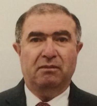 Muhammad Abdul Ghani合作论文数Division of Diabetes, University of Texas Health Science Center at San Antonio;Department of Medicine, University of Texas Health Science Center at San Antonio35
 Nicolas Musi合作论文数Department of Medicine, University of Texas Health Science Center at San Antonio;San Antonio Claude D. Pepper Older Americans Independence Center, University of Texas Health Science Center at San Antonio;Barshop Institute for Longevity and Aging Studies, University of Texas Health Science Center at San Antonio;San Antonio Geriatric Research, Education and Clinical Center, University of Texas Health Science Center at San Antonio32
 Amalia Gastaldelli合作论文数Institute of Clinical Physiology, National Research Council;UT Health San Antonio24
 Geoffrey D. Clarke合作论文数Department of Radiology, Long School of Medicine, The University of Texas Health Science Center at San Antonio22Qin Yuejuan合作论文数21Mariam Alatrach合作论文数19Christina Agyin合作论文数16Estela Wajcberg合作论文数15Gozde Baskoy合作论文数13
 Alexander Jacob Moody合作论文数13Sriwijitkamol Apiradee合作论文数13
 Henri Honka合作论文数Turku PET Centre, University of Turku;Department of Endocrinology (, Turku University Hospital;Faculty of Medicine (N.K.),, Kagawa University;Institute of Biomedical Engineering (A.M.),, National Research Council;and Institute of Clinical Physiology (P.I.),, National Research Council12
 Giuseppe Daniele合作论文数Department of Endocrinology and Metabolism, University of Pisa12Robert Martinez合作论文数11Hussein Al Jobori合作论文数11
 Francisca M Acosta合作论文数Dept Biomed Engn & Chem Engn, Univ Texas San Antonio11
 Siva Neppala合作论文数10Marianella Fernandez合作论文数10Merovci Aurora合作论文数Division of Diabetes (A.Me., University of Texas Health Science Center at San Antonio;and Consiglio Nazionale delle Ricerche Institute of Biomedical Engineering (A.Ma.), 35127 Padova, Italy.10Hansis-Diarte Andrea合作论文数10Christopher P. Jenkinson合作论文数10
 Daniel Hale合作论文数9Patricia Iozzo合作论文数9Robert J. Chilton合作论文数9Alberto O. Chavez合作论文数9
 Olga Lavrynenko合作论文数8Tantiwong Puntip合作论文数8Lynch Jane合作论文数8Ravindranath Duggirala合作论文数7Marjorie Molina-Wilkins合作论文数7Rector Arya合作论文数7Puppala Sobha合作论文数7Farook Vidya S合作论文数National Institute of Diabetes and Digestive and Kidney Diseases, National Institutes of Health7Puckett C合作论文数7
 John Blangero合作论文数Department of Human Genetics, South Texas Diabetes and Obesity Institute, School of Medicine, University of Texas Rio Grande Valley;Genomics Computing Center, University of Texas Rio Grande Valley7Jennifer Schneider合作论文数7Anthony G. Comuzzie合作论文数7
 Sharon P Fowler合作论文数7Devjit Tripathy合作论文数Department of Medicine, University of Texas Health Science Center at San Antonio7Garduño-García José de Jesús合作论文数Departamento de Endocrinología y Metabolismo, Instituto Nacional de Ciencias Médicas y Nutrición Savador Zubiran;Centro de Investigacion en Ciencias Medicas, Universidad Autonoma del Estado de Mexico;Instituto Nacional de Ciencias Médicas y Nutrición Savador Zubiran, Universidad Autonoma del Estado de Mexico6A. Cervera合作论文数6
 Kelly K. Hunt合作论文数Department of Breast Surgical Oncology, Division of Surgery, The University of Texas MD Anderson Cancer Center6Xiaojing Xu合作论文数6
 Sara M. Reyna合作论文数Department of Medicine, University of Texas Health Center at San Antonio6
 Laura Almasy合作论文数Department of Genetics, Perelman School of Medicine, University of Pennsylvania6




