B-cell migration and adhesion are critical to form a germinal center response, the site for B-cell production of high-affinity antibodies. Here, we describe two assays that can be used to examine B-cell cytoskeletal responses needed during the germinal center response: B-cell spreading and homotypic adhesion. Spreading of B cells is dependent on Cdc42, while Rac1 and Rac2 are necessary for homotypic adhesion. These in vitro assays can be used to examine functional responses of B cells mediated by the cell cytoskeleton, for example when comparing B cells from different gene knockout animals.
The Rho GTPases Cdc42, Rac1, and Rac2 coordinate receptor signaling to cell adhesion, migration, and proliferation. Deletion of Rac1 and Rac2 early during B cell development leads to failure in B cell entry into the splenic white pulp. Here, we sought to understand the role of Rac1 and Rac2 in B cell functionality and during the humoral antibody response. To circumvent the migratory deficiency of B cells lacking both Rac1 and Rac2, we took the approach to inducibly delete Rac1 in Rac2-/- B cells in the spleen (Rac1BRac2-/- B cells). Rac1BRac2-/- mice had normal differentiation of splenic B cell populations, except for a reduction in marginal zone B cells. Rac1BRac2-/- B cells showed normal spreading response on antibody-coated layers, while both Rac2-/- and Rac1BRac2-/- B cells had reduced homotypic adhesion and decreased proliferative response when compared to wild-type B cells. Upon challenge with the T-cell-independent antigen TNP-conjugated lipopolysaccharide, Rac1BRac2-/- mice showed reduced antibody response. In contrast, in response to the T-cell-dependent antigen sheep red blood cells, Rac1BRac2-/- mice had increased serum titers of IgG1 and IgG2b. During in vitro Ig class switching, Rac1BRac2-/- B cells had elevated germline γ2b transcripts leading to increased Ig class switching to IgG2b. Our data suggest that Rac1 and Rac2 serve an important role in regulation of the B cell humoral immune response and in suppressing Ig class switching to IgG2b.
We sought to identify genes necessary to induce cytoskeletal change in B cells. Using gene expression microarray, we compared B cells stimulated with interleukin-4 (IL-4) and anti-CD40 antibodies that induce B cell spreading, cell motility, tight aggregates, and extensive microvilli with B cells stimulated with lipopolysaccharide that lack these cytoskeletal changes. We identified 84 genes with 10-fold or greater expression in anti-CD40 + IL-4 stimulated B cells, one of these encoded the guanine nucleotide exchange factor (GEF) dedicator of cytokinesis 10 (Dock10). IL-4 selectively induced Dock10 expression in B cells. Using lacZ expression to monitor Dock10 promoter activity, we found that Dock10 was expressed at all stages during B cell development. However, specific deletion of Dock10 in B cells was associated with a mild phenotype with normal B cell development and normal B cell spreading, polarization, motility, chemotaxis, aggregation, and Ig class switching. Dock10-deficient B cells showed lower proliferation in response to anti-CD40 and IL-4 stimulation. Moreover, the IgG response to soluble antigen in vivo was lower when Dock10 was specifically deleted in B cells. Together, we found that most B cell responses were intact in the absence of Dock10. However, specific deletion of Dock10 in B cells was associated with a mild reduction in B cell activation in vitro and in vivo.
The Rho GTPase Cdc42 coordinates regulation of the actin and the microtubule cytoskeleton by binding and activating the Wiskott-Aldrich syndrome protein. We sought to define the role of intrinsic expression of Cdc42 by mature B cells in their activation and function. Mice with inducible deletion of Cdc42 in mature B cells formed smaller germinal centers and had a reduced Ab response, mostly of low affinity to T cell-dependent Ag, compared with wild-type (WT) controls. Spreading formation of long protrusions that contain F-actin, microtubules, and Cdc42-interacting protein 4, and assumption of a dendritic cell morphology in response to anti-CD40 plus IL-4 were impaired in Cdc42-deficient B cells compared with WT B cells. Cdc42-deficient B cells had an intact migratory response to chemokine in vitro, but their homing to the B cell follicles in the spleen in vivo was significantly impaired. Cdc42-deficient B cells induced a skewed cytokine response in CD4(+) T cells, compared with WT B cells. Our results demonstrate a critical role for Cdc42 in the motility of mature B cells, their cognate interaction with T cells, and their differentiation into Ab-producing cells.
Ig class switching requires cell proliferation and is division linked, but the detailed mechanism is unknown. By analyzing the first switching cells early in the kinetics, our analysis suggested that proliferating B cells had a very short G1 phase (<3.5 h), a total cell cycle time of ∼ 11 h, and that Ig class switching preferentially occurred in the late G1 or early S phase. Inhibition of cyclin-dependent kinases (CDKs) caused dramatic reduction of switching rate within 6 h. This was associated with less targeting of activation-induced cytidine deaminase (AID) to the Igh locus. Interestingly, ectopically expressed nuclear AID in HeLa cells was preferentially found in the early S phase. Furthermore, in CDK2 hypomorphic cells there was reduced nuclear AID accumulation. Thus, our data are compatible with the idea that division-linked Ig class switching is in part due to CDK2-regulated AID nuclear access at the G1/S border.
We describe a spontaneously derived mouse line that completely failed to induce Ig class switching in vitro and in vivo. The mice inherited abolished IgG serum titers in a recessive manner caused by a spontaneous G → A transition mutation in codon 112 of the aicda gene, leading to an arginine to histidine replacement (AID(R112H)). Ig class switching was completely reconstituted by expressing wild-type AID. Mice homozygous for AID(R112H) had peripheral B cell hyperplasia and large germinal centers in the absence of Ag challenge. Immunization with SRBCs elicited an Ag-specific IgG1 response in wild-type mice, whereas AID(R112H) mice failed to produce IgG1 and had reduced somatic hypermutation. The phenotype recapitulates the human hyper-IgM (HIGM) syndrome that is caused by point mutations in the orthologous gene in humans, and the AID(R112H) mutation is frequently found in HIGM patients. The AID(R112H) mouse model for HIGM provides a powerful and more precise tool than conventional knockout strategies.
This is an opinion based on the paper “Cloning of cDNA encoding the murine IgG1 induction factor by a novel strategy using SP6 promoter” (1).
B cells devoid of the Rho GTPase Cdc42 coordinate the actin and microtubule cytoskeleton less effectively and form an extrafollicular antibody response
B cells can be activated by T cell-dependent stimuli, such as CD40 ligation and cytokines, which induce extensive proliferation, class switch recombination and somatic hypermutation.Epstein-Barr virus (EBV) can also induce B cell activation by mimicking T cell help through its main oncoprotein, latent membrane protein 1 (LMP-1). It is regulated by another EBV-encoded protein, EBV nuclear antigen 2 (EBNA-2), which is absent in Hodgkin and Burkitt lymphomas. We have studied LMP-1 induction by cytokines in vitro and shown that LMP-1 is induced through the transcription factor signal transducer and activator of transcription (STAT6) and a newly defined high-affinity STAT6-binding site.When IL-4 is added together with lipopolysaccharide (LPS) or α-CD40 to B cells, it induces homotypic round and tight aggregates in vitro, whereas LPS alone does not induce such morphological changes. I describe here attempts to identify the molecules that regulate these responses.I have shown that the Rho GTPase Cdc42 controls the spreading of B cells, whereas two other molecules in the same family, Rac1 and Rac2, control homotypic adhesion. Further, I have shown by conditional deletion of Cdc42 in B cells that it is important in the humoral immune response. Dock10 is a guanosine nucleotide exchange factor (GEF) for Cdc42. It is expressed through all differentiation stages of B cell development. However, targeted deletion of Dock10 in B cells does not result in an aberrant phenotype. Furthermore, by studying conditional knockout mice for Dock10, Cdc42, Rac1 and Rac2, I have elucidated the mechanism of cytoskeletal changes during B cell activation, leading to adhesion and motility.My results may lead to a better understanding of normal B cell activation and of EBV infection, which is associated with many human tumours and may help to understand cancer development and progression in B cells.
Type I interferons (IFN) exert multiple effects on both the innate and adaptive immune system in addition to their antiviral and antiproliferative activities. Little is known, however about the direct effects of type IFNs on germinal center (GC) B cells, the central components of adaptive B cell responses. We used Burkitt's lymphoma (BL) lines, as a model system of normal human GC B cells, to examine the effect of type I IFNs on the expression of BCL-6, the major regulator of the GC reaction. We show that type I IFNs, but not IFN gamma, IL-2 and TNF alpha rapidly down-regulate BCL-6 protein and mRNA expression, in cell lines derived from endemic, but not from sporadic BL. IFN alpha-induced down-regulation is specific for BCL-6, independent of Epstein-Barr virus and is not accompanied by IRF-4 up-regulation. IFN alpha-induced BCL-6 mRNA down-regulation does not require de novo protein synthesis and is specifically inhibited by piceatannol. The proteasome inhibitor mG132 non-specifically prevents, while inhibitors of alternate type I IFN signaling pathways do not inhibit IFNa-induced BCL-6 protein downregulation. We validate our results with showing that IFN alpha rapidly down-regulates BCL-6 mRNA in purified mouse normal GC B cells. Our results identify type I IFNs as the first group of cytokines that can down-regulate BCL-6 expression directly in GC B cells. (C) 2011 Elsevier Ltd. All rights reserved.
In line with the B-lymphotropic nature of Epstein-Barr virus (EBV), the virus is present in several types of B-cell lymphomas. EBV expresses a different set of latent genes in the associated tumors, such as EBV nuclear antigen 1 (EBNA-1) and latent membrane proteins (LMPs; type II latency) in classical Hodgkin lymphomas (HLs). We previously reported that exposure of in vitro EBV-converted, HL-derived cell line KMH2-EBV to CD40-ligand and interleukin-4 (IL-4) induced the expression of LMP-1. Here, we show that exposure to IL-4 or IL-13 alone induced LMP-1 in the absence of EBNA-2. Induction of LMP-1 by IL-4 and IL-13 was mediated by the signal transducer signal transducer and activator of transcription 6 (STAT6) and a newly defined high-affinity STAT6-binding site in the LMP-1 promoter. IL-4 induced LMP-1 also in Burkitt lymphoma-derived lines and in tonsillar B cells infected with the EBNA-2-deficient EBV strain P3HR-1. Furthermore, coculture of EBV-carrying Burkitt lymphoma cells with activated CD4(+) T cells resulted in the induction of LMP-1 in the absence of EBNA-2. Because Hodgkin/Reed-Sternberg cells are known to secrete IL-13, to have constitutively activated STAT6, and to be closely surrounded by CD4(+) T cells, these mechanisms may be involved in the expression of LMP-1 in EBV-positive chronic HLs.
Agaricus blazei: Murill used for treatment tissue inflammation in alternative medicine was selected for immunopharmacologica1 activity test.The effects of 4-Hydroxy-17-methylincisterol (4-HM; C 21 H 33 O 3 ; M.W. 333) isolated from A. blazei on human peripheral blood mononuclear cells (PBMC) proliferation were determined by tritiated thymidine uptake.The results showed that 4-HM suppressed PBMC proliferation stimulation with phytohemagglutinin (PHA) with an IC 50 3.16 mM.Cell cycle analysis indicated that 4-HM arrested the cell cycle progression of activated PBMC from the G1 transition to the S phase.In an attempt to localize the point in the cell cycle where arrest occurred, a set of key regulatory events including gene expression of interleukin-2 (IL-2), interferon-g (IFN-g) and cyclins, activation of NF-AT, NF-kB, and Ca 2+ mobilization was examined.4-HM suppressed, in activated PBMC, the production and mRNA expression of IL-2 and IFN-g in a dose-dependent manner.Levels of cyclin protein in activated PBMC were reduced by 4-HM.The data indicated that 4-HM blocked NF-kB and NF-AT translocation in PBMC activated with PHA.The Ca 2+ mobilization in PHA treated PBMC was decreased by 4-HM.Thus, the suppressant effects of 4-HM on proliferation of PBMC activated by PHA appeared to be mediated, at least in part, through inhibition of Ca 2+ mobilization, NF-AT, and NF-kB activation, early transcripts of PBMC, especially those of important cytokines, IL-2, IFN-g, and cyclins, and arresting cell cycle progression in the cells.We predict that 4-HM may be an immunomodulator.
Wiskott-Aldrich syndrome is caused by mutations in the WASP gene, which affects the function of most hematopoietic cells and leads to immunodeficiency.[1][1] In this issue of Blood , Morales-Tirado and colleagues show that T helper cells lacking WASp have a selective deficiency in their memory
WAS is an X-linked immunodeficiency disease caused by mutations in WASp.[1][1] Meyer-Bahlburg and colleagues and Westerberg and colleagues in this issue of Blood have investigated lymphocyte subpopulations in WASp+/- mice and found that WASp confers a selective advantage to the most mature T and B
To more precisely identify the B-cell phenotype in Wiskott-Aldrich syndrome (WAS), we used 3 distinct murine in vivo models to define the cell intrinsic requirements for WAS protein (WASp) in central versus peripheral B-cell development. Whereas WASp is dispensable for early bone marrow B-cell development, WASp deficiency results in a marked reduction in each of the major mature peripheral B-cell subsets, exerting the greatest impact on marginal zone and B1a B cells. Using in vivo bromodeoxyuridine labeling and in vitro functional assays, we show that these deficits reflect altered peripheral homeostasis, partially resulting from an impairment in integrin function, rather than a developmental defect. Consistent with these observations, we also show that: (1) WASp expression levels increase with cell maturity, peaking in those subsets exhibiting the greatest sensitivity to WASp deficiency; (2) WASp + murine B cells exhibit a marked selective advantage beginning at the late transitional B-cell stage; and (3) a similar in vivo selective advantage is manifest by mature WASp + human B cells. Together, our data provide a better understanding of the clinical phenotype of WAS and suggest that gene therapy might be a useful approach to rescue altered B-cell homeostasis in this disease.
Germline (GL) transcription is regulated by specific promoters and immunoglobulin heavy chain (IgH) 3' locus enhancers and is necessary for Ig class-switch recombination (CSR). We have generated different transgenic lines containing the GL epsilon promoter, switch (S) epsilon region, and constant (C) epsilon region with or without the DNase I-sensitive regions (HS) 3A-HS1,2 or HS3B-HS4 3' IgH enhancer pairs. The enhancerless construct was expressed in B cells activated by interleukin (IL)-4 and CD40, thus resembling regulation of the endogenous gene. Both enhancer-containing transgenes efficiently increased expression in B cells and were strongly up-regulated by stimuli. In addition, Sepsilon regions of the transgene containing HS3B-HS4 were mutated in activated, sorted B cells. Such mutations are known to precede CSR and are dependent on activation-induced cytidine deaminase (AID). Our findings show that all elements necessary for recruitment of the recombination machinery are present in the transgene containing HS3 and HS4. These enhancers probably provide something more specific than mere increased accessibility of switch regions. We propose that transcription factors binding the enhancers help to target the recombination machinery to the switch regions.
The Wiskott-Aldrich syndrome protein (WASp) is mutated in the severe immunodeficiency disease Wiskott-Aldrich syndrome (WAS). The function of B cells and the physiologic alterations in WAS remain unclear. We show that B cells from WAS patients exhibited decreased motility and had reduced capacity to migrate, adhere homotypically, and form long protrusions after in vitro culture. WASp-deficient murine B cells also migrated less well to chemokines. Upon antigen challenge, WASp-deficient mice mounted a reduced and delayed humoral immune response to both T-cell-dependent and -independent antigens. This was at least in part due to deficient migration and homing of B cells. In addition, the germinal center reaction was reduced in WASp-deficient mice. Thus, WASp is crucial for optimal B-cell responses and plays a pivotal role in the primary humoral immune response.
 S Bergstedt-Lindqvist合作论文数16
 Lisa Westerberg合作论文数Harvard Medical School16
 Natalija Gerasimcik合作论文数10
 Tasuku Honjo (本庶佑)合作论文数Department of Immunology and Genomic Medicine, Graduate School of Medicine, Kyoto University9Paschalis Sideras合作论文数Karolinska Institute, Umeá University9Janet Stavnezer合作论文数Department of Pathology, T.H. Chan School of Medicine, University of Massachusetts;Department of Microbiology and Physiological Systems, T.H. Chan School of Medicine, University of Massachusetts8
 Minghui He合作论文数7Carina Elenström合作论文数7Tatsuo Kinashi合作论文数7Gediminas Greicius合作论文数7G Möller合作论文数6Laurencikiene Jurga合作论文数Lipid Lab, Karolinska Inst6
 Lena Strom合作论文数6Magdalena Lundgren合作论文数6Moon H B合作论文数5Ulla Persson合作论文数5Yoshio Yaoita合作论文数5M. Baptista合作论文数5Carin Dahlberg合作论文数Stockholm South Hospital, Karolinska Institutet5C Azuma合作论文数4toshizumi tanabe合作论文数4
 Warren Strober合作论文数National Institute of Allergy and Infectious Diseases4
 Nils Lycke合作论文数Department of Microbiology and Immunology, University of Gothenburg4
 Lennart Hammarström合作论文数Department of Medical Biochemistry and Biophysics, Karolinska Institutet4
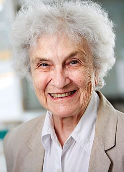 Eva Klein合作论文数Karolinska Institute4Birgitta Clinchy合作论文数4Edward J. Davey合作论文数4K Takatsu合作论文数3
 Noemi Nagy合作论文数3C Heusser合作论文数3Deveikaite V合作论文数3
 Adrian Thrasher合作论文数Great Ormond Street Institute of Child Health, University College London3C. I. Edvard Smith合作论文数3
 George Klein合作论文数Department of Tumour Biology, Karolinska Institute3Daniel Salamon合作论文数3Akira Tominaga合作论文数3
 Lorand L Kis合作论文数3Johan Thyberg合作论文数Department of Cell and Molecular Biology, Karolinska Institutet3Carina Elenström-Magnusson合作论文数3A Rosén合作论文数2Y Noma合作论文数2Takayuki Naito合作论文数2P Björck合作论文数2Ramiro E Verdun合作论文数Univ Miami, Miller Sch Med, Dept Med, Div Gerontol & Geriatr Med, Miami, FL 33136 USA2Azuma C合作论文数2Bergstedt-Lindqvist S合作论文数2
 Elena Cortizas合作论文数2
 Kiyoshi Takatsu合作论文数Toyama Medical and Pharmaceutical University;University of Toyama2




