10053 Background: Disease monitoring in pediatric sarcomas relies on invasive surgical biopsies and radiologic imaging, which often trail disease activity. Circulating tumor DNA (ctDNA) is an attractive potential biomarker, but studies in pediatric sarcomas have reported low detection rates by tracking copy number alterations or recurrent genetic alterations. We aim to establish a personalized ctDNA pipeline in pediatric sarcomas based on identification of tumor-specific single nucleotide variants (SNVs) from whole genome sequencing (WGS) to enhance ctDNA detection. Methods: Patients < 25 years old with new or relapsed sarcoma treated at our institution are eligible to enroll in our study, which is ongoing. Plasma is collected at imaging response evaluations. Matched tumor and germline DNA are collected at baseline and undergo paired WGS to identify tumor-specific SNVs, and personalized hybrid capture panels are designed to track up to 5000 mutations per patient. Cell-free DNA from each time point is analyzed with CAncer Personalized Profiling by deep Sequencing (CAPP-Seq) using duplex sequencing to minimize the background error rate and maximize sensitivity. Results: This approach was piloted in 8 pediatric patients with osteosarcoma (5), Ewings sarcoma (2), and rhabdomyosarcoma (1). A median of 619 tumor-derived SNVs (range 160-4937) were tracked per patient. ctDNA was detected in all patients at baseline with mean allele fractions ranging from 0.000937-27.1. Table 1 summarizes characteristics, number of SNVs tracked, and mean allele fraction for each patient. Seven patients had longitudinal ctDNA samples available for analysis. In all patients, ctDNA broadly correlated with radiologic response with ctDNA becoming undetectable in patients with an imaging response and ctDNA remaining detectable in patients who experienced progression. One patient achieved initial remission with undetectable ctDNA and relapsed with ctDNA re-emergence. Conclusions: Personalized whole genome-based ctDNA analysis may improve ctDNA detection in pediatric sarcomas. In this pilot, ctDNA was detected at baseline in 8/8 patients, and ctDNA levels correlated with treatment response on imaging. A personalized approach for ctDNA detection in pediatric sarcomas has the potential to aid in disease monitoring and treatment selection. Patient characteristics and baseline ctDNA allele fraction. Disease New (N) v Relapsed/ Refractory (R) Localized (L) v Metastatic (M) Sex (M, F) Age (years) SNVs (n) Baseline ctDNA Mean Allele fraction Osteosarcoma N L M 17 692 0.25 Osteosarcoma N L M 14 301 4.29 Osteosarcoma R M M 20 4937 0.00094 Osteosarcoma R M F 11 1684 27.10 Osteosarcoma R M M 12 546 2.70 Ewings sarcoma N L M 5 160 14.11 Ewings sarcoma N L M 6 198 0.78 Rhabdomyosarcoma R M F 19 935 2.79 Each row represents 1 patient with disease and ctDNA detection characteristics displayed.
The FDA approval of T cell receptor-engineered T cells (TCR-T) for synovial sarcoma demonstrates the potential for adoptive T cell therapies (ACTs) in solid tumors. However, the paucity of tumor-associated targets without expression in normal tissues remains a major bottleneck, especially in rare cancer subtypes. We developed a comprehensive computational pipeline called SCAN-ACT that leverages single-cell RNA sequencing and multi-omics data from tumor and normal tissues to nominate and prioritize putative targets for both chimeric antigen receptor (CAR)- and TCR-T cells. For surface membrane targets, SCAN-ACT proposes monospecific targets and potential target pairs for bispecific Boolean logic-gated CAR T cells. For peptide-MHC targets, SCAN-ACT proposes intracellular peptides bound to a diverse set of human leukocyte antigens. Selected targets were validated experimentally by protein expression and for peptide-MHC binding. We applied the SCAN-ACT pipeline to soft tissue sarcoma (STS), analyzing 986,749 single cells to identify and prioritize 395 monospecific CAR-T targets, 14,192 bispecific CAR-T targets, and 5020 peptide-MHC targets for TCR-T cells. Proposed targets and target pairs reflected the mesenchymal, neuronal, and hematopoietic ontogeny of STS. We further validated SCAN-ACT in glioblastoma revealing its versatility. This work provides a robust data repository along with a web-based and user-friendly set of analysis tools to accelerate ACT development for solid tumors ( https://scanact.stanford.edu/ ).
Introduction: Classic Hodgkin lymphoma (cHL) is distinguished by a unique tumor microenvironment (TME), which is comprised of rare Hodgkin-Reed-Sternberg (HRS) cells surrounded by immune cells. Although single cell and spatial techniques have advanced the characterization of the TME in cHL, prior studies lack comprehensive cohorts spanning the age and geographic spectrum. To address this challenge, we aimed to conduct a multimodal characterization of the TME in cHL using international patient cohorts of pediatric and adult cases including from EBV-endemic areas. Methods: We identified a training cohort of 198 patients diagnosed with cHL from 2013-2025 diagnosed at 4 international institutions with either formalin-fixed, paraffin-embedded (FFPE) blocks (n=188) or fresh-frozen excisional biopsies (n=10). FACS purification and single cell RNA-seq (scRNA-seq) were performed on 8 cryopreserved tissues, and 2 were profiled using single nucleus RNA-seq (snRNA-seq). Using sc- and snRNA-seq data, we constructed and benchmarked a cHL-specific signature matrix for use with CIBERSORTx. Subsequently, we performed digital deconvolution of 188 bulk RNA-seq cases. Finally, we applied a machine-learning framework (EcoTyper) to discover transcriptionally-defined cell states and ecosystems. Validation of the EcoTyper model was performed using an independent microarray dataset (n=130) and Visium spatial transcriptomics data (n=4). Results: The median age in the training cohort was 28 years (IQR=14-49), and 75 cases were EBV+ (44% of 169 tested). The median follow-up was 5.9 years (IQR=2.7-8.2). From sc- and snRNA-seq data, we profiled 44,769 cells and identified 16 distinct cell phenotypes including classical and plasmacytoid dendritic cells, gamma-delta T cells, natural killer cells, and HRS cells to derive the cHL-specific signature matrix. We performed benchmarking of the signature matrix using using several methods: pseudo-bulk resampling of scRNA-seq data, correlation of cell abundances from digital cytometry with spatial proteomics, and comparison of tumor genotyping allele frequencies versus HRS abundances. All approaches showed significant positive correlations (P<0.05). After performing digital deconvolution of the 188 FFPE samples, and using EcoTyper, we discovered 28 unique cell states clustered within 2 cellular ecosystems or “Hodgkin lymphoma ecotypes” (HLE). 56% of cases were predominantly HLE1 and were characterized by EBV+ HRS cells overexpressing oncogene BCAS1 and anti-apoptotic IL22RA, fibroblasts promoting endothelial development (VEGF, VCAM1), and abundant B cells and natural killer cells. Conversely, 44% of cases were predominantly HLE2, characterized by EBV- HRS cells expressing ECM remodeling genes (LUM, MMP2, COL1A1, FN1), fibroblasts promoting extracellular matrix reorganization (COL5A2, COL3A1, COL5A1, COL16A1, COL1A1), and macrophages expressing CXCL8 (a tumor migration marker). HLE2 was associated with worse freedom from progression (FFP) in our cohort (HR=3.18, P=0.048) after multivariable adjustment for the age, gender, and clinical stage. We used additional external validation cohorts profiled with Visium spatial transcriptomics (n=4) and RNA microarray (n=130, Steidl et al. NEJM 2010). We recovered the 2 HLE in the validation datasets and found a significant spatial colocalization of the cell states (median Z=25.4) in the Visium dataset, as well as a worse FFP for HLE2 after multivariable adjustment for the age, sex, and disease stage in the RNA microarray cohort (HR=1.81, P=0.02). Conclusions: In a large cohort containing pediatric, elderly, and EBV+ cHL, we leveraged multimodal approaches to identify HRS, immune, and stromal cell signatures found within clinically relevant HLEs. We show that approximately half of cHL cases were predominantly HLE2 characterized by EBV- migratory HRS cells, fibroblasts promoting extracellular remodeling, and abundant macrophages. HLE2 is associated with worse freedom from progression in both training and validation cohorts. Further prospective validation of our HLEs is crucial particularly in the era of checkpoint inhibition and will aid in individualized therapeutic approaches.
Soft tissue sarcomas (STS) represent a paradigm of cancer evolution, where spatial and temporal heterogeneity creates dynamic ecosystems that rapidly adapt to therapeutic pressures. Through multi-region multi-omics profiling of 45 patients across primary, irradiated, and recurrent tumors, we reconstructed evolutionary trajectories using an integrated analytical framework. DNA methylation (RRBS) was processed via Bismark, with CAMDAC deconvolution distinguishing cell type-specific signals from copy number alterations. Transcriptomic data were analyzed using Salmon and edgeR, while copy number landscapes were inferred through TitanCNA and InferCNV. Evolutionary dynamics were reconstructed using PRISM, enabling methylation-based phyloepigenome reconstruction to trace subclonal epigenetic evolution. Our analysis reveals that STS evolution follows a branching phylogenetic pattern, with early “trunk” alterations establishing the foundational epigenome and subsequent “branch” events driving radiation adaptation. PRISM-based methylation phylogenies demonstrated conserved evolutionary trajectories across patients, with epigenetic divergence increasing through treatment. At diagnosis, widespread methylome remodeling systematically rewires cellular identity, with convergent hypermethylation-mediated silencing of tumor suppressive networks (SMARCA4, DICER1, FOXO3) and hypomethylation-driven activation of oncogenic pathways (CCND1/2, CDK6). Across patients, these events were recurrent and enriched for functional gene expression changes, representing positive selection for survival advantages. Under radiotherapy selection pressure, the evolutionary dynamics shift from broad remodeling to targeted adaptation. The epigenome stabilizes, focusing on DNA repair (DDB2), stemness (ZBTB16), and proliferation (CCND1) pathways—evidence of directional selection for resistance mechanisms. Single-cell phylogenetics of >150,000 cells reveals that radiation selects for pre-existing subclones rather than generating novel populations, with overall ecosystem architecture maintained despite cellular turnover. Spatial-temporal mapping demonstrates that evolutionary trajectories vary dramatically both between patients and within individual tumors, generating multi-focal ecosystems where distinct subclones coexist, compete, and occasionally converge on shared adaptive solutions. This evolutionary framework explains radiotherapy resistance as the inevitable outcome of selection acting upon pre-existing heterogeneity. The convergent emergence of DNA repair enhancement, stem-like properties, and immunosuppressive niches across patients represents parallel evolution under common selective pressures. Together, these findings argue for the development of evolution-informed therapeutic strategies that anticipate and intercept adaptive trajectories, with the potential to overcome the formidable challenge of heterogeneity in treatment-resistant STS. Shaghayegh Soudi, Nadia Silvia, Ajay Subramanian, Serey Nouth, Faith Ryu, Taryn Kaneko, Christin New, Deborah Kenney, Raffi Avedian, Robert Steffner, David Mohler, Anusha Kalbasi, Matt van de Rijn, Gregory Charville, Everett Moding. Decoding the evolutionary landscape of soft tissue sarcomas: From multiregion origins to therapy-driven adaptation [abstract]. In: Proceedings of the AACR Special Conference in Cancer Research: Cancer Evolution: The Dynamics of Progression and Persistence; 2025 Dec 4-6; Albuquerque, NM. Philadelphia (PA): AACR; Cancer Res 2025;85(23_Suppl):Abstract nr B018.
Liquid biopsies are shifting the paradigm for diagnosis and treatment of many types of cancer. Circulating tumor DNA (ctDNA), defined as small fragments of tumor DNA shed into circulation from tumor cells, represents a major analyte for liquid biopsy approaches. ctDNA has potentially wide translational applications, including early detection of cancer, monitoring disease response, detection of minimal residual disease (MRD), and biological-sampling of tumor biology noninvasively. Although there is significant interest and a growing body of work in applying ctDNA to the clinic, existing studies are generally limited by the access to patient plasma samples. Importantly, computational models are often useful to simulate data in contexts where actual experiments require such precious resources; however, there are currently no existing computational models of tumor ctDNA shedding. In that context, the current study overcomes this limitation and describes a novel bioinformatic pipeline which is able to model ctDNA shedding during tumor initiation, growth, progression, and metastasis. We developed a novel method for generating simulated ctDNA next-gen sequencing (NGS) data in a variety of tumor scenarios. To accomplish this, we enhanced a previously-characterized spatial model of tumor growth which generates simulated whole exome sequencing data in silico to also generate ctDNA data. Outputs from this spatial model include a list of variants called from the simulated exome data. To generate the simulated ctDNA data, the distribution of these variant allele frequencies (VAF) were split into quantile bins with an associated scale factor. All variants had their mean tumor VAFs divided by their respective scale factors to determine corresponding ctDNA VAFs utilizing a validated “downsampling” technique. First we utilized the novel pipeline to generate simulated ctDNA in the setting of both a non-metastatic primary tumor and a metastatic tumor. We next validated the fidelity of simulated ctDNA data to real-world biologic data using published datasets of ctDNA from both metastatic and nonmetastatic sarcomas. In that validation analysis, we demonstrated a high degree of similarity between the simulated and published ctDNA data. Here we demonstrate a novel computational approach to model the paradigm-shifting technology of liquid biopsy completely in silico. Our ability to generate simulated data with validated biologic fidelity has potential to augment discovery-based ctDNA studies. Furthermore, the potential of this technology is underscored by the emerging potential of AI-based approaches, digital twin technologies, and simulated clinical trial data as viable synergistic approaches to conventional clinical studies. Jasmine R. Alvarez, Ajay Subramanian, Everett J. Moding, Erik S. Blomain. In silico modeling of ctDNA shedding using a novel computational pipeline which models tumorigenesis, progression, and metastasis [abstract]. In: Proceedings of the American Association for Cancer Research Annual Meeting 2025; Part 1 (Regular Abstracts); 2025 Apr 25-30; Chicago, IL. Philadelphia (PA): AACR; Cancer Res 2025;85(8_Suppl_1):Abstract nr 3705.
Abstract Introduction: Definitive chemoradiation therapy (CRT) is essential for controlling locoregionally advanced non-small cell lung cancer (NSCLC); however, up to 30% of patients experience local recurrences within the radiation field. Analyzing favorable responders to treatment could enable identification of genetic alterations associated with radiosensitivity, but standard imaging cannot distinguish residual disease from inflammation and fibrosis during treatment to assess treatment response. We hypothesized that circulating tumor DNA (ctDNA) kinetics during CRT could identify genetic alterations associated with radiosensitivity that could be validated in preclinical models and harnessed to develop new combination therapies with radiotherapy (RT). Methods: We applied cancer personalized profiling by deep sequencing (CAPP-Seq) ctDNA analysis to pre-CRT and mid-treatment (mid-CRT) plasma samples from 61 patients with Stage II-III NSCLC. We identified ctDNA rapid responders as the 10 patients with the largest decrease in ctDNA concentration mid-CRT without local progression and determined the prevalence of genetic alterations in rapid responders versus slow responders using Fisher’s exact tests corrected for multiple hypothesis testing. To investigate the therapeutic potential of targeting PDYN during RT, we performed clonogenic assays on isogeneic PDYN CRISPR-Cas9 knockout and wildtype H1299 and SW1573 human NSCLC cell lines and H1299 cells treated with prodynorphin-neutralizing antibodies. Results: Mid-CRT log-fold change in ctDNA concentration was significantly associated with progression-free survival (P=0.02) in Stage II-III NSCLC. Mutations in PDYN, which encodes the opioid peptide precursor protein prodynorphin, were observed in 30% of ctDNA rapid responders and 0% of ctDNA slow responders (adjusted P=0.04). In NSCLC patients from TCGA treated with RT, the cumulative incidence of local failure was significantly lower in tumors with PDYN mutations (0% vs. 17% at 3 years, P<0.001). PDYN knockout (PDYNNULL) radiosensitized human NSCLC cancer cells by clonogenic assay, and extracellular administration of prodynorphin or its cleavage product dynophin A partially restored radioresistance in PDYNNULL cells. Prodynorphin cleavage products have been reported to signal through opioid receptors and to antagonize N-methyl-D-aspartate receptor (NMDAR) signaling. Although treatment of wildtype H1299 cells with opioid receptor antagonists did not affect radiosensitivity, the NMDAR antagonist MK-801 partially restored RT resistance in PDYNNULL cells. Finally, prodynorphin-neutralizing antibodies significantly reduced clonogenic survival of human NSCLC cells. Conclusions: ctDNA kinetics enable the identification of patients responding favorably to treatment and putative targets to enhance the efficacy of RT. Targeting prodynorphin may enhance the radiosensitivity of NSCLC by increasing NMDAR signaling. Citation Format: Ziwei Wang, Angela B. Hui, Ash A. Alizadeh, Maximilian Diehn, Everett J. Moding.Circulating tumor DNA kinetics identify prodynorphin signaling as a target to radiosensitize non-small cell lung cancer.[abstract]. In: Proceedings of the AACR Special Conference in Cancer Research: Translating Targeted Therapies in Combination with Radiotherapy; 2025 Jan 26-29; San Diego, CA. Philadelphia (PA): AACR; Clin Cancer Res 2025;31(2_Suppl):Abstract nr P017
Background: In September of 2024, the 2nd annual meeting of the Strategic Advances in Sarcoma Science (SASS) convened at the National Institutes of Health. This gathering of national sarcoma experts focused on preclinical studies, clinical trials, opportunities, challenges, and future directions in sarcoma biology and clinical care with a focus on immunotherapy. The Immunology in Sarcoma breakout group conducted a dedicated discussion focused on the current and future implementation of adoptive cellular therapies (ACTs) in sarcomas. The current manuscript summarizes these discussions and provides a comprehensive resource for researchers and clinicians. Results: Adoptive cell therapy (ACT) has shown encouraging results in sarcomas with afami-cel achieving durable responses in synovial sarcoma and early TCR-T trials against NY-ESO-1 and MAGE-A4 demonstrating meaningful response rates. Building on these outcomes will require discovering new targets, selecting optimal cell types, refining conditioning regimens, combining with alternative treatment strategies such as TKIs, and leveraging predictive biomarkers informed by a deeper understanding of the tumor microenvironment. Conclusions: Sarcomas are promising targets for adoptive cell therapy (ACT), as shown by afami-cel’s success in synovial sarcoma, but broader impact requires new target discovery, optimal cell selection, improved conditioning, combination treatments, deeper tumor microenvironment understanding, and predictive biomarkers to achieve more durable responses for more patients.
Small molecules that induce nonapoptotic cell death are of fundamental mechanistic interest and may be useful to treat certain cancers. Here we report that tegavivint, a drug candidate undergoing human clinical trials, can activate a unique mechanism of nonapoptotic cell death in sarcomas and other cancer cells. This lethal mechanism is distinct from ferroptosis, necroptosis and pyroptosis and requires the lipid metabolic enzyme trans-2,3-enoyl-CoA reductase (TECR). TECR is canonically involved in the synthesis of very-long-chain fatty acids but appears to promote nonapoptotic cell death in response to CIL56 and tegavivint via the synthesis of the saturated long-chain fatty acid palmitate. These findings outline a lipid-dependent nonapoptotic cell death mechanism that can be induced by a drug candidate currently being tested in humans.
Radiotherapy is an integral component in the treatment of many types of cancer, with approximately half of patients with cancer receiving radiotherapy. Systemic therapy applies pressure that can select for resistant tumor subpopulations, underscoring the importance of understanding how radiation impacts tumor evolution to improve treatment outcomes. We integrated temporal genomic profiling of 120 spatially distinct tumor regions from 20 patients with undifferentiated pleomorphic sarcomas (UPS), longitudinal circulating tumor DNA analysis, and evolutionary biology computational pipelines to study UPS evolution during tumorigenesis and in response to radiotherapy. Most unirradiated UPSs displayed initial linear evolution, followed by subsequent branching evolution with distinct mutational processes during early and late development. Metrics of genetic divergence between regions provided evidence of strong selection pressures during UPS development that further increased during radiotherapy. Subclone abundance changed after radiotherapy with subclone contraction tied to alterations in calcium signaling, and inhibiting calcium transporters radiosensitized sarcoma cells. Finally, circulating tumor DNA analysis accurately measured subclone abundance and enabled noninvasive monitoring of subclonal changes. These results demonstrate that radiation exerts selective pressures on UPSs and suggest that targeting radioresistant subclonal populations could improve outcomes after radiotherapy.Significance: Radiotherapy mediates tumor evolution by leading to the expansion of resistant subclonal cancer cell populations, indicating that developing approaches to target resistant subclones will be crucial to improve radiotherapy response.
Small molecules that induce non-apoptotic cell death are of fundamental mechanistic interest and may be useful to treat certain cancers. Here, we report that tegavivint, a drug candidate undergoing human clinical trials, can activate a unique mechanism of non-apoptotic cell death in sarcomas and other cancer cells. This lethal mechanism is distinct from ferroptosis, necroptosis and pyroptosis and requires the lipid metabolic enzyme trans-2,3-enoyl-CoA reductase (TECR). TECR is canonically involved in the synthesis of very long chain fatty acids but appears to promote non-apoptotic cell death in response to CIL56 and tegavivint via the synthesis of the saturated long-chain fatty acid palmitate. These findings outline a lipid-dependent non-apoptotic cell death mechanism that can be induced by a drug candidate currently being tested in humans.
Purpose: Synovial sarcoma (SS) is a rare, aggressive soft tissue malignancy that is divided into biphasic and monophasic histologic subtypes. In addition to surgical resection, radiation therapy (RT) improves local control in patients at higher risk of recurrence. This study aimed to investigate the impact of histologic subtype on radiation response and survival outcomes in patients treated with RT as part of definitive management. Methods and Materials: We retrospectively identified patients with SS treated with RT and surgical resection from 1997 to 2020 at Stanford Medical Center. We assessed the association between histologic subtypes (biphasic vs monophasic) and response to preoperative RT based on imaging and pathology. Volumetric response was calculated using the pre-RT and post-RT/preoperative postcontrast T1-weighted magnetic resonance imaging images. Progression-free survival (PFS) and overall survival (OS) were estimated using the Kaplan-Meier method. Univariable and multivariable analyses were conducted using Cox regression models. Variables for univariable and multivariable analyses included age, histologic subtypes, tumor location, tumor size, margin status, chemotherapy, and performance status. Results: In our study, 50 patients met the inclusion criteria. The median age was 34.8 years at diagnosis, and 36% (n = 18) received concurrent chemotherapy. Biphasic (n = 18, 36%) and monophasic (n = 32, 64%) tumors exhibited significant differences in negative margin status (94% vs 66%, P = .036). Of the 22 patients who underwent preoperative RT, 15 patients had pre-RT and post-RT imaging to assess volumetric changes. Biphasic tumors demonstrated less necrosis at the time of surgical resection but a significantly greater volumetric decrease with preoperative RT (42% vs 5%, P = .004). PFS and OS were superior in biphasic tumors (P = .003 and P = .009, respectively). Multivariable analyses identified histologic subtypes (monophasic vs biphasic) as a significant factor impacting PFS (HR, 5.65; 95% CI, 1.78-17.91; P = .003). Conclusions: Biphasic tumors exhibit an improved volumetric response to preoperative RT and improved outcomes. These findings underscore the importance of considering histology when tailoring treatment for patients with SS.
Cytokines and their receptors enable precise tuning of T cell function. Leveraging this biology holds tremendous promise for optimizing antitumor immunity. Arming T cells with a synthetically orthogonal interleukin (IL)-9 receptor (o9R), for instance, permits facile engraftment and potent anti-tumor functions. Exploiting the paucity of wild-type IL-9R expression and the safety of high doses of IL-9, here, we showed that, compared with o9R, T cells engineered with wild-type IL-9R exhibited superior tissue infiltration, stemness, and anti-tumor activity. These qualities were consistent with a stronger Janus kinase (JAK)/signal transducer and activator of transcription (STAT) signal, which included canonically IL-12-driven STAT4 in addition to STAT1/3/5. IL-9R T cells were exquisitely sensitive to perturbations of proximal signaling, including structure-guided attenuation, amplification, and rebalancing of JAK/STAT signals. Biased IL-9R mutants showed that STAT1 acts as a rheostat between stem-like and effector states. In summary, we identify IL-9/IL-9R as a naturally orthogonal cytokine-receptor pair with an optimal JAK/STAT signaling profile for engineered T cell therapy.
The complementarity and clinical utility of combining liquid biopsies and radiomic image analysis has not been demonstrated. ctDNA minimal residual disease after chemoradiotherapy (CRT) for non-small cell lung cancer (NSCLC) is highly prognostic, but on-treatment biomarkers are needed to enable response-adapted therapies. In this study, we analyzed 418 patients with NSCLC undergoing CRT to develop and validate a novel dynamic risk model that accurately predicts ultimate progression-free survival during treatment. We optimize tissue-free variant calling from plasma samples to facilitate ctDNA monitoring and demonstrate the importance of accounting for persistent clonal hematopoiesis variants. We show that mid-CRT ctDNA concentration is prognostic for disease progression and integrate additional pre-CRT risk factors, including radiomics, into a combined model that improves outcome prediction. Our results suggest that tumor features, radiomics, and mid-CRT ctDNA analysis are complementary and can identify patients at high and low risk of progression to potentially enable response-adapted therapies. SIGNIFICANCE:This study demonstrates that combining tumor features, radiomics, and ctDNA analysis improves outcome prediction in NSCLC treated with CRT therapy. Our integrated model could enable personalized and response-adapted therapies to reduce toxicity and improve outcomes in patients. See related commentary by Anagnostou and Aggarwal, p. 1534.
Nodular lymphocyte-predominant Hodgkin lymphoma (NLPHL) is a rare cancer, and few studies have comprehensively investigated the immune microenvironment and rare lymphocyte-predominant (LP) cells. Here we develop a NLPHL specific lymphocyte-predominant ecotype (LPE) model to identify 34 distinct cell states across 14 cell types that co-occur within 3 LPEs for 171 cases. LPE1 and LPE2 were characterized by immunosuppressive microenvironments with high expression of B2M on LP cells, CD8 T-cell exhaustion, immune checkpoint genes expressed by follicular T-cells, and an improved freedom from progression compared to LPE3 in training (n = 109, with 65% LPE1/2) and validation cohorts (n = 62, with 61% LPE1/2). We validate the co-occurrence and co-localization of cell states using spatial transcriptomics. Protein expression of HLA-I and HLA-II on LP cells and SSTR2 on dendritic cells was predictive of LPE1 (C-statistic=0.69), LPE2 (C-statistic=0.79), and LPE3 (C-statistic=0.60). This study establishes a clinically relevant biologic categorization for NLPHL.
PURPOSE Small cell lung cancer (SCLC) is characterized by rapid progression after platinum resistance. Circulating tumor (ctDNA) dynamics early in treatment may help determine platinum sensitivity. MATERIALS AND METHODS Serial plasma samples were collected from patients receiving platinum-based chemotherapy for SCLC on the first 3 days of cycle one and on the first days of subsequent cycles with paired samples collected both before and again after infusions. Tumor-informed plasma analysis was carried out using CAncer Personalized Profiling by deep Sequencing (CAPP-Seq). The mean variant allele frequency (VAF) of all pretreatment mutations was tracked in subsequent blood draws and correlated with radiologic response. RESULTS ctDNA kinetics were assessed in 122 samples from 21 patients. Pretreatment VAF did not differ significantly between patients who did and did not respond to chemotherapy (mean 22.5% v 4.6%, P = .17). A slight increase in ctDNA on cycle 1, day 1 immediately post-treatment was seen in six of the seven patients with available draws (fold change from baseline: 1.01-1.44), half of whom achieved a response. All patients who responded had a >2-fold decrease in mean VAF on cycle 2 day 1 (C2D1). Progression-free survival (PFS) and overall survival (OS) were significantly longer in patients with a >2-fold decrease in mean VAF after one treatment cycle (6.8 v 2.6 months, log-rank P = .0004 and 21.7 v 6.4 months, log rank P = .04, respectively). CONCLUSION A >2-fold decrease in ctDNA concentration was observed by C2D1 in all patients who were sensitive to platinum-based therapy and was associated with longer PFS and OS.
Purpose: Although there is a theoretical risk of skin seeding during surgical resection of soft tissue sarcomas (STSs), current consensus guidelines recommend against routine use of bolus during radiation therapy (RT). However, the risk of skin recurrence has not been systematically assessed. We aimed to assess the patterns of local recurrence (LR) in patients with STS treated with surgery with or without RT. Methods and Materials: We performed a retrospective analysis of adults with STSs evaluated at our institution between 2007 and 2021. For patients who developed LR, the depth was evaluated. Progression -free survival and overall survival were analyzed from time of first LR using the Kaplan -Meier method. Cumulative incidence of distant metastasis was calculated with competing risk analysis from date of LR. Results: Of the 206 patients evaluated, 20 had LR (9.7%). Among patients with LR, 5 patients (25.0%) were treated with surgery alone and 15 patients (75.0%) with surgery and RT. In patients treated with RT, 46.7% had preoperative RT, 53.3% had postoperative RT, and bolus was used in 46.7%. Surgical margins were close (<1 mm) in 4 patients (20.0%) and positive in 10 patients (50.0%). LR occurred in the deep subfascial tissue in 9 patients (45%), subcutaneous tissue in 10 patients (50.0%), and skin in 1 patient (5.0%). The patient with a skin recurrence was treated with surgery alone, and the tumor involved the skin at presentation. In patients treated with RT, LR occurred within the RT field in 13 patients (86.7%). At 1 year after LR, progression -free survival was 70.3%, overall survival was 81.7%, and cumulative incidence of distant metastasis was 5.9%. Conclusions: Skin recurrences were rare after surgical resection of STSs and only occurred in a tumor that involved the skin at initial presentation. These findings support current recommendations against routine use of bolus in STSs not involving the skin at presentation. (c) 2023 American Society for Radiation Oncology. Published by Elsevier Inc. All rights reserved.
Introduction: Classic Hodgkin lymphoma (cHL) is a unique cancer distinguished by its tumor microenvironment (TME), which is comprised by rare Hodgkin-Reed-Sternberg (HRS) cells (1-5%) surrounded by an abundance of immune cells. Comprehensive characterization of the malignant and immune cell states in cHL is challenging due to the scarcity of HRS cells and the intrinsic heterogeneity of the TME. To address this challenge, we aimed to characterize the cell states and ecosystems of cHL microenvironment using advanced experimental and bioinformatic approaches. Methods: We identified 111 patients diagnosed with cHL from 2013-19 managed at participating institutions with either formalin-fixed, paraffin-embedded (FFPE, n = 106) or cryopreserved cell suspensions prepared from excisional biopsies (n = 5). FACS purification and single cell RNA-seq (scRNA-seq) were performed on the cryopreserved tissues, resulting in the annotation of 13 distinct cell types within the scRNA-seq data. Next, we employed CIBERSORTx to construct a cHL-specific signature matrix encompassing the features of the 13 annotated cell types. For our FFPE samples, we performed bulk RNA-seq followed by digital deconvolution using the cHL-specific signature matrix, allowing us to estimate the cellular fractions of the 13 cell types within the 106 tumors. Finally, we employed a machine-learning framework (EcoTyper) to discover transcriptionally-defined cell states and ecosystems. Validation of the EcoTyper model was performed in additional scRNA-seq data (n = 3) and Visium Spatial Platform (n = 4). Results: Patients with FFPE biopsies had a median age of 35, were predominantly male (53%), and the majority had stage IV disease (32%). From the scRNA-seq data (n = 5), we identified 13 distinct cell phenotypes including the minute classical and plasmacytoid dendritic cells, gamma delta T cells, natural killer cells, and HRS cells; then developed a gene expression signature matrix allowing for the deconvolution of bulk transcriptomes from 106 cHL biopsies. Among patients with Variant Allele Frequencies (VAFs) data from tumor and plasma biopsies, we observed a positive correlation between the HRS cell proportion and VAFs from tumor or cell-free DNA sequencing (R = 0.44 and 0.94, respectively). From EcoTyper, we discovered 28 unique cell states and 3 conserved cellular communities or “Hodgkin Lymphoma ecotypes” (HLEs) in the 106 bulk transcriptomes. HLE2 was enriched in elderly patients (Age > 45), H2 phenotype (Alig, Nature, 2024), and EBV infection (P < 0.01). In HLE2, we discovered the enrichment of an immune-suppressing CD8 T cell state expressing LAG3 (P = 1.6 * 10-11), as well as an HRS cell state enriched in cell cycle pathways (P = 3.41 * 10-6). Moreover, HLE2 was associated with worse progression-free survival in our cohort (HR = 3.18, P = 0.048) after multivariable adjustment for the disease stages. To validate our EcoTyper model, we employed additional scRNA-seq on 3 validation samples (n = 31,195 cells) and spatial transcriptomics using Visium 10X (n = 4 tumors). We recovered the established 28 cell states and 3 HLEs in these datasets and found a significant spatial autocorrelation of the HLEs (median Z = 25.4). Conclusions: Our study represents a significant advancement in unraveling the complexities of the cHL TME. By leveraging scRNA-seq, digital deconvolution, and EcoTyper framework, we have shed light on key cell states and discovered a unique HLE associated with clinical parameters and prognostic significance. Further validation in additional cohorts may allow HLEs to risk stratify patients at diagnosis and aid in identification of poor performing subgroups within current low, intermediate, and high-risk groups.
Characterization of the diverse malignant and stromal cell states that make up soft tissue sarcomas and their correlation with patient outcomes has proven difficult using fixed clinical specimens. Here, we employed EcoTyper, a machine-learning framework, to identify the fundamental cell states and cellular ecosystems that make up sarcomas on a large scale using bulk transcriptomes with clinical annotations. We identified and validated 23 sarcoma-specific, transcriptionally defined cell states, many of which were highly prognostic of patient outcomes across independent datasets. We discovered three conserved cellular communities or ecotypes associated with underlying genomic alterations and distinct clinical outcomes. We show that one ecotype defined by tumor-associated macrophages and epithelial-like malignant cells predicts response to immune-checkpoint inhibition but not chemotherapy and validate our findings in an independent cohort. Our results may enable identification of patients with soft tissue sarcomas who could benefit from immunotherapy and help develop new therapeutic strategies. Subramanian et al. use the EcoTyper machine-learning framework to characterize the tumor, immune and stromal cell states and ecosystems that comprise sarcomas.
 Maximilian Diehn合作论文数Department of Radiation Oncology, School of Medicine, Stanford University32
 David Kirsch合作论文数Department of Radiation Oncology, University of Toronto31
 Ash A. Alizadeh合作论文数Stanford Cancer Institute, Stanford University31
 Jake Chabon合作论文数Foresight Diagnostics Inc.21
 Ajay Subramanian合作论文数21
 Nabet Barzin Y合作论文数Abramson Family Canc Res Inst, Univ Penn21
 Chang Lung Lee合作论文数Department of Radiation Oncology, Duke University School of Medicine;Duke Cancer Institute, Duke University19
 Heather Wakelee合作论文数Division of Oncology, Department of Medicine, Stanford University19
 Joel Neal合作论文数Division of Oncology, Department of Medicine, School of Medicine, Stanford University;Stanford Health Care;Stanford Cancer Institute;Stanford Cancer Center18
 Sukhmani Kaur Padda合作论文数Lewis Katz School of Medicine, Temple University16
 Michael S Binkley合作论文数Department of Radiation Oncology, School of Medicine, Stanford University16
 Matt Van De Rijn合作论文数Maternal & Child Health Research Institute, School of Medicine, Stanford University;Stanford Cancer Institute, Stanford University15Angela B Hui合作论文数Stanford Univ, Stanford Canc Inst, Stanford, CA 94305 USA14
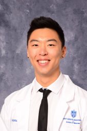 Christopher H Yoo合作论文数Saint Louis University School of Medicine14
 Billy W Loo Jr合作论文数Department of Radiation Oncology, School of Medicine, Stanford University14Ma Yan合作论文数13
 David Kurtz合作论文数Department of Medicine, School of Medicine, Stanford University;Foresight Diagnostics Inc.13Diego Almanza合作论文数12
 Emily G Hamilton合作论文数Program Canc Biol, Stanford Sch Med12
 Das Millie合作论文数Medicine Department, Stanford University12Ramchandran Kavitha合作论文数12
 Raffi S. Avedian合作论文数Department of Orthopaedic Surgery, Stanford University;Stanford Medicine Outpatient Center, Stanford University12
 Robert Steffner合作论文数Department of Orthopaedic Surgery, Stanford University12
 Ryan Ko合作论文数11
 Henning Stehr合作论文数Department of Pathology, Stanford University11Chih Long Liu合作论文数11
 Susan Hiniker合作论文数Department of Radiation Oncology, School of Medicine, Stanford University10
 Anusha Kalbasi合作论文数Dept Radiat Oncol, Univ Calif Los Angeles10
 Aadel A. Chaudhuri合作论文数Division of Biology, California Institute of Technology10David Mohler合作论文数9Rene F. Bonilla合作论文数Dept Biol, Univ Nevada9Neda Nemat-Gorgani合作论文数Stanford University9Gojenola Linda合作论文数9
 Kristen N Ganjoo合作论文数Division of Oncology, Department of Medicine, School of Medicine, Stanford University9Young-Jun Jeon合作论文数Dept. of Computer Science & Engineering, University of Incheon9
 Alexander Chin合作论文数Department of Radiation Oncology, School of Medicine, Stanford University8
 Bob T. Li合作论文数AstraZeneca8Nesselbush Monica合作论文数Personal Genome Diagnostics8
 Mohammad Shahrokh Esfahani合作论文数School of Medicine, Stanford University8
 Mark Francis Berry合作论文数Department of Cardiothoracic Surgery, School of Medicine, Stanford University8Anish Somani合作论文数7Eniola Oladipo合作论文数7Christin New合作论文数7
 Aaron Newman合作论文数Department of Biomedical Data Science, School of Medicine, Stanford University;Institute for Stem Cell Biology and Regenerative Medicine, School of Medicine, Stanford University;Stanford Cancer Institute7
 Matthew Hellmann合作论文数Memorial Sloan Kettering Cancer Center7
 Charles M. Rudin合作论文数David H. Koch Center for Cancer Care, Memorial Sloan Kettering Cancer Center7
 Joseph Shrager合作论文数Division of Thoracic Surgery, School of Medicine, Stanford University;Palo Alto Veterans Affairs Health Care System7
 Dieter Saur合作论文数School of Medicine and Health, Technical University of Munich;Division of Translational Cancer Research, German Cancer Research Center;Institute of Experimental Cancer Therapy, Technical University of Munich7
 Steven H. Lin合作论文数Department of Experimental Radiation Oncology, The University of Texas MD Anderson Cancer Center/Department of Radiation Oncology, The University of Texas MD Anderson Cancer Center7




