Treatment of the tumor and dural margin with surgery and sometimes radiation are cornerstones of therapy for meningioma. Molecular classifications have provided insights into the biology of disease; however, response to treatment remains heterogeneous. In this study, we used retrospective data on 2,824 meningiomas, including molecular data on 1,686 tumors and 100 prospective meningiomas, from the RTOG-0539 phase 2 trial to define molecular biomarkers of treatment response. Using propensity score matching, we found that gross tumor resection was associated with longer progression-free survival (PFS) across all molecular groups and longer overall survival in proliferative meningiomas. Dural margin treatment (Simpson grade 1/2) prolonged PFS compared to no treatment (Simpson grade 3). Molecular group classification predicted response to radiotherapy, including in the RTOG-0539 cohort. We subsequently developed a molecular model to predict response to radiotherapy that discriminates outcome better than standard-of-care classification. This study highlights the potential for molecular profiling to refine surgical and radiotherapy decision-making.
Abstract BACKGROUND Despite multimodal treatment (resection, radiation therapy, alkylating chemotherapy and/or tumor-treating fields), the treatment of progressive CNS WHO Grade 3 astrocytoma and glioblastoma remains challenging. Re-irradiation (Re-RT) might be considered in the course of the disease. Optimal fractionation/dose prescription schedules are still under investigation, underlining a gap in clinical data in this regard. Here, we retrospectively analyzed post-progression survival (PPS) and progression-free survival (PFS) in a single center cohort. MATERIAL AND METHODS Patients receiving Re-RT with 20 Gy in 5 fractions (4 Gy per fraction; corresponding to 35 Gy in an equivalent dose normalized to 2 Gy (EQD2)) upon progression of recurrent astrocytoma glioblastoma or grade 3 astrocytoma (12/2020-12/2022) were included. Planning target volume for Re-RT was gross tumor volume = (T1 with contrast) + 5 mm. Clinical endpoints included PPS, PFS since start of Re-RT, therapy in case of further progression, bevacizumab therapy, MGMT promotor methylation (MGMT meth), and IDH status. RESULTS To date, 40 patients were analyzed (27 male); median age 62.25 a, range 31-83 a; 34 GBM IDHwt, 6 other), 15 of who showed MGMT meth (24 w/o MGMT meth, 1 not determined). Primary therapy was 60 Gy with concomitant temozolomide (TMZ), TMZ + lomustine, or study medication in 30, 54.0 Gy + adjuvant TMZ in 3, and 40.05 Gy + TMZ in 7 patients. Patients received heterogenous systemic therapies and 0-3 treatment lines prior to Re-RT (median 1 prior treatment modality). Re-RT was considered in localized non-resectable progression. Median time between first line therapy and first progression was 10.8 months and between first line therapy and Re-RT 16.7 months, respectively. Seven patients received an integrated boost of up to 30 Gy. Median PFS was 2.6 months (range <1-10.4 months); with 7,6 months PPS (range <1 - 22.4 months). 19 patients (47.5%) received bevacizumab during further course of the disease. Following Re-RT, 16 patients (40%) received further tumor-directed therapy. CONCLUSION This is a heterogenous single-center cohort. PFS after Re-RT was 2.6 months, and PPS was 7.6 months, respectively, published data reporting 6-12 months PPS after re-irradiation (EQD2 ranging between 48-60 Gy) despite a comparatively low and conservative cumulative dose of 20 Gy (35 Gy EQD2). Treatment time was only 5 days. Thus, re-irradiation with 4 Gy x 5 could be an option in a palliative setting and could be included in interdisciplinary treatment discussions for progressive CNS WHO grade 3 astrocytoma and glioblastoma.
Background: In meningiomas, CDKN2A/B deletions are associated with poor outcomes but are rare in most cohorts (1-5%). Large molecular datasets are therefore required to explore these deletions and their relationship to other prognostic CDKN2A alterations. Methods: We utilized multidimensional molecular data of 560 meningiomas from 5 independent cohorts to comprehensively interrogate the spectrum of CDKN2A alterations through DNA methylation, copy number variation, transcriptomics, and proteomics using an integrated molecular approach. Results: Meningiomas with either CDKN2A/B deletions (partial or homozygous loss) or an intact CDKN2A gene locus but elevated mRNA expression (CDKN2A high ) both had poor clinical outcomes. Increased CDKN2A mRNA expression was a poor prognostic factor independent of CDKN2A deletion. CDKN2A expression and p16 protein increased with tumor grade and more aggressive molecular and methylation groups. CDKN2A high meningiomas and meningiomas with CDKN2A deletions were enriched for similar cell cycling pathways dysregulated at different checkpoints. p16 immunohistochemistry was unreliable in differentiating between meningiomas with and without CDKN2A deletions, but increased positivity was associated with increased mRNA expression. CDKN2A high meningiomas were associated with gene hypermethylation, Rb-deficiency, and lack of response to CDK inhibition. Conclusions: These findings support the role of CDKN2A mRNA expression as a biomarker of clinically aggressive meningiomas with potential therapeutic implications.
Aims Previous data suggest that expression of transcription factors FoxG1 and Olig‐2 can separate hotspot histone H3 family member 3A (H3F3A)‐mutant tumours in paediatric glioma. We evaluated their prognostic potential and feasibility for identifying H3F3A‐mutant tumours among IDH‐mutant/wild‐type gliomas. Methods Immunohistochemistry of FoxG1/Olig‐2 and α‐thalassaemia/mental‐retardation‐syndrome‐X‐linked gene (ATRX) in 471 cases of diffuse gliomas and molecular determination of IDH, H3F3A, MGMT and 1p/19 codeletion status. Results Mean percentage of FoxG1‐positive tumour cells increased from 17% in WHO grade II to over 21% in grade III to 37% in grade IV tumours, whereas mean Olig‐2 indices decreased from 29% to 28% to 17% respectively. FoxG1 indices were similar in astrocytic and oligodendroglial tumours, whereas Olig‐2 indices were increased in oligodendrogliomas compared to astrocytic tumours ( n = 451, P < 0.0001). FoxG1‐positive nuclei were significantly reduced in IDH and H3F3A K27‐mutant tumours, whereas Olig‐2‐positive nuclei were significantly reduced in IDH‐wild‐type and H3F3A G34‐mutant tumours. Among IDH‐mutant tumours, mean Olig‐2 index was significantly higher in 1p/19q codeleted tumours (mean: 43%) compared to IDH‐mutant tumours with ATRX loss (mean: 23%, P < 0.0001). A significantly better outcome was first suggested for FoxG1 low tumours ( n = 212, log rank P = 0.0132) and Olig‐2 high tumours ( n = 203, log‐rank P = 0.0011) based on classification and regression tree determined cutoffs, but this was not confirmed by multivariate analysis including IDH mutation, WHO grade, ATRX status and age. Conclusions While the combined FoxG1/Olig‐2 profile may discriminate H3F3A K27‐ and G34‐mutant tumours and define a prognostically favourable subset in IDH‐mutant gliomas, our data show that labelling indices of these transcription factors overlap with adult IDH‐mutant and wild‐type tumour classes.
 G. Tabatabai合作论文数8Jens Schittenhelm合作论文数8
 Benjamin Bender合作论文数5
 Friedrich Paulsen合作论文数Institut für Funktionelle und Klinische Anatomie, Medizinische Fakultät, Friedrich-Alexander-Universität;Institut für Anatomie, Medizinische Fakultät, Friedrich-Alexander-Universität5E. Hoffmann合作论文数4
 Marco Skardelly合作论文数4
 Franziska Eckert合作论文数4M Tatagiba合作论文数3J Honegger合作论文数3Felix Sahm合作论文数2Ramneet Kaloti合作论文数2Rebecca Yakubov合作论文数2
 Jill S. Barnholtz-Sloan合作论文数Winship Cancer Institute, Emory University;Department of Biostatistics and Bioinformatics, Rollins School of Public Health, Emory University2
 Farshad Nassiri合作论文数Division of Neurosurgery,Department of Surgery,University of Toronto/MacFeeters Hamilton Center for Neuro-Oncology,Princess Margaret Cancer Center2Zips Daniel合作论文数2
 Vikas Patil合作论文数From the Department of Microbiology and Cell Biology, Indian Institute of Science2
 Mirjam Renovanz合作论文数2
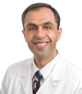 Aaron A. Cohen-Gadol合作论文数Goodman Campbell Brain and Spine, Indiana University2
 Marcos Tatagiba合作论文数Klinik für Neurochirurgie, Universitätsklinikum Tübingen2
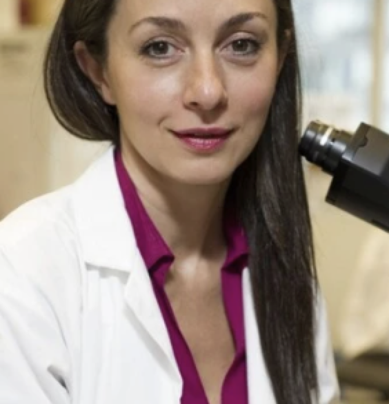 Gelareh Zadeh合作论文数Department of Surgery, Temerty Faculty of Medicine, University of Toronto;Toronto Western Hospital, University Health Network2M. Tatagiba合作论文数1M. S. Tatagiba合作论文数1Andrew Ajisebutu合作论文数1Hans Boesmueller合作论文数1KD Aldape合作论文数1Ghazaleh Tabatabai合作论文数1Zeel Patel合作论文数1Chloe Gui合作论文数1Jz Wang合作论文数1
 Leland Rogers合作论文数1
 Justin Z. Wang合作论文数1
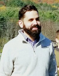 Damian A Almiron Bonnin合作论文数Human Biology Division, Fred Hutchinson Cancer Center1Vlatko Potkrajcic合作论文数1J. Ehlers合作论文数1Helin Dogan合作论文数Univ Hosp Heidelberg, Inst Pathol, Dept Neuropathol, Heidelberg, Germany1Christopher D Wilson合作论文数Indiana University1B. Frey合作论文数1
 Alexander Landry合作论文数1
 Alexander D Rebchuk合作论文数Div Neurosurg, Univ British Columbia1
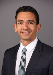 Mohamed Zaazoue合作论文数Dept Neurol Surg, Indiana Univ Sch Med1Serge Makarenko合作论文数Department of Surgery, Faculty of Medicine, The University of British Columbia1Olli Saarela合作论文数Biostatistics Division, Dalla Lana School of Public Health, University of Toronto1
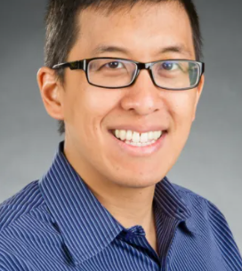 Derek S. Tsang合作论文数Department of Radiation Oncology, Temerty Faculty of Medicine, University of Toronto1
 Paul Boutros合作论文数Jonsson Comprehensive Cancer Center, University of California, Los Angeles;Paul Boutros Lab, Institute of Urologic Oncology, University of California, Los Angeles;Department of Human Genetics and Urology, David Geffen School of Medicine, University of California, Los Angeles;Eli and Edythe Broad Center of Regenerative Medicine and Stem Cell Research1
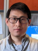 Andrew Gao合作论文数Department of Laboratory Medicine and Pathobiology, Temerty Faculty of Medicine, University of Toronto1
 Craig M Horbinski合作论文数Department of Pathology, Feinberg School of Medicine, Northwestern University1
 Kenneth Aldape合作论文数Laboratory of Pathology, Center for Cancer Research, National Cancer Institute, National Institutes of Health1




