Purpose: Parent worry about torsional or angular anomalies of the lower limbs of their children is widespread. The relationship between a child’s postural habits and torsional anomalies of the lowe...
Purpose: Ligament laxity is a common feature of trisomy 21 and is incriminated in most of the orthopaedic disorders observed. Early diagnosis and management is essential. C1-C2 instability is a rec...
Dysplasia epiphysealis hemimelica (DEH) or Trevor's disease is a rare disorder affecting the epiphyses and short bones of the limbs, and characterized by a benign overgrowth of the medial-half of the epiphysis resembling osteochondroma. Upper limb involvement is very rare and most commonly affects the carpal bones. Only five established cases of DEH around the elbow were found in the orthopedic literature. We herein report an additional case of Trevor's disease of the elbow in a child with a symptomatic ulnar nerve compression and its management. To the best of our knowledge, this is the first case of DEH of the elbow presenting with nerve compromise.
BACKGROUND:Percutaneous adductor longus tenotomy (PAT) is a frequently used procedure, yet no study has ever compared its effectiveness and safety with those of open adductor longus tenotomy (OAT). We conducted this prospective study to describe the effects of PAT and to compare them with those of OAT.METHODS:This consisted of a cross-over randomized controlled trial including 50 consecutive hips from 27 patients with cerebral palsy scheduled for adductor tenotomy in the setting of multilevel tendon lengthening/release procedures or hip surgery (femoral or Dega osteotomy) in a university hospital. A pediatric orthopaedic surgeon conducted a PAT. Another surgeon extended the wound to explore what had been cut during the PAT, and completed the tenotomy if necessary. Hip abduction (HA) was assessed by a third surgeon immediately before PAT, after PAT, and then after OAT, using a goniometer, in a standardized reproducible manner. All 3 surgeons were blinded to the others' findings. Primary end-points included the percentage of tendon/muscle portion sectioned percutaneously, and the HA measure. Comparison between HA after PAT and OAT was done using a paired t-test with a 95% confidence interval. The influence of anatomic variants of adductor longus origin was also assessed.RESULTS:Mean HA (hips flexed) measured 40.36 degrees preoperatively and increased to 50.04 degrees after PAT (P<0.0001). After OAT, HA averaged 53.32 degrees with no statistical gain compared with that observed after PAT (P=0.2). The tendinous portion of adductor longus was cut to an average of 98% by PAT (completely in 46 cases and more than 75% in only 4 cases). The muscular portion of adductor longus origin was cut to an average of 83.7% (completely in only 15 cases, cut to more than 75% in 26 cases, and approximately 50% in 9 cases). The gain in HA positively correlated with the extent of the tendinous portion divided (P=0.03) but not with the extent of muscular portion divided. Results were independent of the anatomic variants of adductor longus origin. Partial section of adductor brevis after PAT was encountered in 6 cases. No major iatrogenic lesion was observed (obturator nerve, major vessels).CONCLUSIONS:This is the only prospective study concerning the effects of PAT. The anatomic factor associated with gain in HA seems to be the extent of the section of the tendinous portion of adductor longus origin, which was found to be cut to more than 90% in all cases after PAT. The extent of muscular portion section does not seem to influence the gain in HA. The researchers detail the technique of percutaneous adductor tenotomy and show that when done correctly, PAT is a fast and simple procedure, as reliable and effective as the open release and without any major risks.LEVEL OF EVIDENCE:Level II therapeutic study-prospective comparative study.
Purpose To evaluate the efficacy and safety of lateral shelf acetabuloplasty in Legg–Calvé–Perthes (LCP) hips classically associated with poor prognosis. Methods A retrospective study was conducted on 30 consecutive pediatric patients (average age 8.6 years) presenting with a severe and progressive form of LCP disease, with (16 hips) or without (14 hips) femoral varus osteotomy (FVO), and treated by lateral shelf acetabuloplasty. Shelf was done on hips presenting an aspherical incongruency with flattening, subluxation, and lack of femoral head coverage, as demonstrated on pre-operative radiographs and arthrography. All patients were reexamined at an average follow-up of 9.5 years (range 5.2–12 years). Clinical, radiological, and computed tomography scan evaluations were undertaken. Stulberg and Mose classifications were applied as radiological indicators of prognosis. Statistical analysis was performed using Student's t test and the Pearson correlation test with variance analysis for repetitive measures. Results At the last follow-up, all patients were pain free and had normal or almost normal hip motion. Twenty-seven patients are now able to walk normally or with a slight limp. Eighteen hips are classified as Stulberg 1 or 2, and 18 hips are classified as Mose 1 or 2. The average neck-shaft angle is 127°. A statistically significant improvement in the majority of radiographic parameters was found. There was no statistically significant worsening of leg length discrepancy following the procedure. The scanographic study found neither offset nor migration of the shelf in any of the hips. The average coronal and anteroposterior length of the shelf was 9.9 and 25 mm, respectively. A borderline positive correlation was found between Wiberg angle improvement and young age at the time of surgery. There was no statistically significant difference between hips that underwent shelf procedures alone and those in which it was combined with FVO. Conclusion Lateral shelf acetabuloplasty improves the outcome of hips with severe LCP. The combined procedure insures a better and lasting coverage and remodeling of the femoral head, while preserving acetabular roof growth.
A retrospective review was conducted on 50 children with cerebral palsy, having 89 femoral varus osteotomy at an average age of 7.4 years, trying to identify risk factors for complications, particularly redislocation and avascular necrosis. Among the well-reduced hips, new subluxation developed in 12 cases; postoperative radiographic measurements showed a mean neck-shaft angle of 135° and an acetabular slope of 32°. Main risk factors for secondary dislocation seem to be insufficient correction of preexisting valgus and uncorrected acetabular dysplasia. Avascular necrosis was observed in 33 hips (37%): 26 minor and seven severe. Older age at surgery and high preoperative Reimer's migration index seem statistically significant risk factors.
Background: Radiofrequency energy is being used more and more frequently in orthopaedics, mainly in the treatment of bone tumors. We postulated that radiofrequency ablation may produce growth plate lesions similar to those observed in the bone and conducted this study to see whether radiofrequency may be used as a technique for producing epiphysiodesis.Methods: We randomized 60 8-week-old female New Zealand white rabbits into 3 groups. Group A was destined for a total epiphysiodesis at 60 degrees C, group B was destined for a total epiphysiodesis at 90 degrees C, and group C for a lateral hemiepiphysiodesis at 90 degrees C. Radiofrequency energy was delivered in 1 minute in all 3 groups. Using fluoroscopic imaging, radiofrequency was applied percutaneously to the left proximal tibial physis whereas the right growth plate received a sham procedure. A bicortical pin was used to evaluate the longitudinal growth rate at every monthly radiologic control, beginning 8 weeks after the procedure. Comparisons between the right and left side and between groups A and B were achieved using a paired t test. A histopathologic study was conducted in parallel to the radiographic study.Results: In a radiograph at the 8-week point, pin migration was 4.74 mm on the left side compared with 9.72 mm on the right (P < 0.0001), in group A. In group B, pin migration on the left was 1.37 mm compared with 5.49 mm on the right (P < 0.0001). In group C, mean angular deviation was 11.6 degrees on the left compared with 1.9 degrees on the right (P = 0.0001). These differences were maintained until the end of growth. Pathology specimens revealed cellular anarchy, loss of columnar stratification, and height of the physis on the left side, which occurred earlier and were more pronounced in group B than in group A. In group C, these changes involved only the lateral half of the left physis whereas its medial counterpart remained normal. There was no evidence of articular cartilage damage.Conclusions: This experimental study shows that radiofrequency can efficiently and rapidly achieve epiphysiodesis. It is one of many methods that can be used for this purpose. The development of new electrodes suitable for use on human growth plates and the elaboration of specific utilization protocols may lead to its use in children. Its simplicity and precision may lead to a quick and efficient growth arrest with little pain and postoperative disability in addition to reduced costs.Clinical Relevance: Radiofrequency has proved to be effective in producing growth arrest in rabbits with no complications. Technical improvements and adaptations may allow its use for pediatric limb inequalities or angular deformities in the near future.
Visual estimation (VE) is an essential tool for evaluation of range of motion. Few papers discussed its validity in children orthopedics' practice. The purpose of our study was to assess validity and reliability of VE for passive range of motions (PROMs) of children's lower limbs.
Congenital pseudarthrosis of the ulna (CPU) is an extremely rare disorder that has been treated with various traditional methods with poor results on long-term deformity correction and bony union. Free vascularized fibular grafting (FVFG) has been used to treat congenital pseudarthroses with improved success rates. We report herein 2 cases of CPU treated with FVFG in which bone union was obtained without major difficulty. The distal ulnar hypoplastic physis and epiphysis were spared during excision of the pseudarthrosis in both cases leading to continuous ulnar growth following bone healing. The main challenge concerned the stability of the radiocapitellar joint. There was no complication at the donor site. Free vascularized fibula is a safe and effective method for the treatment of congenital pseudarthrosis of the ulna.
Fibrodysplasia ossificans progressiva (FOP) is a rare disorder of the connective tissue associating malformation of the great toes and progressive heterotopic ossifications. Diagnosis is mostly clinical and currently no treatment is available. We report on the case of an 8 year old boy having an unusual form of FOP. We discuss clinical aspects of the disease and insist on the importance of early diagnosis, avoiding harmful events and therapeutic pitfalls.
Multifocal skeletal Ewing sarcoma is very rare. A 10-year-old boy presented with multiple independent lesions involving the first phalanx of the middle toe, the second metatarsal bone, the cuneiform and cuboid bones, and the talus. Diagnosis of Ewing sarcoma was confirmed by open biopsy of 2 nonadjacent bones. Chest computed tomography disclosed the presence of multiple lung metastases. The patient underwent chemotherapy, below-knee amputation, and lung irradiation. To the best of the authors' knowledge, no similar case has been previously reported in the literature.
Bicipitoradial bursitis is a rare condition: we found 36 cases reported in the Literature, none in children. The main manifestation is a painful tumefaction. We report a case observed in an adolescent whose magnetic resonance imaging findings were compatible with a malignant tumor. Surgical biopsy enabled the correct diagnosis of inflammatory synovium without signs of malignancy compatible with bicipitoradial bursitis. Complete cure was achieved without resection. (c) 2008 Elsevier Masson SAS.
PURPOSE OF THE STUDY Appropriate management of degenerative joint disease in patients with dysplasic hips is a controversial issue. Non-surgical treatment is generally insufficient to achieve good long-term results and, in young patients, indications for total hip replacement, arthrodesis, or salvage procedures (Chiari, bone block) are limited. The most physiological option is to re-orient the acetabulum to cover the femoral head with a sufficient amount of joint cartilage. Several types of redirection osteotomy have been described. In the present work, we assess the clinical and radiological outcome after Ganz periacetabular osteotomy in patients with dysplastic hips who developed degenerative joints. We searched for factors influencing outcome. MATERIAL AND METHODS This retrospective analysis was conducted in 57 patients with hip dysplasia who had undergone 64 peri-acetabular osteotomies for joint degeneration. Mean age at surgery was 31 years. The preoperative diagnosis was primary osteochondritis of the hip in 9 patients. The Merle d'Aubigne scores and VCE, VCA, HTE angles on standard x-rays were obtained before surgery and at last follow-up. The Tonnis grade for the severity of the joint degeneration was determined. In 19 patients, inter-trochanteric femoral osteotomy was performed to improve joint congruency in addition to peri-acetabular osteotomy. Clinical and radiological results were assessed at a mean follow-up of 3.5 years (range 2-6 years). The effects of age, cause of dysplasia, surgical history of the hip, severity of joint degeneration, association with femoral osteotomy, and learning curve were studied. Complications were recorded. RESULTS On the average, the Merle d'Aubigne score improved from 13 points preoperatively to 16.5 points at last follow-up. Limping was observed for a longer period after surgery in patients who had an associated inter-trochanteric osteotomy. At last follow-up, the overall score was lower in grade 3 hips than in grade 1 and 2 hips. Serious complications were observed in the first 15 patients. There was no statistical relationship between age at surgery or surgical history of the hip and final outcome. Comparing the preoperative x-rays with those obtained at last follow-up demonstrated an improvement in the mean value for VCE (2 degrees preoperatively to 25 degrees at last follow-up), VCA (- 6 degrees to 39.5 degrees ), and HTE (25 degrees to 15 degrees ). Radiologically, the severity of joints degeneration was improved or remained unchanged in 83% of the hips (especially for grade 1 and 2 hips). DISCUSSION In our hands, peri-acetabular osteotomy has provided good results, especially in terms of improved function and control of joint degeneration. Although this procedure is basically indicated for young patients with hip dysplasia free of joint degeneration, it would also be a useful alternative for those who have developed only moderately extensive joint degeneration (Tonnis grades 1 and 2) and a moderately excentric femoral head. Success in these patients depends on surgical experience, correct indications, and careful attention to technical details. Nevertheless, complications are not exceptional and the learning curve may be long. In our series, the main factor predictive of outcome was the Tonnis grade of joint degeneration.
PURPOSE OF THE STUDY:Conventional techniques proposed for total knee arthroplasty (TKA), necessarily require an acceptable alignment of the lower limb. Computer-assisted surgery is becoming increasingly popular in order to improve the precision of the component alignment, an essential element for good long-term results. The purpose of this prospective study was to present our preliminary results with computer-assisted implantation of TKA. MATERIAL AND METHODS:This was a prospective study of 55 patients (60 knees) included at random for computer-assisted TKA between April 2004 and September 2005. Mean age was 70.5 years. The preoperative assessment noted genu varum in 56 knees and genu valgum in four knees. Three knees with unilateral degenerative disease presented a post traumatic tibia malunion. The same surgeon performed all of the operations using the same prosthesis and navigation system (P.F.C. Sigma). Lower limb alignment and orientation of the prosthetic implants were assessed with standard pre- and postoperative gonometry. Sagittal alignment was measured on the standard X-rays (lateral and anteroposterior view). RESULTS:Knee alignment improved from 8.1+/-4.5 degrees varus (10 degrees valgus to 18 degrees varus) preoperatively, to 0.4+/-0.6 degrees varus (1 degrees valgus to 2 degrees varus) postoperatively. In the frontal plane, the mean angle of the femoral component on the anteroposterior (ap) view was 89.7+/-0.7 degrees (88-91 degrees). The mean angle of the tibial component on the ap view was 89.9+/-0.7 degrees (88.5-91 degrees). The femorotibial mechanical axis was within +/-2 degrees for all prostheses. In the sagittal plane, the mean angle of the femoral component on the lateral view was 4.8 degrees (3-6.5 degrees). The mean tibial slope was 2.7 degrees (1-4 degrees) for the prostheses with a fixed tibial plateau and 0.2 degrees (-1 degrees to +1 degrees) for those with a rotating plateau. The mean operative time was 135 min (110-180 min) and was inversely proportional to experience. There was one conversion to conventional surgery due to software dysfunction. There were no complications related to the operative technique. DISCUSSION:The best outcome, particularly in terms of aseptic loosening, is reported for knees with a valgus or varus angle within 3 degrees . The improved accuracy of computer-assisted implantation has enabled better orientation of the components in the frontal, sagittal and horizontal planes with implantations well within this range.
Purpose. - To evaluate the use of radiation therapy for the prevention of heterotopic ossification.Patients and methods. - Twenty-two patients, 20 to 76 years old were irradiated for the prevention of heterotopic ossification. All patients received a postoperative irradiation consisting of 700 cGy delivered by 18 MV photons using a linear accelerator. The delay between surgery and irradiation was 24 hours for 20 patients, 48 hours for three patients, 72 hours for two patients and 96 hours for one patient. The follow-up of all patients was based on radiologic findings (Brooker score) as well as the clinical status of the articulation before and after surgery. Of the 14 cases for which follow-up data was available, seven received primary prophylaxis and seven received secondary prophylaxis.Results. - Heterotopic ossification prophylaxis using radiation therapy was radiologically successful in 71.5% of patients: 85.7% success for patients receiving primary prophylaxis and 57.1% success for patients receiving secondary prophylaxis. No functional failure was reported. No patient developed radiation related side effects.Conclusion. - Radiation therapy is proven to be effective in preventing heterotopic ossification. A larger study group is needed in order to compare our results with those seen in the literature. (c) 2008 Elsevier Masson SAS. Tous droits reserves.
Here we report the case of a 13-year-old girl with a history of massive left knee arthroplasty after excision of a proximal tibial osteosarcoma with subsequent leg length inequality. Owing to her short stature and her parents' refusal of surgical shortening by epiphysiodesis of the right lower limb, tibial lengthening over the prosthesis stem using an Ilizarov apparatus was undertaken and achieved 5 cm good quality regenerate but with a secondary infection leading to prosthesis removal. To the best of the authors' knowledge, no similar case has been previously reported in the literature. Although the goal of bone lengthening was achieved, this procedure should be avoided because of the high risk of secondary infection.
Cervical spine instability in children is rare but not exceptional and may be due to many factors. Although it mostly occurs at the upper cervical spine, all vertebrae from the occiput to T1 may be involved. It may be acute or chronic, occurring secondary to trauma or due to congenital anomaly, skeletal or metabolic dystrophy or rheumatoid arthritis. It can be isolated or associated with other musculoskeletal or visceral anomalies. A thorough knowledge of embryology, anatomy, physiology and physiopathology of the cervical spine in children is essential to avoid pitfalls, recognize normal variants and identify children at risk of developing cervical spine instability and undertake the appropriate treatment.
Purpose The purpose of this study was to describe the skeletal manifestations of primary hyperoxaluria type 1 (PH1), the most common of the primary hyperoxalurias.Methods We clinically and radiographically reviewed 12 consecutive patients diagnosed with PH1, aged between 2 and 17 years. All patients had evidence of some type of renal involvement, 4 of whom were at end-stage renal disease (ESRD) and were under dialysis.Results The main symptom was skeletal pain and was present only in the 4 severely involved patients and appeared during the second year of dialysis. The 2 most severely involved patients had evidence of pathological fractures. Radiological signs were present in patients with or without symptoms. These radiological signs were of two distinct types: those almost specific of oxalosis, such as dense and radiolucent metaphyseal bands and vertebral osteocondensations, which are found mainly in the severely involved individuals, and those less specific, such as signs of renal osteodystrophy, which are also found in less severely involved patients. Interestingly, our study revealed the presence of spondylolysis in 25% of cases. This latter finding is unique and has not previously been reported in the literature.Conclusions The skeletal manifestations of PH1 include specific and less specific radiological signs, with some patients being asymptomatic, and others presenting with bone pain and pathological fractures, as well as spondylolysis.
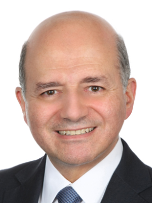 Ismat Ghanem合作论文数Hôtel-Dieu de France;ENSAM;Faculty of Medicine, St Joseph University47
 Khalil Kharrat合作论文数44
 Samer Elhage合作论文数8Khalife Rami合作论文数5E. Samaha合作论文数4
 R. El Rachkidi合作论文数4n aoun合作论文数3Rachid Haidar合作论文数3F. El Masri合作论文数3Antoine Checrallah合作论文数3Ph Wicart合作论文数3A. Yatim合作论文数3Ibrahim Kalouche合作论文数2S. El Hage合作论文数2Samer El-Hage合作论文数2Romanos Elie合作论文数2Haddad E合作论文数2Abiad R合作论文数2
 Ronald Moussa合作论文数2Elie Samaha合作论文数2
 Andre Megarbane合作论文数Department of Human Genetics, Gilbert and Rose-Marie Chagoury School of Medicine, Lebanese American University2A Ghossain合作论文数1V H Nassar合作论文数1I S Salti合作论文数1M Ghossain合作论文数1
 Nabil Okais合作论文数1Georges Aftimos合作论文数1Gabi Kreichati合作论文数1Chebel Mourani合作论文数1D. Nehme Nasr合作论文数1G Nohra合作论文数1
 N Waked合作论文数Faculty of Medicine, Saint Joseph University of Beirut1
 David Atallah合作论文数Hôtel-Dieu de France University Hospital, Saint Joseph University1Azoury Fares合作论文数1Okais Nabil合作论文数1Maalouf Antoine合作论文数1
 Samir Mallat合作论文数Department of Nephrology, Hôtel-Dieu de France - Saint Joseph University1Djambas Khayat C合作论文数Pediatric Department, Hotel Dieu de France Hospital1
 Aline Khazzaka合作论文数1
 Khalil Ghoussoub合作论文数1Diab Michel G合作论文数College of Physicians and Surgeons, Columbia University1Frayha R A合作论文数1
 Paul Henri Nassib Torbey合作论文数1Saliba Elie合作论文数1Naim Choucair合作论文数1E. Haddad合作论文数1Noel Aoun合作论文数1Sabbagh Sandra合作论文数1C. Razzouk合作论文数1




