Importance:Treatment decisions in drug-resistant focal epilepsy remain largely empirical, as direct comparative evidence among newer antiseizure medications (ASMs) is limited. Real-world data can complement randomized clinical trials by providing insights into long-term effectiveness and safety across diverse populations. Objective:To compare effectiveness and safety of brivaracetam, cenobamate, lacosamide, and perampanel as adjunctive therapies in adults with drug-resistant focal epilepsy. Design, Setting, and Participants:This was a multicenter pooled analysis of 4 previously conducted retrospective real-world medical record-review studies (January 2017-January 2024). Included were adult patients (aged ≥16 years) with drug-resistant focal epilepsy, as defined by the International League Against Epilepsy. Participants were recruited from 71 epilepsy centers. Exposures:Add-on treatment with brivaracetam, cenobamate, lacosamide, or perampanel. Main Outcomes and Measures:The primary outcome was the responder rate at 6 months, defined as greater than or equal to 50% seizure frequency reduction from baseline. Secondary outcomes included 12-month responder rate, seizure freedom (≥3 months at 6 months and ≥6 months at 12 months), and 12-month ASM retention. Safety was assessed by incidence of adverse effects. Generalized linear mixed models adjusted for demographic and clinical covariates were used to compare treatment outcomes, with cenobamate as reference ASM. Results:Of 2386 ASM prescriptions screened, 1993 prescriptions from 1949 patients (1036 of 1947 female [53.2%]; sex information was missing in 0.1% of prescriptions) with a median (IQR) age of 42 (29-55) years at ASM prescription, met inclusion criteria and were included in the pooled analysis. Brivaracetam accounted for 953 prescriptions (47.8%), followed by perampanel (607 [30.5%]), lacosamide (241 [12.1%]), and cenobamate (192 [9.6%]). After adjustment, cenobamate demonstrated significantly higher odds of 50% or greater response at 6 months compared with brivaracetam (odds ratio [OR], 0.18; 95% CI, 0.12-0.28; P < .001), perampanel (OR, 0.26; 95% CI, 0.16-0.42; P < .001), and lacosamide (OR, 0.29; 95% CI, 0.17-0.49; P < .001). Results were consistent for secondary effectiveness outcomes at 12 months, with cenobamate outperforming other ASMs in terms of 50% or greater response and seizure freedom. Cenobamate was associated with the highest rate of adverse effects during follow-up (111 [57.8%]), and lacosamide was associated with the lowest (35 [14.8%]). Cenobamate was associated with a higher likelihood of treatment retention at 12 months compared with brivaracetam (OR, 0.43; 95% CI, 0.26-0.69; P < .001) and perampanel (OR, 0.56; 95% CI, 0.32-0.99; P = .047), with no significant difference vs lacosamide (OR, 0.81; 95% CI, 0.41-1.59; P = .53). Conclusions and Relevance:These study findings suggest superior effectiveness of cenobamate over brivaracetam, lacosamide, and perampanel in adults with drug-resistant focal epilepsy in a large real-world setting.
OBJECTIVES:Polymorphous low-grade neuroepithelial tumor of the young (PLNTY) was recognized as a new histologic entity in the 2021 World Health Organization (WHO) classification of central nervous system tumors. It represents a distinct epileptogenic tumor, with about 80 cases reported to date across all age groups. The aim of this study is to describe the features of a cohort of PLNTY patients with a multimodal approach. METHODS:Patients diagnosed with PLNTY from 2014 to 2023 at our Institute were retrospectively reviewed to collect clinical, EEG, neuroimaging, histopathological, and molecular data. RESULTS:We identified 14 surgically treated patients (median age 21.5 years, range 12-46), with drug-resistant seizures in 78.6%. Seizure onset preceded surgery by a median period of 4 years (range .5-27). Bilateral interictal epileptiform EEG discharges and/or mislateralizing/mislocalizing ictal EEG discharge and/or semiological signs were found in 85.7% of patients, compared to 19.2% in a group of 26 non-PLNTY low-grade epilepsy-associated tumors (LEATs) (p < .001). All PLNTYs were supratentorial solid or solid-cystic cortical mass, involving the temporal (n = 8), temporo-occipital (n = 4), frontal (n = 1), or parietal lobe (n = 1). They featured an infiltrative growth pattern, oligodendroglioma-like cells, strong and often diffuse CD34 immunostaining, and frequent calcifications. Focal cortical dysplasia was associated in 14.3% of cases. Proliferation activity (MIB-1 labeling index) was very low (1%), except for one case (3%). BRAFV600E was found in 50% (n = 6/12) of cases, fibroblast growth factor receptor 3 (FGFR3) protein expression in 7 cases, 30.7% (n = 4/13) carried FGFR3 fusion. No fibroblast growth factor receptor 2 (FGFR2) fusion (n = 0/6) was identified. Postsurgical seizure outcome was excellent (Engel class I) in all cases. SIGNIFICANCE:This study confirms that, despite its name, PLNTY is not limited to pediatric patients. Findings underscore the highly epileptogenic nature of PLNTY and its recognizable electroclinical features, potentially related to its distinctive neuropathology. Most PLNTYs show mitogen-activated protein kinase (MAPK) pathway activating alterations, demonstrated by BRAFV600E mutation and FGFR3 fusion.
OBJECTIVE:The spatio-temporal propagation of the Interictal Epileptiform Discharges (IEDs) can reveal mesial temporal lobe (mTL) involvement in focal temporal seizures. We investigated whether simultaneous recording with magnetoencephalography (MEG), electroencephalography (EEG), and Foramen Ovale Electrodes (FOE) recording improve the definition of the Epileptogenic Zone (EZ) in temporal lobe epilepsies with uncertain lateralization. METHODS:Sixteen patients with drug-resistant epilepsy, suspected mTL involvement, and inconclusive lateralizing signs underwent simultaneous MEG, EEG, and FOE recordings. IEDs were localized using sub-averaging and hierarchical clustering of source maps to identify consistent spatiotemporal propagation patterns. Based on MEG data, patients were classified as having "good" (unilateral, localized EZ) or "poor" surgical candidacy. These classifications were compared to FOE-EEG assessments. MEG accuracy was then evaluated in relation to surgical outcomes (Engel class > I = poor outcome). RESULTS:All patients showed mTL involvement. In seven patients, MEG revealed primary mTL activation; in nine, mTL involvement was secondary. The agreement between MEG-based and FOE-based surgical eligibility in the patients with good or poor sbad surgical eligibility and in the entire sample was 87.5%, 75.0% and 81.3%, respectively. The accuracy of MEG-based analysis in identifying suitable candidates for the surgery was 85.7%. SIGNIFICANCE:Simultaneous MEG/EEG/FOE recordings enhance characterization of IED propagation, offering complementary insight into mTL involvement. This multimodal approach improves localization of the EZ and helps predict surgical outcomes in drug-resistant mTLE, particularly in cases with ambiguous lateralization.
We describe the case of a 29-year-old woman with focal drug-resistant epilepsy and an unusual Focal Cortical Dysplasia (FCD) IIa with lipofuscin accumulation in dysmorphic neurons. We discuss the commonalities and discrepancies between the present case and FCDIIa cases without lipofuscin accumulation at the clinical, histopathological and proteomic levels. (a) Coronal 3D-FLAIR imaging demonstrates an unusually deep F1-F2 sulcus associated with subtle signal alteration (arrowhead). (b) Coronal FDG-PET coregistered with T1 3D MRI reveals marked hypometabolism of the same sulcus (arrowhead). (c-f) Histological images of the surgical specimen show numerous dysmorphic neurons (DN), a typical hallmark of FCD type IIa. Notably, DN (green arrows) exhibit cytoplasmic PAS-positive granular material identified as lipofuscin (c), neurofilament accumulation with peculiar 'ring-like' aspect (d,e) and occasional ubiquitin-binding protein p62 labelling (f). (g-h) Matrix-assisted laser desorption/ionisation imaging mass spectrometry (MALDI-IMS) further supports differences at proteomic level between the present case and classic FCDIIa without lipofuscin accumulation.
Objective Corpora amylacea (CA), now called wasteosomes, are basophilic inclusions associated with aging, neurodegeneration, and impaired glymphatic waste clearance. Their presence is well described in temporal lobe epilepsy with hippocampal sclerosis (TLE-HS) and related to hippocampal neuronal loss and longer epilepsy duration. Here, we assessed CA deposition in TLE patients submitted to epilepsy surgery with any histological diagnosis and explored its relationship with clinical and neuropsychological variables. Histological signs of the glymphatic system were evaluated.Methods Qualitative and quantitative evaluation of CA was performed on neocortical and hippocampal surgical specimens and correlated with age at onset, duration of epilepsy, type of seizure at surgery, antecedents, psychiatric symptoms, and seizure outcome. Podoplanin (PDPN) expression was assessed in a subgroup of patients with high and low CA presence as a histological marker of lymphatic elements. Comparison with nonepileptic tissues was also performed.Results Fifty consecutive patients were included with the following histological diagnosis: 18 patients with HS only, 10 patients with HS and associated focal cortical dysplasia (type IIIa), seven patients with encephalocele, five cryptogenic patients with no abnormalities on resected brain specimens (no lesion), and 10 patients with other histological findings. Patients with HS were vulnerable to CA accumulation compared to other etiologies. There was a significant association between CA density and duration of epilepsy, independent of age at surgery or other clinical parameters, and the presence of CA was associated with a worse preoperative cognitive profile. Preliminary histological observations from a few cases suggested enlarged perivascular spaces and altered PDPN vascular expression in patients with higher deposition of CA.Significance These findings provide evidence that CA accumulation correlates with epilepsy duration and suggest a contribution of altered brain clearance mediated by the glymphatic system to cortical dysfunction and cognitive deficits. Future research on glymphatic alterations and their therapeutic modulation may improve management of TLE and other epileptic or neurodegenerative conditions.
Musicogenic epilepsy (ME) is characterized by seizures triggered by music. The epileptogenic focus in this rare reflex epilepsy is often in the temporal lobe, although the precise localization is still unclear. A correlation between ME and the presence of GAD65 antibodies indicates a potential immunological pathogenic mechanism. We evaluated a 32-year-old woman with drug-resistant temporal lobe epilepsy as a candidate for epilepsy surgery. In the absence of clear clinical lateralizing signs, video-EEG monitoring with intracranial electrodes inserted through the foramen ovale was performed to record from the amygdalo-hippocampal regions. The foramen ovale electrodes revealed bilateral, asynchronous, and independent seizure onsets in the mesial temporal regions triggered by music. Testing for GAD65 antibodies confirmed high-titer positivity. The efficacy of epilepsy surgery in antiGAD65-positive ME patients remains limited. We highlight the use of semi-invasive recording with foramen ovale electrodes in ME, as it can reveal bilateral seizures of mesial origin that contraindicate surgery and support the consideration of immunotherapy options. PLAIN LANGUAGE SUMMARY: Musicogenic epilepsy is a type of epilepsy in which music triggers seizures. Our understanding of its origin and cause is still limited. We assessed a patient with music-induced seizures to see if surgery was an option. Since noninvasive tests before surgery were not clear, we used a minimally invasive method with electrodes inserted through a small opening in the skull called the foramen ovale to record the seizures. Thus, we found that the seizures started independently from both temporal lobes, contraindicating epilepsy surgery. We also found high levels of GAD65 antibodies indicating an immunological pathogenic mechanism.
Temporal lobe epilepsy with isolated amygdala enlargement (TLE-AE) still lacks a definite characterization and controversies exist. We conducted a retrospective study identifying brain MRI scans with isolated AE between 2015 and 2021. We collected clinical and paraclinical data of patients with TLE-AE and evaluated the outcome. Forty-one subjects were included (20 males; AE: right 13; left 24; bilateral 4). A strong correlation was found between AE and MRI T2-hyperintensity (right: p < 0.005; left: p < 0.003). There was no history of febrile seizures; 85,4
Autoimmune encephalitis (AIE) consists of a heterogeneous group of inflammatory disorders affecting the central nervous system. Since several conditions, including primary brain tumors, can mimic AIE, diagnosis may be challenging. We report the case of a 69-year-old woman initially diagnosed and treated for a suspected AIE, who later received a histological diagnosis of glioblastoma. Based on this case, we performed a systematic review of the literature to identify cases of high-grade gliomas (HGG) with an AIE-like presentation. Data were collected from each article to characterize patient demographics, clinical manifestations, cerebrospinal fluid (CSF) results, antibody profiling, neuroradiological and other findings, treatment options and outcome. Overall, 15 studies that described 21 patients were included in the literature revision. Median age was 59 years (range 32–86). Seizures were present in 17 patients. The first brain MRI did not reveal typical HGG features in any case. CSF analysis showed pleocytosis in 6 patients, increased protein content in 4, CSF-restricted oligoclonal bands (OCBs) in 1 patient. Autoantibodies were detected in 8 patients. Four patients retrospectively met the 2016 criteria for a definite AIE diagnosis by Graus et al. The median diagnostic delay to the later diagnosis of glioma was 3 months (range 1–24). One patient was diagnosed post-mortem. HGG may uncommonly have an AIE-like presentation, showing potentially initial overlapping clinical and radiological features. Moreover, HGG may present with CSF pleocytosis and elevated protein count, CSF-restricted OCBs, positive autoantibodies and transitory response to immunotherapy. Patients who initially meet AIE diagnostic criteria must be followed closely over time, as distinctive oncologic features may emerge later during the disease course.
Objective Type II focal cortical dysplasias (FCDII) present a clear-cut anatomo-electro-clinical profile and are associated with optimal surgical outcome when completely resected. Alongside presurgical planning and neuroimaging, intraoperative electrocorticography (ECoG) can aid in delineating FCDII boundaries. We report outcomes from patients undergoing FCDII resection using 3D-ECoG with an intracerebral electrode guided by intraoperative ultrasound (ioUS). Methods Patients with suspected FCDII underwent 3D-ECoG during surgery to record intracerebral interictal epileptiform discharges (IEDs) classified as: 1) rhythmic spikes (RS), and 2) periodic bursts of polyspikes (PBOP). Results Ten patients (5 male, 5 female; median age 19.5 years, median epilepsy duration 16 years) were included. Bottom-of-sulcus dysplasia (BOSD) was found in 60 %. 3D-ECoG identified RS in 30 % and PBOP in 70 %. Total IED removal was achieved in 60 %. Histopathology revealed FCDII in 80 %, while 20 % had a diagnosis of “no definite FCD on histopathology”. After a median 24-month follow-up, 90 % achieved ILAE class 1 outcome (seizure free), 10 % had class 2 (only auras). No major complications occurred. Conclusions IoUS-assisted 3D-ECoG is a safe procedure for intraoperative delineation of FCDII, supporting complete resection. Significance Integrating IoUS with 3D-ECoG can offer substantial benefits for surgical management of FCDII-related epilepsy.
Autoimmune polyglandular syndromes (APS) encompass a broad spectrum of disorders. APS III is characterized by autoimmune thyroiditis (AT) plus any autoimmune disorder, excluding Addison's disease. This study explores the link between GAD65 related-temporal lobe epilepsy (GAD-TLE) and APS III. We retrospectively analyzed 29 GAD-TLE patients. Eighteen patients (62
OBJECTIVE:Drug-resistant focal epilepsy is commonly dichotomized based on magnetic resonance imaging (MRI) lesion visibility into positive (MRI-pos) and negative (MRI-neg). Yet, the criteria used to ascribe such categorization are variable. We used a systematic review and meta-analysis to synthesize evidence for the designation of MRI-neg status. METHODS:In accordance with Preferred Reporting Items for Systematic reviews and Meta-Analyses (PRISMA) guidelines, the systematic review (1990-2025) across Embase, Cochrane, and Medline databases identified cohorts with MRI-neg epilepsy. Unsupervised clustering stratified studies based on co-occurrence of imaging modalities. Within identified classes, we assessed the consistency of reporting MRI parameters, rater expertise, post-processing, and stereo-electroencephalography (SEEG). Meta-analyses evaluated the effects of post-processing on diagnostic yield and MRI-neg status on post-surgical outcome. RESULTS:We screened 2622 records and assessed the eligibility of 448 full-text articles, 246 of which met the inclusion criteria for systematic review: 108 (44%) provided data only on MRI-neg and 138 (56%) on mixed adult cohorts, for a total of 10.463 MRI-neg and 7436 MRI-pos patients. Compared to MRI-pos, MRI-neg patients underwent SEEG more frequently (75% vs 54%, p < 0.05), underwent surgery less frequently (73% vs 84%; odds ratio [OR] = 1.14, p < 0.001), and had less favorable outcomes (61% vs 72%, p < 0.05). Clustering identified three classes: MRI-dominant, typified by consistent reporting of MRI parameters (ORs >3.11, p < 0.001), rater-expertise (ORs >9.94, p < 0.001), and post-processing (ORs >3.38, p < 0.03), as opposed to Limited-MRI (χ2 = 41.08, p < 0.001); MRI-and-nuclear-imaging class was typified by use of SEEG (ORs >3.33, p < 0.02). Meta-analyses showed a 39% gain in diagnostic yield after post-processing (11.10, 95% confidence interval [CI] 7.45-16.53) and a higher proportion of favorable surgical outcome in MRI-pos compared to MRI-neg (75% vs 58%; χ2 = 19.10, p < 0.001). Time-based sensitivity analyses did not affect results. SIGNIFICANCE:The designation of MRI-neg is ambiguous, with most studies lacking details on imaging parameters and reader expertise. Given a 39% gain in diagnostic yield, MRI post-processing should be performed systematically as part of a modern multimodal approach to epilepsy surgery before ascribing MRI-neg status.
OBJECTIVE:Autoimmune encephalitis (AE) is characterized by inflammatory processes in the central nervous system and frequently presents with seizures. Even though an ictogenic potential has been shown for some antibodies against neuronal surface antigens (NSAbs), AE pathophysiology is complex, and NSAbs-independent mechanisms are likely to contribute to seizures. We investigated whether the secretome released by peripheral blood mononuclear cells (PBMCs) from AE patients contributes to seizure generation independently of NSAbs. METHODS:PBMCs were isolated from 19 patients with AE (including both those with and those without detectable NSAbs) and 13 healthy volunteers. After 6 h in culture, the PBMC supernatant (secretome) was infused into a heterologous in vitro whole guinea pig brain preparation. Neurophysiological activity was monitored in the isolated in vitro guinea pig brain preparation during coperfusion of PBMC-derived supernatant (secretome) with the endotoxin lipopolysaccharide and human recombinant serum albumin, to induce and mimic, respectively, a mild functional blood-brain barrier impairment. Morphological analysis of ionized calcium-binding adapter molecule 1-positive glial cells was performed in these brains after the electrophysiological experiment. Secretome obtained after 6 h in culture was analyzed with the Multiplex ELLA array system for inflammatory mediator detection. RESULTS:Electrophysiological recordings and immunofluorescence analyses revealed that the secretome from PBMCs derived from AE patients induced seizurelike events and microglial activation in our in vitro brain preparation. Multiplex ELLA immunoassay analysis showed significantly lower concentration of interleukin (IL)-10, IL-1Ra, tumor necrosis factor-α, IL-2, and IL-6 in the secretome of PBMCs derived from AE patients compared to healthy subjects. IL-1β levels were comparable in the secretome of PBMCs derived from AE and healthy subjects. SIGNIFICANCE:Our findings suggest, for the first time, that peripheral inflammatory mediators could represent a trigger factor for seizure activity in AEs, beyond a possible antibody-mediated mechanism.
The identification of the epileptogenic zone (EZ) boundaries is crucial for effective focal epilepsy surgery. We verify the value of a neurophysiological biomarker of focal ictogenesis, characterized by a low-voltage fast-activity ictal pattern (chirp) recorded with intracerebral electrodes during invasive presurgical monitoring (stereoelectroencephalography [SEEG]). The frequency content of SEEG signals was retrospectively analyzed with semiautomatic software in 176 consecutive patients with focal epilepsies that either were cryptogenic or presented with discordant anatomoelectroclinical findings. Fast activity seizure patterns with the spectrographic features of chirps were confirmed by computer-assisted analysis in 95.4% of patients who presented with heterogeneous etiologies and diverse lobar location of the EZ. Statistical analysis demonstrated (1) correlation between seizure outcome and concordance of sublobar regions included in the EZ defined by visual analysis and chirp-generating regions, (2) high concordance in contact-by contact analysis of 68 patients with Engel class Ia outcome, and (3) that discordance between chirp location and the visually outlined EZ correlated with worse seizure outcome. Seizure outcome analysis confirms the fast activity chirp pattern is a reproducible biomarker of the EZ in a heterogeneous group of patients undergoing SEEG.
OBJECTIVE:Epilepsy surgery is a treatment option for patients with seizures that do not respond to pharmacotherapy. The histopathological characterization of the resected tissue has an important prognostic value to define postoperative seizure outcome in these patients. However, the diagnostic classification process based on microscopic assessment remains challenging, particularly in the case of focal cortical dysplasia (FCD). Imaging mass spectrometry is a spatial omics technique that could improve tissue phenotyping and patient stratification by investigating hundreds of biomolecules within a single tissue sample, without the need for target-specific reagents. METHODS:An in situ proteomic technique called matrix-assisted laser desorption/ionization mass spectrometry imaging (MALDI-MSI) is here investigated as a potential new tool to expand conventional diagnosis on standard paraffin brain tissue sections. Unsupervised and region of interest-based MALDI-MSI analyses of sections from 10 FCD type IIb (FCDIIb) cases were performed, and the results were validated by immunohistochemistry. RESULTS:MALDI-MSI identified distinct histopathological features and the boundaries of the dysplastic lesion. The capability to visualize the spatial distribution of well-known diagnostic markers enabling multiplex measurements on single tissue sections was demonstrated. Finally, a fingerprint list of potential discriminant peptides that distinguish FCD core from peri-FCD tissue was generated. SIGNIFICANCE:This is the first study that explores the potential application of MALDI-MSI in epilepsy postsurgery fixed tissue, by utilizing the well-characterized FCDIIb features as a model. Extending these preliminary analyses to a larger cohort of patients will generate spectral libraries of molecular signatures that discriminate tissue features and will contribute to patient phenotyping.
BACKGROUND AND OBJECTIVE:Patients with presumed nonlesional focal epilepsy-based on either MRI or histopathologic findings-have a lower success rate of epilepsy surgery compared with lesional patients. In this study, we aimed to characterize a large group of patients with focal epilepsy who underwent epilepsy surgery despite a normal MRI and had no lesion on histopathology. Determinants of their postoperative seizure outcomes were further studied. METHODS:We designed an observational multicenter cohort study of MRI-negative and histopathology-negative patients who were derived from the European Epilepsy Brain Bank and underwent epilepsy surgery between 2000 and 2012 in 34 epilepsy surgery centers within Europe. We collected data on clinical characteristics, presurgical assessment, including genetic testing, surgery characteristics, postoperative outcome, and treatment regimen. RESULTS:Of the 217 included patients, 40% were seizure-free (Engel I) 2 years after surgery and one-third of patients remained seizure-free after 5 years. Temporal lobe surgery (adjusted odds ratio [AOR]: 2.62; 95% CI 1.19-5.76), shorter epilepsy duration (AOR for duration: 0.94; 95% CI 0.89-0.99), and completely normal histopathologic findings-versus nonspecific reactive gliosis-(AOR: 4.69; 95% CI 1.79-11.27) were significantly associated with favorable seizure outcome at 2 years after surgery. Of patients who underwent invasive monitoring, only 35% reached seizure freedom at 2 years. Patients with parietal lobe resections had lowest seizure freedom rates (12.5%). Among temporal lobe surgery patients, there was a trend toward favorable outcome if hippocampectomy was part of the resection strategy (OR: 2.94; 95% CI 0.98-8.80). Genetic testing was only sporadically performed. DISCUSSION:This study shows that seizure freedom can be reached in 40% of nonlesional patients with both normal MRI and histopathology findings. In particular, nonlesional temporal lobe epilepsy should be regarded as a relatively favorable group, with almost half of patients achieving seizure freedom at 2 years after surgery-even more if the hippocampus is resected-compared with only 1 in 5 nonlesional patients who underwent extratemporal surgery. Patients with an electroclinically identified focus, who are nonlesional, will be a promising group for advanced molecular-genetic analysis of brain tissue specimens to identify new brain somatic epilepsy genes or epilepsy-associated molecular pathways.
The interaction between basic science epilepsy researchers and clinical epileptologists is a longstanding issue. Efforts to provide opportunities for a dialogue between preclinical and clinical epilepsy professionals are crucial to reduce the knowledge gap between them and improve the translational success of neurobiology-based research. The International League Against Epilepsy (ILAE) Research and Innovation Task Force circulated a survey to investigate the need for an update on new clinical epilepsy concepts within the basic science community. The 336 respondents included basic scientists (BS), preclinical scientists (PCSs), and/or clinical scientists (CSs). The majority of the 237 BSs/PCSs were engaged in preclinical studies in translational epilepsy research and declared translational research as a priority research interest. Fewer respondents from low-middle-income countries than from upper-middle or high-income countries (40.7% vs 65%) considered translational research a critical aspect of their research. A broad understanding of both clinical and neurobiological aspects of epilepsy was declared by 48% of BSs/PCSs; 96% of CSs declared a superficial knowledge of neurobiology of epilepsy. Most BSs/PCSs were aware that epilepsy is a complex condition that should be investigated with the help of clinical epileptologists, even though concerns were expressed on the relationship with clinicians. A focused training program on emerging clinical epileptological aspects tailored for BSs/PCSs was recommended by 81% of the participants; the majority of respondents preferred either 1- or 2-week in-presence tutoring or continuous online training coordinated by ILAE at the regional/national level. The survey also underscored the value of educational programs on neurobiology of epilepsy targeting CSs and low-middle-income countries (LMIC) investigators.
 Giuseppe Didato合作论文数60
 Flavio Villani合作论文数Clinical Epileptology and Experimental Neurophysiology Unit, Fondazione Istituto Neurologico “Carlo Besta”53
 Marco de Curtis合作论文数40
 Roberta Di Giacomo合作论文数30
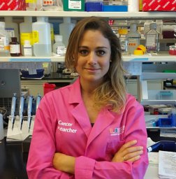 Chiara Pastori合作论文数Neurology Residency School, University of Milano-Bicocca29Rita Garbelli合作论文数26Roberto Spreafico合作论文数23Andrea Stabile合作论文数21
 Laura Tassi合作论文数Department of Epilepsy Surgery, Niguarda Hospital20Tringali Giovanni合作论文数17Giovagnoli Anna Rita合作论文数Fdn IRCCS Ist Neurol C Besta12Gianluca Marucci合作论文数11
 Ludovico D'Incerti合作论文数11
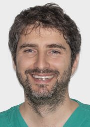 Michele Rizzi合作论文数Department of Neurosurgery, Fondazione IRCCS Istituto Neurologico “C. Besta”,11Rossini Laura合作论文数10Dominese Ambra合作论文数9Parente Annalisa合作论文数8Elena Freri合作论文数Department of Pediatric Neuroscience, Istituto Nazionale Neurologico “C Besta”,8A. Parente合作论文数7S. Matricardi合作论文数7
 Fabio M. Doniselli合作论文数Dept Biomed Sci Hlth, Univ Milan7Bramerio Manuela合作论文数7
 Angelo Del Sole合作论文数Hospital San Paolo Institute of Radiological Sciences, University of Milan7Laura Canafoglia合作论文数7Valeria Cuccarini合作论文数7
 Roberto Mai合作论文数“C. Munari” Epilepsy Surgery Center, Niguarda Hospital7Dalia De Santis合作论文数6
 Rui Quintas合作论文数6
 Pappalardo Irene合作论文数Clinical Epileptology and Experimental Neurophysiology Unit, Foundation IRCCS "Carlo Besta"Neurological Institute;Clinical Epileptology and Experimental Neurophysiology Unit, Foundation IRCCS "Carlo Besta" Neurological Institute6Giorgio Lo Russo合作论文数6
 Tiziana Granata合作论文数Unità Operativa di Neurofisiopatologia ed Epilettologia Diagnostica, Fondazione Istituto Neurologico “C. Besta”,6Elisa Visani合作论文数Department of Neurophysiology, Fondazione IRCCS Istituto Neurologico Carlo Besta6
 Connie Marras合作论文数Institute of Health Policy, Management and Evaluation, University of Toronto;Department of Neurology, University of Toronto5
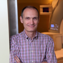 Andrea Bernasconi合作论文数Department of Neurology and Neurosurgery, Faculty of Medicine and Health Sciences, McGill University5Rossi Sebastiano Davide合作论文数5
 Simona Lattanzi合作论文数Department of Clinical and Experimental Medicine, Marche Polytechnic University5
 Francesco Cardinale合作论文数Clinical Epidemiology, Istituto Nazionale per la Ricerca sul Cancro5Ferruccio Panzica合作论文数5
 Maria Grazia Bruzzone合作论文数Neuroradiology Department, Fondazione IRCCS Istituto Neurologico Carlo Besta5
 Vadym Gnatkovsky合作论文数5Francesca Andreetta合作论文数5Francesca Ragona合作论文数5
 Neda Bernasconi合作论文数5
 Carmen Barba合作论文数4Alessandra Burini合作论文数4Nadia Colombo合作论文数4
 Ravnoor S. Gill合作论文数McGill Univ, Montreal Neurol Inst & Hosp, Neuroimaging Epilepsy Lab, Montreal, PQ, Canada4
 Boris Bernhardt合作论文数Department of Neurology and Neurosurgery, Faculty of Medicine and Health Sciences, McGill University;Multimodal Imaging and Connectome Analysis Lab, McConnell Brain Imaging Centre, Montreal Neurological Institute, McGill University4Gozzo Francesca合作论文数4




