Developmental and/or epileptic encephalopathy with spike-and-wave activation in sleep (D/EE-SWAS) represents a rare but severe group of childhood onset epilepsies characterized by sleep-potentiated epileptiform activity, seizures, and developmental stagnation or regression affecting cognition, language, and behavior. Once considered a self-limited electroencephalographic (EEG) phenomenon, D/EE-SWAS is now recognized as a disorder of brain network dysfunction in which persistent epileptiform discharges during non-rapid eye movement sleep disrupt synaptic plasticity, sleep-dependent memory consolidation, and neurodevelopmental trajectories. This review synthesizes recent advances in clinical phenotyping, genetics, neurophysiology, and therapeutics. Etiologically, D/EE-SWAS is highly heterogeneous, with pathogenic variants identified in nearly half of affected individuals, including copy number variants and single-gene disorders involving ion channels, synaptic proteins, and transcriptional regulators. GRIN2A is the most frequently implicated gene, although marked intrafamilial and interfamilial variability underscores the role of modifying genetic and network-level factors. Structural lesions-particularly those affecting thalamocortical circuits-represent another major disease substrate and are critical for treatment stratification. At the mechanistic level, abnormal thalamocortical oscillations, impaired sleep architecture, and disruption of slow-wave and spindle activity provide a pathophysiological framework linking EEG abnormalities to cognitive and behavioral deterioration. Neuroimaging and EEG-functional magnetic resonance imaging studies support a model of widespread network inhibition and disconnection extending beyond the primary epileptogenic zone. Therapeutically, corticosteroids currently represent the most effective first-line treatment, demonstrating superior cognitive outcomes compared with benzodiazepines, although relapse after tapering is common, and optimal dosing strategies remain undefined. Precision medicine approaches, including N-methyl-D-aspartate receptor-targeted therapies for GRIN variants and channel-specific treatments such as primidone for TRPM3 gain of function, offer promising avenues toward disease modification. Epilepsy surgery should be considered early in children with unilateral structural etiologies, where it can provide substantial neurodevelopmental benefit. Future priorities include standardized outcome measures, integration of sleep-based biomarkers, refinement of steroid protocols, and international collaborative trials to improve long-term neurodevelopmental outcomes in this vulnerable population.
BACKGROUND:Before diagnostic whole exome sequencing, monogenic/chromosomal developmental and epileptic encephalopathies (DEEs) were frequently misdiagnosed as mitochondrial disorders (MDs) with epilepsy, due to overlapping clinical and biochemical features. Assessing muscle functional assays in patients with a genetic diagnosis and epilepsy offers a unique opportunity to explore mitochondrial dysfunction in monogenic/chromosomal DEEs, in comparison to the mitochondrial dysfunction observed in genetically confirmed MDs. METHODS:In this retrospective cohort study, clinical and biochemical data were extracted from patients suspected of MD with epilepsy who underwent muscle/fibroblast biopsy (2005-2015). Patients were classified into four groups based on the final diagnosis. Mitochondrial Disease Criteria scores were assigned. Statistical analyses were conducted using Fisher's exact, analysis of variance, and Kruskal-Wallis tests. RESULTS:Of 27 included participants, eleven (40.7%) had DEEs, four (14.8%) had genetically confirmed MDs, eight (29.6%) were suspected MD cases without genetic confirmation, and four (14.8%) had nonmitochondrial metabolic diseases. Mitochondrial dysfunction was common across all groups; 85% of participants met probable/definite Mitochondrial Disease Criteria, over 70% had elevated plasma lactate (>2.5 mmol/L), and 92% exhibited impaired adenosine triphosphate production. Surprisingly, moderate to severe complex dysfunction was observed in all groups except genetically confirmed MDs. CONCLUSIONS:Our findings indicate that mitochondrial dysfunction is prevalent in nonmitochondrial DEEs. Patients previously diagnosed with an MD based only on muscle/fibroblast biopsy may benefit from whole exome sequencing to identify genetic variants, for which targeted therapy may be available. Future research should explore whether treatment or prognosis of nonmitochondrial DEEs should be tailored to improve mitochondrial function.
PURPOSE:The course of epilepsy in patients with tuberous sclerosis complex (TSC) is highly variable. Consistency of the epileptogenic zone is considered important in presurgical evaluation. The authors aimed to (1) assess consistency of ictal epileptogenic foci over time in patients with TSC, (2) evaluate concordance between interictal epileptiform abnormalities and ictal epileptogenic foci, and (3) identify determinants of ictal consistency. METHODS:This single-center retrospective, observational study included children with definite TSC and epilepsy with ≥2 ictal presurgical EEGs with an interval ≥1 year. In all EEGs, the regional localization of interictal and ictal epileptiform abnormalities was scored based on the EEG reports. All ictal epileptogenic foci were classified with an "ictal focus consistency score," according to one of the following subcategories: "single focus, consistent," "from unclear localization/diffuse or generalized to unifocal," "≥2 or more foci, each consistent," "≥2 foci of which at least one is consistent," "starting multifocal but at last EEG unifocal," and "multifocal changing otherwise." The primary end point was unifocal or multifocal consistency of ictal foci. The secondary end point was concordance between ictal epileptogenic foci and localization of interictal epileptiform abnormalities. The third end point involved an exploratory assessment of potential determinants of ictal consistency, including TSC variant, age at epilepsy onset, epileptic spasms, multiple seizure types, interictal multifocality, and tuber load. RESULTS:The authors included 45 patients with TSC with a median of 3 (interquartile range 2-4) ictal and 8 (interquartile range 5-10) interictal EEGs per patient. Median time between first and last ictal EEG was 28.4 months (19.0-60.3). Ictal foci were consistent over time in 11 patients (24.4%), of whom 9 had 1 consistent ictal focus. Nine other patients had a single consistent ictal focus on the first EEGs, but developed a second focus at the median age of 67.7 months. Of the 13 patients who later underwent surgery, 3 had a single focus, and only 2 of 13 had consistent ictal EEGs. The same three patients with a single focus over time were part of the seven postoperative seizure-free patients. All six nonseizure-free children had ictal multifocality. Interictal and ictal epileptogenic foci were discordant in 42 of 45 patients (93.3%). Exploratory analyses of potential determinants of ictal consistency suggested a negative association with interictal multifocality (risk ratio 0.32, 95% confidence interval 0.06-0.97, p = 0.04), which was not significant after adjustment for monitoring duration. CONCLUSIONS:Ictal epileptogenic foci are consistent in only 25% of the patients with TSC. Interictal epileptogenic foci are often discordant with ictal foci. This instability in the ictal epileptogenic focus might be explained by the presence of larger epileptogenic networks in TSC that undergo evolution over time. SIGNIFICANCE:The findings in this study could have important implications for presurgical evaluation in patients with TSC and might help to understand the cause of (late) surgical failure.
Objective N-of-1 trials aim to determine the therapeutic effect for a single individual. This individualized approach necessitates collecting multiple data points over time through repeated alternating periods of active treatment and a comparator or control condition. The extended duration of the treatment periods may increase patient burden, prolong placebo exposure, and increase the likelihood of study discontinuation. In theory, treatment responders (or non-responders) can be identified early during the trial if the therapeutic effect is strong (or completely lacking). There are no theoretical constraints to evaluate treatment efficacy more regularly-instead of only after a predetermined number of treatment periods. Regularly updating estimates on treatment effects allows clinicians to accelerate clinical decision-making regarding N-of-1 study termination. This study examined the value of continuous treatment effect estimation using Bayesian hypothesis testing in N-of-1 trials to accelerate and nuance clinical decision-making.Methods An N-of-1 trial with severe epilepsy was simulated and three N-of-1 trials in neurological conditions were (re-)analyzed continuously with consecutive data points using Bayesian hypothesis testing and/or a minimally clinically important threshold (30% seizure frequency reduction). Trial duration based on Bayesian testing with strong evidence for treatment effects was compared to original trial duration.Results Original trial duration could be reduced between 9.5% and 35% of the trial length by using continuous outcome estimation in two of the analyzed trial examples. The moment that strong evidence supporting beneficial treatment effects using Bayesian hypothesis testing and a significant probability of minimally clinically important differences are achieved during the trial may differ. Obtaining additional data points and alternating interventions over time improve certainty of the estimates of treatment effects.Significance Treatment efficacy decisions can be expedited when outcome estimation is performed continuously rather than delayed until the end of the trial. Clinical significance of N-of-1 trial outcome can be improved combining both Bayesian hypothesis testing and a minimally clinically important threshold.
OBJECTIVES:Timely referral and efficient presurgical evaluation are important to optimize postoperative seizure and developmental outcomes in epilepsy surgery patients. We aimed to identify determinants of time intervals from epilepsy diagnosis to referral to specialized centers and epilepsy surgery in children with malformations of cortical development (MCDs) and low-grade epilepsy-associated tumors (LEATs). METHODS:We performed a retrospective cohort study in 3 European centers, including children with MCD or LEAT who underwent epilepsy surgery between 2010 and 2020. Primary endpoints were (1) time interval between epilepsy diagnosis and referral for presurgical evaluation, (2) duration of presurgical evaluation trajectory, and (3) time from diagnosis to surgery. The secondary endpoint was postoperative seizure outcome. In our primary analysis, we performed a multivariable Cox proportional hazards analysis and used histopathological diagnosis, age at first seizure, presence of developmental delay, 1st seizure classification, initial EEG and MRI findings, duration until drug resistance, and family history as covariates. RESULTS:Of the 507 children included, 259 (51%) had an MCD and 248 (49%) had a LEAT. Earlier drug resistance and the presence of an MR-visible epileptogenic lesion were associated with shorter times from diagnosis to referral and surgery, and later onset of seizures was associated with a shorter time to surgery. LEAT was associated with a shorter presurgical evaluation and shorter time to surgery. An older age at surgery, shorter time from diagnosis to surgery, and a histopathological diagnosis of FCDII and LEAT were all independently associated with better postoperative seizure outcome. SIGNIFICANCE:Epilepsy surgery was performed earlier in children with MR-visible lesions and early drug resistance, particularly those with LEAT, and a shorter duration to epilepsy surgery was associated with better seizure outcomes. Many patients were initially considered MRI-negative in the referring center, highlighting the importance of early referral, epilepsy-dedicated MRI protocols, and experienced neuroradiologists. Increasing awareness of timely referral and surgery benefits is crucial for improving outcomes. PLAIN LANGUAGE SUMMARY:Children with severe focal structural epilepsy who have not responded to medication often benefit from surgery, especially when they are referred early. This study of 507 children found that certain factors-such as visible brain abnormalities on the first MRI, especially a specific type of brain tumor (LEAT), early signs of drug resistance, and seizures starting later in childhood-were linked to shorter times from diagnosis to surgery and that shorter time to surgery was linked to better seizure outcomes. Many children whose initial MRIs appeared to be normal were later found to have an epileptogenic brain lesion when examined at specialized centers. Early referral, advanced imaging, and expert evaluation are key to improving postsurgical outcomes.
Somatostatin (SST), a neuropeptide primarily synthesized by GABAergic interneurons, modulates neuronal excitability and synaptic transmission through its interaction with somatostatin receptors (SSTRs). Dysregulation of SST signaling has been implicated in neurodevelopmental disorders, including tuberous sclerosis complex (TSC). However, its precise role in these pathologies remains incompletely understood. We investigated SST and SSTR expression across diverse brain cell types in control and TSC cortical samples using single-cell RNA sequencing (scRNA-seq). We conducted functional assessments of SST signaling using electrophysiological recordings in Xenopus laevis oocytes microtransplanted with human brain membranes. We pharmacologically modulated SST receptor activity to elucidate receptor-specific effects on GABAergic transmission. scRNA-seq analysis revealed that SST expression is predominantly confined to GABAergic interneurons, while SSTR1 and SSTR2 exhibit strong expression in both glutamatergic and GABAergic neuronal populations. In TSC samples, SSTR5 was upregulated in GABAergic neurons, SSTR2 in glutamatergic neurons, while SSTR3 was downregulated in both glutamatergic neurons and microglia. Functional experiments demonstrated that SST enhances GABAergic currents in control tissues through a receptor-mediated mechanism involving protein kinase C activation. In contrast, SST application in TSC samples resulted in a significant suppression of GABAergic currents. Pharmacological inhibition of SSTR3 further exacerbated this effect, suggesting a compensatory role for this receptor subtype. Our findings reveal a disruption of SST signaling in TSC, contributing to altered coordination of excitatory-inhibitory activity and epileptogenesis. Targeting SST signaling may represent a therapeutic strategy for restoring inhibitory network function in TSC and related disorders.
Malformations of cortical development are manifestations of mTORopathies, including tubers in context of Tuberous Sclerosis Complex (TSC) cortical tubers and Focal Cortical Dysplasia (FCD), and are associated with epilepsy, often accompanied by comorbidities such as autism spectrum disorder (ASD). This study aims to investigate the cell-type-specific transcriptional alterations and disrupted intercellular communication networks in mTORopathies, focusing on their implications for cortical network dysfunction. Using single-cell RNA sequencing, we identified 33 transcriptionally distinct cell clusters across control and pathological samples, including neuronal, glial, and endothelial populations. Our analysis revealed disease-specific changes, such as the loss of certain glutamatergic and microglial clusters in cortical tubers (TSC), MTOR_FCD and DEPDC5_FCD, and the presence of a unique endothelial cluster in pathological samples. Pathway enrichment analysis highlighted the critical role of synaptic signaling, axonogenesis, and neuroimmune regulation in these disorders. Additionally, cell-cell communication network analysis demonstrated disrupted interactions between neuron-astrocyte, astrocyte-OPC, and microglia-neuron across mTORopathies. We found that the neurexins-neuroligins (NRXN-NLGN) signaling pathway, crucial for synapse formation and stability, was altered in both glutamatergic and GABAergic neurons, reflecting dysregulated synaptic plasticity and impaired neuron-glia communication. These findings provide novel insights into the molecular underpinnings of mTORopathies and suggest potential therapeutic targets to restore cellular communication and synaptic function in these disorders.
Die Internationale Liga gegen Epilepsie (ILAE) hat die operationale Klassifikation epileptischer Anfälle 2017 auf der Grundlage der damals entwickelten Rahmenbedingungen aktualisiert. In diese Überarbeitung wurden die publizierten Erfahrungen mit der Umsetzung der 2017er-Klassifikation einbezogen. Eine 37 Personen zählende Arbeitsgruppe wurde vom ILAE-Exekutivausschuss eingesetzt. Die internationalen Expertinnen und Experten aus allen ILAE-Regionen wandten ein modifiziertes Delphi-Verfahren an, bei dem für jeden Vorschlag ein Konsens von mehr als zwei Dritteln erforderlich war. Nach Veröffentlichung auf der ILAE-Homepage mit dem Ersuchen, Kommentare zu dem Entwurf einzugeben, ernannte der Exekutivausschuss 7 zusätzliche Sachverständige für die Arbeitsgruppe zur Überarbeitung des Positionspapieres, um die eingegangenen Kommentare zu diskutieren und gegebenenfalls einzubeziehen. Die aktualisierte Klassifikation behält die Hauptklassen von Anfällen bei: fokal, generalisiert, unbekannt (ob fokal oder generalisiert) und nicht klassifiziert. Taxonomische Regeln unterscheiden zwischen Klassifikatoren, die biologische Klassen widerspiegeln und sich direkt auf die klinische Behandlung auswirken, und Deskriptoren, die andere wichtige Anfallsmerkmale angeben. Fokale Anfälle und Anfälle unbekannten Ursprungs werden darüber hinaus nach dem Bewusstseinszustand des Patienten während des Anfalls klassifiziert, je nachdem ob eine Bewusstseinsstörung vorliegt oder nicht. Die Bewertung, ob eine Bewusstseinsstörung vorliegt, wird durch Gewahrsein (engl. „awareness“) und Reaktionsfähigkeit (engl. „responsiveness“) während eines Anfalls und erhaltene Erinnerung (engl. „recall“) nach einem Anfall klinisch operationalisiert. Wenn der Bewusstseinszustand nicht bestimmbar ist, wird der Anfall unter dem übergeordneten Begriff, d. h. der Hauptanfallsklasse (fokaler Anfall oder Anfall unbekannten Ursprungs) klassifiziert. Generalisierte Anfälle werden in Absencen, generalisierte tonisch-klonische Anfälle und andere generalisierte Anfälle eingeteilt, wobei jetzt auch der negative Myoklonus als Anfallstyp beschrieben wird. Anfälle werden in der Grundversion als solche mit oder ohne beobachtbare Manifestationen beschrieben, während eine erweiterte Version die chronologische Abfolge der Anfallssemiologie verwendet. Diese aktualisierte Klassifikation umfasst 4 Hauptklassen und nur noch 21 Anfallstypen (und nicht mehr 63 wie in der 2017 Klassifikation; Anm. d. Übersetzer). Besonderer Wert wurde auf die Übersetzbarkeit in andere Sprachen jenseits von Englisch gelegt. Ziel ist es, eine gemeinsame Sprache für alle im Bereich Epilepsie tätigen Gesundheitsfachkräfte zu schaffen – von ressourcenarmen Gebieten bis hin zu hoch spezialisierten Zentren – und leicht zugängliche Begriffe für Patientinnen und Patienten sowie Betreuungspersonen bereitzustellen.
Precision treatments for monogenic epilepsies, i.e. treatments that can at least partially reverse the biochemical consequences of a pathogenic gene variant, have been gradually emerging over the years. To date, however, information on the efficacy of these treatments is mostly based on case-reports and retrospective studies. As a result, utilisation of precision treatments often lack consistency and a pre-defined outcome monitoring plan. N-of-1 strategies in clinical care are pre-defined, individually tailored, repeated challenge-withdrawal therapeutic trials designed to assess the value of a treatment of interest for an individual. Despite their potential to improve clinical decision-making, N-of-1 strategies have been hampered by limited guidance on their implementation and lack of consensus on oversight procedures. To improve treatment selection for rare monogenic epilepsies, the PINPOINT initiative (Precision Treatments In MoNogenic EPilepsies: Observational Registry And N-of-1 Trial Recommendations) was set up as a collaborative effort within the European Reference Network for Rare and Complex Epilepsies. PINPOINT aims to develop recommendations for the design of N-of-1 strategies with off-label precision treatments for monogenic epilepsies. Using available N-of-1 trial manuals, different components of N-of-1 design were tailored to the context of epilepsy and oversight procedures were outlined. These efforts resulted in this guidance document—or blueprint for N-of-1 strategies for monogenic epilepsies in clinical care. This blueprint defines the characteristics of treatments and patients that would be suitable for N-of-1 strategies. Key principles for outcome measure selection, period duration and statistical analysis are defined. Consideration is given to interim assessment rules, which establish whether proceeding onto an additional treatment cycle is likely to provide significant advantages. Procedures for ethical oversight are proposed. This blueprint for N-of-1 strategies can be used as a basis for master protocols to optimise individualised clinical care in a standardised and consistent manner. We are confident that this document will provide physicians with the building blocks needed to elevate precision treatments for rare monogenic epilepsies out of their current landscape of inadequate evidence.
The International League Against Epilepsy (ILAE) has updated the operational classification of epileptic seizures, building upon the framework established in 2017. This revision, informed by the implementation experience, involved a working group appointed by the ILAE Executive Committee. Comprising 37 members from all ILAE regions, the group utilized a modified Delphi process, requiring a consensus threshold of more than two thirds for any proposal. Following public comments, the Executive Committee appointed seven additional experts to the revision task force to address and incorporate the issues raised, as appropriate. The updated classification maintains four main seizure classes: Focal, Generalized, Unknown (whether focal or generalized), and Unclassified. Taxonomic rules distinguish classifiers, which are considered to reflect biological classes and directly impact clinical management, from descriptors, which indicate other important seizure characteristics. Focal seizures and those of unknown origin are further classified by the patient's state of consciousness (impaired or preserved) during the seizure, defined operationally through clinical assessment of awareness and responsiveness. If the state of consciousness is undetermined, the seizure is classified under the parent term, that is, the main seizure class (focal seizure or seizure of unknown origin). Generalized seizures are grouped into absence seizures, generalized tonic-clonic seizures, and other generalized seizures, now including recognition of negative myoclonus as a seizure type. Seizures are described in the basic version as with or without observable manifestations, whereas an expanded version utilizes the chronological sequence of seizure semiology. This updated classification comprises four main classes and 21 seizure types. Special emphasis was placed on ensuring translatability into languages beyond English. Its aim is to establish a common language for all health care professionals involved in epilepsy care, from resource-limited areas to highly specialized centers, and to provide accessible terms for patients and caregivers.
This paper provides a practical guide to applying the updated seizure classification in clinical settings. The updated classification, published by the International League Against Epilepsy in 2025, builds on the operational classification introduced in 2017. It aims to enhance clarity, clinical relevance, and consistency in seizure terminology across various healthcare settings. The classification system distinguishes between four main seizure classes: focal, generalized, unknown whether focal or generalized, and unclassified. The basic version allows for application even in resource-limited or primary care environments, while the expanded version offers detailed semiological descriptors and supports advanced diagnostic and surgical decision-making. Consciousness, defined through awareness (recall) and responsiveness, is now considered a classifier. The paper also highlights the importance of distinguishing observable from non-observable features and integrates semiology into the expanded classification with attention to temporal sequence and somatotopic detail. Generalized seizures are categorized into seizure types having a direct impact on syndrome diagnosis and treatment decisions. Epileptic spasms are given special attention because of their unique presentation and the urgency of early intervention, particularly in infants. To support clinical implementation, this paper includes a comprehensive table of semiological descriptors, definitions of generalized seizure types, and a series of real-world case vignettes illustrating the application of the updated seizure classification. Supplementary figures and videos further support the educational aims of the paper. This practical guide is intended for healthcare professionals managing patients with epilepsy, providing a clear, structured approach to seizure classification that is adaptable to varying levels of clinical expertise and diagnostic resources.
Identification of the epileptogenic lesion is challenging in tuberous sclerosis complex as multiple lesions might represent the seizure onset zone. A combination of dysplastic MRI features has diagnostic value in pre-surgical evaluation. However, these radiological characteristics may be difficult to identify and have not been studied on early unmyelinated brain MRI in tuberous sclerosis complex infants. Our study aimed to assess the diagnostic accuracy of T2-hypointense lesions on unmyelinated MRI in identifying the epileptogenic lesion. We included children with tuberous sclerosis complex who underwent resective or disconnective epilepsy surgery in the Motol University Hospital Prague and the University Medical Center Utrecht with available (i) unmyelinated MRI (before the age of 9 months), (ii) pre- and post-operative brain MRI and (iii) at least 2 years follow-up post-surgery. We identified T2-hypointense lesions and highly dysplastic lesions on unmyelinated or myelinated MRI, assessing their diagnostic accuracy in epileptogenic lesion identification by comparing seizure free to non-seizure free patients. Twenty-seven patients met inclusion criteria. We identified 54 T2-hypointense lesions in 24 patients, 30 were already highly dysplastic on unmyelinated MRI, showing cortical thickening and transmantle sign in most cases, while calcifications appeared later. Diagnostic accuracy of T2-hypointense (70.8%) was superior to the presence of the most dysplastic features (55.6%) in epileptogenic lesion identification. Positive predictive value for complete resection of all T2-hypointense lesions was 63.6%, compared to 50.0% for highly dysplastic lesions. Seizure recurrence was high (negative predictive value 76.9%) when T2-hypointense lesions remained outside the resected area. Assessing T2-hypointense lesions on unmyelinated brain MRI has important diagnostic value in identifying the epileptogenic lesion in pre-surgical work-up in infants with tuberous sclerosis complex and drug-resistant epilepsy. Unmyelinated brain MRI deserves a more important position in pre-surgical evaluation in infants with tuberous sclerosis complex and drug-resistant epilepsy.
Developmental and epileptic encephalopathy with spike wave activation in sleep (DEE-SWAS) and epileptic encephalopathy with spike wave activation in sleep (EE-SWAS) are rare but well-known childhood epileptic disorders. Steroids are one of the first line treatment options, but a variety of steroid regimens exists. The aim of this survey was to evaluate the practices of steroid therapy in the treatment of (D)EE-SWAS across European centers. An online survey was conducted (via ‘SurveyMonkey’ Europe) among European epilepsy centers. Questions asked included: characteristics of replying center, applied definition of DEE-SWAS, existence of regional/national guidelines regarding diagnostic and therapeutic management. Particular attention was paid to the indication/contraindication of steroids and treatment regimens used. Responses were obtained from 60 centers across 18 countries. Only 15
BACKGROUND:A pathogenic variant in SCN1A can result in a spectrum of phenotypes, including Dravet syndrome (DS) and genetic epilepsy with febrile seizures plus (GEFS + ) syndrome. Dravet syndrome (DS) is associated with refractory seizures, developmental delay, intellectual disability (ID), motor impairment, and challenging behavior(1,2). GEFS + is a less severe phenotype in which cognition is often normal and seizures are less severe. Challenging behavior largely affects quality of life of patients and their families. This study describes the profile and course of the behavioral phenotype in patients with SCN1A-related epilepsy syndromes, explores correlations between behavioral difficulties and potential risk factors. METHODS:Data were collected from questionnaires, medical records, and semi-structured interviews. Behavior difficulties were measured using the Adult/Child Behavior Checklist (C/ABCL) and Adult self-report (ASR). Other questionnaires included the Pediatric Quality of Life Inventory (PedsQL), the Functional Mobility Scale (FMS) and the Sleep Behavior Questionnaire by Simonds & Parraga (SQ-SP). To determine differences in behavioral difficulties longitudinally, paired T-tests were used. Pearson correlation and Spearman rank test were used in correlation analyses and multivariable regression analyses were employed to identify potential risk factors. RESULTS:A cohort of 147 participants, including 107 participants with DS and 40 with genetic epilepsy with febrile seizures plus (GEFS + ), was evaluated. Forty-six DS participants (43.0 %) and three GEFS + participants (7.5 %) showed behavioral problems in the clinical range on the A/CBCL total problems scale. The behavioral profile in DS exists out of withdrawn behavior, aggressive behavior, and attention problems. In DS patients, sleep disturbances (β = 1.15, p < 0.001) and a lower age (β = -0.21, p = 0.001) were significantly associated with behavioral difficulties. Between 2015 and 2022, behavioral difficulties significantly decreased with age (t = -2.24, CI = -6.10 - -0.15, p = 0.04) in DS participants aging from adolescence into adulthood. A decrease in intellectual functioning (β = 3.37, p = 0.02) and using less antiseizure medications in 2022 than in 2015, (β = -1.96, p = 0.04), were identified as possible risk factors for developing (more) behavioral difficulties. CONCLUSIONS:These findings suggest that, in addition to epilepsy, behavioral difficulties are a core feature of the DS phenotype. Behavioral problems require personalized management and treatment strategies. Further research is needed to identify effective interventions.
AbstractObjectiveNew‐onset seizure‐like events (SLEs) are common in children, but differentiating between epilepsy and its mimics is challenging. This study provides an overview of the clinical characteristics, diagnoses, and corresponding etiologies of children evaluated at a first seizure clinic (FSC), which will be helpful for all physicians involved in the care of children with SLEs.MethodsWe included 1213 children who were referred to the FSC of a Dutch tertiary children's hospital over a 13‐year period and described their clinical characteristics, first routine EEG recording results, and the distribution and specification of their eventual epilepsy and non‐epilepsy diagnoses. The time interval to correct diagnosis and the diagnostic accuracy of the FSC were evaluated.Results“Epilepsy” was eventually diagnosed in 407 children (33.5%), “no epilepsy” in 737 (60.8%), and the diagnosis remained “unclear” in 69 (5.7%). Epileptiform abnormalities were seen in 60.9% of the EEG recordings in the “epilepsy” group, and in 5.7% and 11.6% of the “no epilepsy” and “unclear” group, respectively. Of all children with final “epilepsy” and “no epilepsy” diagnoses, 68.6% already received their diagnosis at FSC consultation, and 2.9% of the children were initially misdiagnosed. The mean time to final diagnosis was 2.0 months, and 91.3% of all children received their final diagnosis within 12 months after the FSC consultation.SignificanceWe describe the largest pediatric FSC cohort to date, which can serve as a clinical frame of reference. The experience and expertise built at FSCs will improve and accelerate diagnosis in children with SLEs.Plain language summaryMany children experience events that resemble but not necessarily are seizures. Distinguishing between seizures and seizure mimics is important but challenging. Specialized first‐seizure clinics can help with this. Here, we report data from 1213 children who were referred to the first seizure clinic of a Dutch children's hospital. One‐third of them were diagnosed with epilepsy. In 68.8% of all children—with and without epilepsy—the diagnosis was made during the first consultation. Less than 3% were misdiagnosed. This study may help physicians in what to expect regarding the diagnoses in children who present with events that resemble seizures.
Background Many rare genetic neurodevelopmental disorders (RGNDs) are characterized by intellectual disability (ID), severe cognitive and behavioral impairments, potentially diagnosed as a comorbid autism spectrum disorder or attention-deficit hyperactivity disorder. Quality of life is often impaired due to irritability, aggression and self-injurious behavior, generally refractory to standard therapies. There are indications from previous (case) studies and patient reporting that cannabidiol (CBD) may be an effective treatment for severe behavioral manifestations in RGNDs. However, clear evidence is lacking and interventional research is challenging due to the rarity as well as the heterogeneity within and between disease groups and interindividual differences in treatment response. Our objective is to examine the effectiveness of CBD on severe behavioral manifestations in three RGNDs, including Tuberous Sclerosis Complex (TSC), mucopolysaccharidosis type III (MPS III), and Fragile X syndrome (FXS), using an innovative trial design. Methods We aim to conduct placebo-controlled, double-blind, block-randomized, multiple crossover N -of- 1 studies with oral CBD (twice daily) in 30 patients (aged ≥ 6 years) with confirmed TSC, MPS III or FXS and severe behavioral manifestations. The treatment is oral CBD up to a maximum of 25 mg/kg/day, twice daily. The primary outcome measure is the subscale irritability of the Aberrant Behavior Checklist. Secondary outcome measures include (personalized) patient-reported outcome measures with regard to behavioral and psychiatric outcomes, disease-specific outcome measures, parental stress, seizure frequency, and adverse effects of CBD. Questionnaires will be completed and study medication will be taken at the participants’ natural setting. Individual treatment effects will be determined based on summary statistics. A mixed model analysis will be applied for analyzing the effectiveness of the intervention per disorder and across disorders combining data from the individual N -of- 1 trials. Discussion These N -of- 1 trials address an unmet medical need and will provide information on the effectiveness of CBD for severe behavioral manifestations in RGNDs, potentially generating generalizable knowledge at an individual-, disorder- and RGND population level. Trial registration EudraCT: 2021-003250-23, registered 25 August 2022, https://www.clinicaltrialsregister.eu/ctr-search/trial/2021-003250-23/NL .
OBJECTIVE:Previous retrospective studies have reported vigabatrin-associated brain abnormalities on magnetic resonance imaging (VABAM), although clinical impact is unknown. We evaluated the association between vigabatrin and predefined brain magnetic resonance imaging (MRI) changes in a large homogenous tuberous sclerosis complex (TSC) cohort and assessed to what extent VABAM-related symptoms were reported in TSC infants. METHODS:The Dutch TSC Registry and the EPISTOP cohort provided retrospective and prospective data from 80 TSC patients treated with vigabatrin (VGB) before the age of 2 years and 23 TSC patients without VGB. Twenty-nine age-matched non-TSC epilepsy patients not receiving VGB were included as controls. VABAM, specified as T2/fluid-attenuated inversion recovery hyperintensity or diffusion restriction in predefined brain areas, were examined on brain MRI before, during, and after VGB, and once in the controls (at approximately age 2 years). Additionally, the presence of VABAM accompanying symptoms was evaluated. RESULTS:Prevalence of VABAM in VGB-treated TSC patients was 35.5%. VABAM-like abnormalities were observed in 13.5% of all patients without VGB. VGB was significantly associated with VABAM (risk ratio [RR] = 3.57, 95% confidence interval [CI] = 1.43-6.39), whereas TSC and refractory epilepsy were not. In all 13 VGB-treated patients with VABAM for whom posttreatment MRIs were available, VABAM entirely resolved after VGB discontinuation. The prevalence of symptoms was 11.7% in patients with VABAM or VABAM-like MRI abnormalities and 4.3% in those without, implicating no significant association (RR = 2.76, 95% CI = .68-8.77). SIGNIFICANCE:VABAM are common in VGB-treated TSC infants; however, VABAM-like abnormalities also occurred in children without either VGB or TSC. The cause of these MRI changes is unknown. Possible contributing factors are abnormal myelination, underlying etiology, recurrent seizures, and other antiseizure medication. Furthermore, the presence of VABAM (or VABAM-like abnormalities) did not appear to be associated with clinical symptoms. This study confirms that the well-known antiseizure effects of VGB outweigh the risk of VABAM and related symptoms.
OBJECTIVE:Defined as prospective single-patient crossover studies with repeated paired cycles of active and control intervention, N-of-1 trials have gained attention as an option to obtain high-quality evidence of efficacy, particularly for patients with rare epilepsies in whom conduction of well-powered randomized controlled trials can be challenging. The objective of this systematic review is to provide an appraisal of the literature on N-of-1 trials in individuals with epilepsy. METHODS:We searched PubMed and Embase on January 12, 2024, for studies meeting the following criteria: prospectively planned, within-patient, multiple-crossover design in individuals with epilepsy and outcomes related to comorbidities. Information on design, outcome measurements, intervention, and analyses was retrieved. Risk of bias assessment was performed using the Risk of Bias in N-of-1 Trials (RoBiNT) scale. We highlighted methodological aspects of the N-of-1 trials identified and discuss future recommendations. RESULTS:Five studies met our inclusion criteria. An additional multiple-crossover trial that evaluated treatment effects exclusively at group level was also included because of its relevance to N-of-1 study methodology. The studies enrolled individuals with focal seizures, absences or cognitive impairement and electrographic discharges. Treatments included established or investigational antiseizure medications, off-label medications, neurostimulation or lifestyle intervention. Three of the five N-of-1 trials reported on individual cases. The studies' strengths were the use of individualized treatment dosages and symptom-specific patient-reported outcomes. Limitations were related to minimal reporting of baseline characteristics and seizure burden. SIGNIFICANCE:The trials identified by our search exemplify how the N-of-1 design can be applied to assess interventions in individuals with epilepsy-related disorders. Future N-of-1 trials of antiseizure interventions should take into account baseline seizure frequency, should apply statistical models suited to capture seizure frequency changes reliably and make predefined interim assessments. Non-seizure outcome measures evaluable over short periods should be considered. Tailored N-of-1 methodology could pave the way to evidence-based, treatment selection for patients with rare epilepsies.
We studied the distribution of germline and somatic variants in epilepsy surgery patients with (suspected) malformations of cortical development (MCD) who underwent surgery between 2015 and 2020 at University Medical Center Utrecht (the Netherlands) and pooled our data with four previously published cohort studies. Tissue analysis yielded a pathogenic variant in 203 of 663 (31%) combined cases. In 126 of 379 (33%) focal cortical dysplasia (FCD) type II cases and 23 of 37 (62%) hemimegalencephaly cases, a pathogenic variant was identified, mostly involving the mTOR signaling pathway. Pathogenic variants in 10 focal epilepsy genes were found in 48 of 178 (27%) FCDI/mild MCD/mMCD with oligodendroglial hyperplasia and epilepsy cases; 36 of these (75%) were SLC35A2 variants. Six of 69 (9%) patients without a histopathological lesion had a pathogenic variant in SLC35A2 (n = 5) or DEPDC5 (n = 1). A germline variant in blood DNA was confirmed in all cases with a pathogenic variant in tissue, with a variant allele frequency (VAF) of ~50%. In seven of 114 patients (6%) with a somatic variant in tissue, mosaicism in blood was detected. More than half of pathogenic somatic variants had a VAF < 5%. Further analysis of the correlation between genetic variants and surgical outcomes will improve patient counseling and may guide postoperative treatment decisions.
BACKGROUND AND OBJECTIVE:Patients with presumed nonlesional focal epilepsy-based on either MRI or histopathologic findings-have a lower success rate of epilepsy surgery compared with lesional patients. In this study, we aimed to characterize a large group of patients with focal epilepsy who underwent epilepsy surgery despite a normal MRI and had no lesion on histopathology. Determinants of their postoperative seizure outcomes were further studied. METHODS:We designed an observational multicenter cohort study of MRI-negative and histopathology-negative patients who were derived from the European Epilepsy Brain Bank and underwent epilepsy surgery between 2000 and 2012 in 34 epilepsy surgery centers within Europe. We collected data on clinical characteristics, presurgical assessment, including genetic testing, surgery characteristics, postoperative outcome, and treatment regimen. RESULTS:Of the 217 included patients, 40% were seizure-free (Engel I) 2 years after surgery and one-third of patients remained seizure-free after 5 years. Temporal lobe surgery (adjusted odds ratio [AOR]: 2.62; 95% CI 1.19-5.76), shorter epilepsy duration (AOR for duration: 0.94; 95% CI 0.89-0.99), and completely normal histopathologic findings-versus nonspecific reactive gliosis-(AOR: 4.69; 95% CI 1.79-11.27) were significantly associated with favorable seizure outcome at 2 years after surgery. Of patients who underwent invasive monitoring, only 35% reached seizure freedom at 2 years. Patients with parietal lobe resections had lowest seizure freedom rates (12.5%). Among temporal lobe surgery patients, there was a trend toward favorable outcome if hippocampectomy was part of the resection strategy (OR: 2.94; 95% CI 0.98-8.80). Genetic testing was only sporadically performed. DISCUSSION:This study shows that seizure freedom can be reached in 40% of nonlesional patients with both normal MRI and histopathology findings. In particular, nonlesional temporal lobe epilepsy should be regarded as a relatively favorable group, with almost half of patients achieving seizure freedom at 2 years after surgery-even more if the hippocampus is resected-compared with only 1 in 5 nonlesional patients who underwent extratemporal surgery. Patients with an electroclinically identified focus, who are nonlesional, will be a promising group for advanced molecular-genetic analysis of brain tissue specimens to identify new brain somatic epilepsy genes or epilepsy-associated molecular pathways.
 Kees Braun合作论文数Division Neuroscience, University Medical Center Utrecht100
 Eleonora Aronica合作论文数Amsterdam University Medical Center;Stichting Epilepsie Instellingen Nederland65
 Martha Feucht合作论文数38Sergiusz Jóźwiak合作论文数Department of Neurology and Epileptology, The Children's Memorial Health Institute37
 Pavel Krsek合作论文数Department of Paediatric Neurology, Second Faculty of Medicine, Charles University35Lieven Lagae合作论文数34
 Wim Spliet合作论文数Department of Pathology, University Medical Center Utrecht30
 Katarzyna Kotulska合作论文数29Anink Jasper J合作论文数28Eva H Brilstra合作论文数Univ Med Ctr Utrecht, Dept Genet, Utrecht, Netherlands27Mühlebner Angelika合作论文数27
 Paolo Curatolo合作论文数University of Rome Tor Vergata27
 Anna Jansen合作论文数26
 James D. Mills合作论文数23
 Theresa Scholl合作论文数22
 B.P.C. (Bobby) Koeleman合作论文数University Medical Center Utrecht19
 van Diessen Eric合作论文数Rudolf Magnus Institute of Neuroscience, University Medical Center Utrecht18
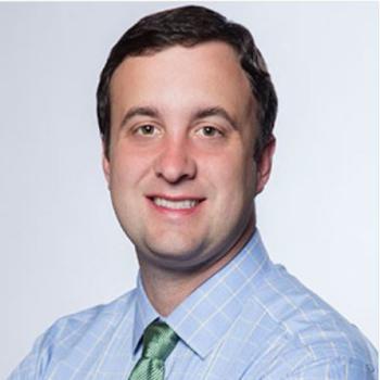 David M. Kwiatkowski合作论文数Cardiovascular Institute, Stanford University;Lucile Packard Children's Hospital18
 Helen Cross合作论文数Great Ormond Street Institute of Child Health, University College London17
 Anand Iyer合作论文数17
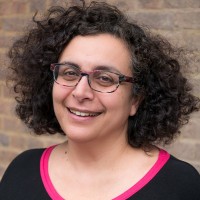 Rima Nabbout合作论文数Université Paris Cité;Greater Paris University Hospitals;Imagine Institut Des Maladies Genetiques, Necker Enfants Malades17Willem M. Otte合作论文数16
 Catherine (Kate) Riney合作论文数16van Campen Jolien S合作论文数14Romina Moavero合作论文数14J. Hainfellner合作论文数Division of Neuropathology and Neurochemistry, Department of Neurology, Medical University of Vienna14
 Erwin A Van Vliet合作论文数14M. (Mark) Nellist合作论文数Erasmus MC14Barbora Benova合作论文数13
 Christoph Hertzberg合作论文数13van Scheppingen Jackelien合作论文数13O. Van Nieuwenhuizen合作论文数13
 Hilgo Bruining合作论文数13
 Anika Bongaarts合作论文数12
 Wim Van Hecke合作论文数Icometrix12Weschke Bernhard合作论文数12
 Frans Leijten合作论文数University Medical Center Utrecht12
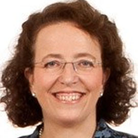 Ingrid Scheffer合作论文数Department of Medicine, The University of Melbourne;Faculty of Medicine, Dentistry and Health Sciences, The University of Melbourne;Florey Institute of Neuroscience and Mental Health;Department of Neurology, Royal Children's Hospital Melbourne;Austin Health12Peter C. Van Rijen合作论文数11
 M. (Marian) Joëls合作论文数Faculty of Medical Sciences, University of Groningen11Caroline Mijnsbergen合作论文数10Diede W M Broekaart合作论文数10Till S Zimmer合作论文数10Buccoliero Anna Maria合作论文数10
 Jackelien van Scheppingen合作论文数10
 Edouard Hirsch合作论文数Université de Strasbourg;University Hospital in Strasbourg10
 Josef Zamecnik合作论文数Department of Pathology and Molecular Medicine, Second Faculty of Medicine, Charles University10Hanna M Hulshof合作论文数9Maurits W C B Sanders合作论文数9




