Objective The decline of the perinatal demise rate is slowing and demises are often unexplained. Significant research has been done regarding diagnostic yield and genetic causes of demise, but little is known about how Geneticist involvement impacts outcomes. The goal of the study was to evaluate post-mortem genetic testing practices and effects of the geneticists involvement. Methods Retrospective data from 111 perinatal demise cases was examined, including rates of prenatal genetic counseling, post-delivery genetics consult, genetic testing, and autopsy investigation. Results In this cohort 54% received genetic testing and 25% received a genetics consult. When compared to those without, cases with genetic specialist involvement were associated with significant increases in testing uptake (p=0.007), diagnostic yield (p<0.001), and patient education (p<0.001). Second trimester stillbirths and those with fewer ultrasound (US) abnormalities were less likely to receive genetic testing (both p values <0.001) and consults (p<0.001, p=0.020). Conclusion Though it was not possible to avoid ascertainment bias, this data demonstrates that geneticist involvement correlates with a higher rate of testing, greater diagnostic yield, and more thorough counseling. These findings underscore the importance of integrating genetics providers into perinatal postmortem healthcare teams. ### Competing Interest Statement The authors have declared no competing interest. ### Funding Statement The study did not receive any funding ### Author Declarations I confirm all relevant ethical guidelines have been followed, and any necessary IRB and/or ethics committee approvals have been obtained. Yes The details of the IRB/oversight body that provided approval or exemption for the research described are given below: The IRB of the University of California Irvine gave ethical approval for this work. I confirm that all necessary patient/participant consent has been obtained and the appropriate institutional forms have been archived, and that any patient/participant/sample identifiers included were not known to anyone (e.g., hospital staff, patients or participants themselves) outside the research group so cannot be used to identify individuals. Yes I understand that all clinical trials and any other prospective interventional studies must be registered with an ICMJE-approved registry, such as ClinicalTrials.gov. I confirm that any such study reported in the manuscript has been registered and the trial registration ID is provided (note: if posting a prospective study registered retrospectively, please provide a statement in the trial ID field explaining why the study was not registered in advance). Yes I have followed all appropriate research reporting guidelines, such as any relevant EQUATOR Network research reporting checklist(s) and other pertinent material, if applicable. Yes All data produced in the present study are available upon reasonable request to the authors
Introduction Chronic myeloid leukemia (CML) is a clonal myeloproliferative neoplasm characterized by t(9;22) that results in the generation of BCR::ABL1 fusion oncogene. The majority of CML patients have fusion of BCR exon 13 or 14 to ABL1 exon 2 (e13a2 or e14a2, p210), while approximately 80% of patients with Ph+ B-cell lymphoblastic leukemia have fusion of BCR exon 1 to ABL1 exon 2 (e1a2, p190). Other less common fusions from outside the usual breakpoints of both BCR and ABL1 genes have also been reported. Here we present a CML case with a rare atypical BCR::ABL1 fusion gene with breakpoints between BCR exon 8 and ABL1 exon 2 (e8a2). Case Presentation A 71 year-old male with history of CML presented with fatigue, subjective fever, and whole body pain. He had discontinued imatinib therapy in late 2020 after reportedly being in molecular remission on TKI therapy for >3 years. Complete blood count showed high leukocytes of 168 x 103/mm3 with prominent left shift. Diagnostic Workup and Management Bone marrow biopsy showed a markedly hypercellular marrow for age (90%) with myeloid hyperplasia, basophilia, and megakaryocytic hyperplasia with micro-megakaryocytes, without increased blasts (2-3%), consistent with CML in chronic phase. Conventional karyotype and FISH studies identified translocation (9;22)/BCR::ABL1 fusion in 20 cells and 93% of the nuclei, respectively. However, quantitative RT-PCR testing for p210 BCR::ABL1 transcripts was negative. A next-generation sequencing (NGS) test detected an atypical BCR::ABL1 fusion with a portion of BCR exon 8 fused to ABL1 exon 2 and a three base pair (bp) insertion (GAG) between the BCR and ABL1 sequences. The patient was restarted on imatinib. After 1.5 months, FISH demonstrated 60% BCR::ABL1 and 22% at 2.5 months. Discussion and Conclusion e8a2 BCR::ABL1 fusions are very rare and only have been reported in a few cases. Interestingly, the majority of such fusions have insertions (15-150 bp) between BCR and ABL1 sequences. In our case, the breakpoint is within BCR exon 8 and has a three-bp insertion, which has not been reported previously. Such atypical BCR::ABL1 fusions cannot be detected by conventional BCR::ABL1 PCR assays as the primers are not situated within the regions with these atypical breakpoints of BCR and ABL1. More aggressive phenotypes have been reported in patients with e8a2 BCR::ABL1 fusion, but while more patients need to be evaluated to fully assess prognosis, in general they still respond to TKI treatment as was the case with this patient. In conclusion, we report a CML case with rare e8a2 BCR::ABL1 fusion, which cannot be detected by a standard BCR::ABL1 quantitative PCR assay. NGS is pivotal in detecting atypical BCR-ABL1 fusion transcripts, underscoring the importance of a comprehensive workup involving NGS for CML patients. This approach facilitates accurate diagnosis and enables effective monitoring of treatment outcomes.
Fluorescence in situ hybridization (FISH) remains the gold-standard clinical assay to detect genetic abnormalities in multiple myeloma (MM). However, FISH panel design, use of conventional chromosome banding analysis and reporting practices have been reported to vary among laboratories. Therefore, standardization in FISH testing and reporting practices is needed to improve report clarity and avoid misinterpretation. The recommendations in this paper represent a consensus of our Cancer Genomics Consortium Plasma Cell Neoplasm Working Group, comprising a joint panel of cytogenetic laboratory directors and clinical investigators with expertise in the diagnosis, risk stratification, and treatment of multiple myeloma. Prior to developing these consensus recommendations, we performed a full literature review and conducted a survey of 102 oncologists to assess current variations and challenges in MM cytogenetic/FISH testing and reporting. Our guidelines establish best practices for the optimization of FISH panel selection, and recommendations for standardized reporting of cytogenetic results to align with the 2025 International Myeloma Society (IMS)/International Myeloma Working Group (IMWG) Updated Risk Stratification.
The CGC program Compendium of Cancer Genome Aberrations (www.ccga.io) is a freely accessible companion resource to the WHO Classification of Tumours (WCT) books. The aim is to include the most updated genetic/genomic content for each cancer entity in collaboration with IARC (the publisher of the book series). The pages are Wiki-style, allowing for real-time updating and community curation by users and editors. The CCGA describes the clinically significant knowledge for gene- and chromosome-level genetic abnormalities in each type of defined cancer entity, with links to other relevant online resources. Google analytics (1/1/24-12/31/24) for the CCGA site showed 6.2 K active users (average 558 per month) from the United States, Canada, Europe, Asia and Australia. Content is currently being created for five of the WCT books. Multiple recent improvements have also been implemented to streamline contribution to the project and improve site navigation such as:•Implemented a system for converting disease pages as new WHO book versions are released•Updated and simplified a somatic disease template by removing non-genetics content sections, automating the generation of a portion of the content, and including additional tables with examples into the genetics sections•Created a specific template for the genetic tumor syndrome pages•Created an openly accessible authorship opportunities page for each book for people to volunteer to author content•Redesigned and simplified the homepage, including adding non-copyrighted book images leading directly to relevant content•Continued expansion of a library of short instructional videos on specific topics such as how to insert an image and more•Significant progress has been made in the infrastructure of this resource and the continued support from CGC members who provide input and expertise will ensure the success of CCGA. Additional volunteers are welcome!
Birt-Hogg-Dubé syndrome (BHDS) is characterized by autosomal dominant alterations in the Folliculin (FLCN) gene, resulting in cutaneous, pulmonary, and renal abnormalities. In particular, affected individuals are susceptible to the development of renal cell carcinoma (RCC), which most frequently present as chromophobe tumors or hybrid cancers with features of oncocytoma and chromophobe RCC. Type 1 and type 2 papillary neoplasms have rarely been described in the setting of BHDS, and we present two additional cases. Utilizing next-generation sequencing, the patients were found to harbor germline FLCN variants that are not well-documented in the medical literature. While RCCs associated with BHDS are thought to portend a better prognosis compared to their non-syndromic counterparts, the patients described here experienced variable clinical outcomes—one developed locally aggressive disease, distant metastases, and quickly succumbed to his disease.
This study analyzed whether differences in cancer reporting exist between different ethno-racial groups in pedigrees from a prenatal genetic counseling setting. Data were collected from 446 charts at University of California, Irvine from January 1, 2015 to August 31, 2020. A total of 795 pedigrees meeting inclusion criteria were analyzed from four ethno-racial groups: White, Hispanic/Latinx, Asian, and African American/Black. The total number of first- and second-degree relatives affected with cancer was analyzed using contingency tables, nonparametric tests, and Poisson regression. Cancer reporting in first- and second-degree relatives was highest among the White group and lower in the Latinx, Asian, and Black groups. Ethno-racial group, presence or absence of the prospective father, and medical interpreter use were significant factors in predicting the number of relatives reported to have cancer in a Poisson regression model. Controlling for the total number of relatives in the pedigree, cancer reporting rates for the Latinx, Black, and Asian groups were 36.3%, 50.2%, and 65.5%, respectively, of that in the White pedigrees. Cancer reporting rates observed in Asian pedigrees (in comparison to White pedigrees) were similar to the rates reported by the Centers for Disease Control and Prevention, but reporting in the Latinx and Black pedigrees was less than would be expected based on population incidence. This suggests that cancer histories in some people of color (POC) may be truncated. Healthcare professionals should recognize that certain patient populations may have limited knowledge of their family cancer history, or that such information may not be disclosed due to nuances of cultural differences or possible provider bias. In order to provide appropriate risk assessment, interventions addressing structural barriers should be undertaken in prenatal clinics to reduce existing health disparities and improve health outcomes in POC, since this may be the only opportunity to obtain a comprehensive family health history.
The occurrence of additional specific chromosomal abnormalities in Philadelphia chromosome-positive (pH+) cells is associated with disease progression in chronic myeloid leukemia (CML), and is considered a marker that defines the accelerated/blast phase of CML. Both DEK::NUP214 fusion and CBFB rearrangement are extremely rare in CML, and their prognostic significance is unknown. Here we present two CML cases, with one case having concurrent translocation 6;9 [t(6;9)] leading to DEK::NUP214 fusion, and the other one presenting with concurrent inversion 16 -inv(16)- leading to CBFB::MYH11 fusion. The t(6;9) is usually associated with poor prognosis in acute myeloid leukemia (AML) patients. In this case, the patient failed to achieve remission and expired. This suggests that the t(6;9) is also associated with poor prognosis in CML. However, more data is needed to verify this association. Detection of inv(16) by karyotyping is technically challenging. In addition, FISH for CBFB is not usually run in the context of CML, thus raising the possibility that similar cases may have been underdiagnosed. With the routine use of next-generation sequencing (NGS) for gene fusion detection, more patients like this one should be diagnosed and treated accordingly. These cases illustrate the importance of a comprehensive molecular/cytogenetic diagnostic work-up.
Chronic myeloid leukemia (CML) with p190 BCR::ABL1 transcript is rare but when present, it is usually associated with increased monocytes. IKZF1, a gene that encodes the lymphoid transcription factor IKAROS, is commonly deleted in B-lymphoblastic leukemia (B-ALL). Here, we describe a 66-year-old male with 2-weeks history of myalgias, night sweats, malaise, and fatigue, and white blood cells of 177K with 90% circulating blasts. At our institute, bone marrow examination showed ∼56% B-lymphoblasts, ∼3% myeloblasts, and increased monocytes (21%). Aberrant CD13 and CD25 expression was noted, which can be seen in B-ALL with BCR::ABL1 fusion (BAF). FISH leukemia panels detected 2-3 BAF, in 94.5% and 4% of the cells, consistent with an extra Ph+, and loss of IKZF1 locus in 91% of cells. RT-PCR showed BAF p190 breakpoint. The initial diagnosis was a B-ALL with BAF but given the presence of increased monocytes and left-shifted granulocytes, a preceding CML could not be ruled out. Subsequently, an abnormal karyotype with two clones was detected; one with an interstitial deletion of 7p leading to IKZF1 deletion, and t(9;22). Clone two, exhibited an extra Ph+, plus t(9;22); both clones were consistent with the proportion of abnormal cells detected by FISH 46,XY,del(7)(p15p11.2),t(9;22)(q34;q11.2)[19]/47,XY,t(9;22),+der(22)t(9;22)[1]. The immunophenotype obtained by flow cytometry/immunohistochemistry and RT-PCR was supportive of B-ALL. The morphologic picture along with the correlation of the karyotype, which detected two distinct cell populations, supported by FISH IKZF1/ BCR::ABL1 results led to a diagnosis of a preceding CML presenting in lymphoid blast crisis. Patient is undergoing initial
Purpose Multiple myeloma (MM) remains an incurable plasma cell malignancy with recurrent primary and secondary cytogenetic abnormalities having prognostic and therapeutic implications. Fluorescence in situ hybridization (FISH) is the gold-standard assay to detect these genetic abnormalities. However, FISH testing for MM is heterogeneous among clinical laboratories, with differences in plasma cell isolation, FISH panel design, and reporting practices. Methods The CGC Plasma Cell Neoplasm workgroup conducted a survey targeting the international MM clinician community on utilization of FISH and result reporting/interpretation. Results There were 102 survey responses representing 14 countries. Most (74%) MM clinicians utilize their own in-house FISH testing service with 81% reporting plasma cell enrichment was performed by their lab. 90% of respondents desired FISH at diagnosis, 72% during disease progression and 40% for treatment/response assessment. The most-requested FISH probes included: TP53 (99%), t(4;14) (92%), 1q gain/amplification (91%), t(14;16) (90%), t(11;14) (85%), t(14;20) (76%), 1p deletion (67%), while FISH for ploidy status, deletion 13q/-13, t(6;14), MYC rearrangement, and other rare IG rearrangements were ranked lower in importance (10-50%). About 40% of respondents were dissatisfied with clarity, summary, and interpretation of FISH reports. When challenged to interpret a FISH report, only 2% of responders interpreted results correctly and the majority were either unsure or misinterpreted the report. Conclusion Our study showed that significant improvements are needed by clinical lab directors in MM FISH report clarity to benefit both the clinician and patient. We propose standardization of best MM FISH practices and reporting.
Delineation of structural variants (SVs) at sequence resolution in highly repetitive genomic regions has long been intractable. The sequence properties, origins, and functional effects of classes of genomic rearrangements such as ring chromosomes and Robertsonian translocations thus remain unknown. To resolve these complex structures, we leveraged several recent milestones in the field, including (1) the emergence of long-read sequencing, (2) the gapless telomere-to-telomere (T2T) assembly, and (3) a tool (BigClipper) to discover chromosomal rearrangements from long reads. We applied these technologies across 13 cases with ring chromosomes, Robertsonian translocations, and complex SVs that were unresolved by short reads, followed by validation using optical genome mapping (OGM). Our analyses resolved 10 of 13 cases, including a Robertsonian translocation and all ring chromosomes. Multiple breakpoints were localized to genomic regions previously recalcitrant to sequencing such as acrocentric p-arms, ribosomal DNA arrays, and telomeric repeats, and involved complex structures such as a deletion-inversion and interchromosomal dispersed duplications. We further performed methylation profiling from long-read data to discover phased differential methylation in a gene promoter proximal to a ring fusion, suggesting a long-range position effect (LRPE) with heterochromatin spreading. Breakpoint sequences suggested mechanisms of SV formation such as microhomology-mediated and non-homologous end-joining, as well as non-allelic homologous recombination. These methods provide some of the first glimpses into the sequence resolution of Robertsonian translocations and illuminate the structural diversity of ring chromosomes and complex chromosomal rearrangements with implications for genome biology, prediction of LRPEs from integrated multi-omics technologies, and molecular diagnostics in rare disease cases.
PURPOSE:High costs of applying to genetic counseling graduate programs (GCGPs) are likely a barrier to workforce diversification. We sought to determine application costs and assess differences between individuals of historically underrepresented racial and ethnic backgrounds in medicine (hURM) and non-hURM applicants. METHODS:Applicants to GCGPs between 2005 to 2020 were surveyed about application history, related expenses, volunteer hours, and financial resources; 383 responses were analyzed. RESULTS:Median total application costs (MTAC) were $2634, $4762, and $5607 (1, 2, and 3 or more application cycles, respectively). Interview-related items (which includes travel) had the highest median cost (1 application cycle: $879). Among those who applied to multiple cycles, hURM respondents had higher MTAC than those of non-hURM ($6713 versus $4762, P = .03) and lower median total volunteer hours (246 versus 381, P = .03). Parental education level differed by hURM status (P = .04). Median financial contribution from parents with and without advanced degrees varied significantly (60% versus 2%, P = .0009). CONCLUSION:Significant costs are incurred during the GCGP application process, but notable differences in costs and resources were observed between hURM and non-hURM applicants. Stakeholders within the profession should implement strategies to reduce financial barriers and the resulting inequities in the application process.
Objective: Recurrent deletions involving 17q12 are associated with a variety of clinical phenotypes, including congenital abnormalities of the kidney and urinary tract (CAKUT), maturity onset diabetes of the young, type 5, and neurodevelopmental disorders. Structural and/or functional renal disease is the most common phenotypic feature, although the prenatal renal phenotypes and the postnatal correlates have not been well characterized.Method: We reviewed pre- and postnatal medical records of 26 cases with prenatally or postnatally identified 17q12/HNF1B microdeletions (by chromosomal microarray or targeted gene sequencing), obtained through a multicenter collaboration. We specifically evaluated 17 of these cases (65%) with reported prenatal renal ultrasound findings.Results: Heterogeneous prenatal renal phenotypes were noted, most commonly renal cysts (41%, n = 7/17) and echogenic kidneys (41%), although nonspecific dysplasia, enlarged kidneys, hydronephrosis, pelvic kidney with hydroureter, and lower urinary tract obstruction were also reported. Postnatally, most individuals developed renal cysts (73%, 11/15 live births), and there were no cases of end-stage renal disease during childhood or the follow-up period.Conclusion: Our findings demonstrate that copy number variant analysis to assess for 17q12 microdeletion should be considered for a variety of prenatally detected renal anomalies. It is important to distinguish 17q12 microdeletion from other etiologies of CAKUT as the prognosis for renal function and presence of associated findings are distinct and may influence pregnancy and postnatal management.
As integration of genomic medicine continues to evolve across clinical specialties, health professionals will be required to stay abreast of genetic discoveries and technologies. The field of genetics and genomics are expanding at a remarkable pace. Translation of this scientific knowledge into clinical practice is becoming more routine, with genomic information now being applied to preventative, diagnostic, and therapeutic decisions. Therefore, medical and health professional school genetics curriculum must be periodically updated to ensure future clinicians have the knowledge and tools necessary to provide optimal care for their patients.
Activation-induced cytidine deaminase (AID) is a B cell-specific base editor required during class switch recombination and somatic hypermutation for B cell maturation and antibody diversification. However, it has also been implicated as a factor in the etiology of several B cell malignancies. Evaluating the AID-induced mutation load in patients at-risk for certain types of blood cancers is critical in assessing disease severity and treatment options. Here, we have developed a digital PCR (dPCR) assay that allows us to track the mutational landscape resulting from AID modification or DNA double-strand break (DSB) formation and repair at sites known to be prone to DSBs. Implementation of this new assay showed that increased AID levels in immature B cells increases genome instability at loci linked to translocation formation. This included the CRLF2 locus that is often involved in chromosomal translocations associated with a subtype of acute lymphoblastic leukemia (ALL) that disproportionately affects Latin Americans (LAs). To support this LA-specific identification of AID mutation signatures, we characterized DNA from immature B cells isolated from the bone marrow of ALL patients. Our ability to detect and quantify these mutation signatures will potentiate future risk identification, early detection of cancers, and reduction of associated cancer health disparities.
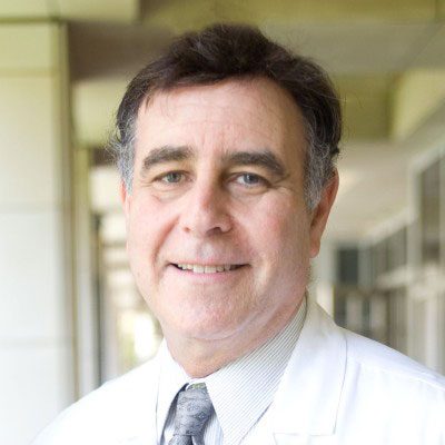 Wayne Grody合作论文数David Geffen School of Medicine, University of California, Los Angeles12
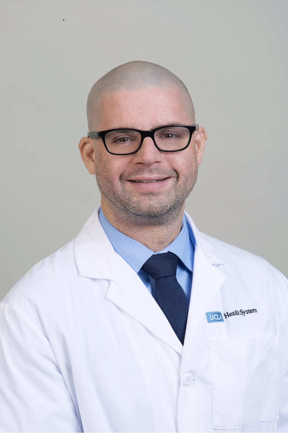 Julian Martinez合作论文数Department of Human Genetics, David Geffen School Of Medicine, University of California12
 Stanley F. Nelson合作论文数David Geffen School of Medicine, University of California, Los Angeles12
 James Francis Gusella合作论文数Department of Genetics, Blavatnik Institute, Harvard Medical School;Center for Genomic Medicine, Massachusetts General Hospital11
 Cynthia Casson Morton合作论文数Division of Medical Sciences, Harvard Medical School;Brigham and Women’s Hospital10
 Katrina M. Dipple合作论文数Department of Medical Genetics, Indiana University School of Medicine9
 Joshua L. Deignan合作论文数Department of Pathology and Laboratory Medicine, University of California, Los Angeles8Dorrani Naghmeh合作论文数8
 Elaine H. Zackai合作论文数Children's Hospital of Philadelphia;Perelman School of Medicine, University of Pennsylvania8Hane Lee合作论文数David Geffen School of Medicine, University of California8Weining Lu合作论文数8
 Richard Maas合作论文数Division of Genetics, Brigham and Women’s Hospital;Department of Medicine, Harvard Medical School;International Center for Genetic Disease, Harvard Medical School7Kantarci Sibel合作论文数6
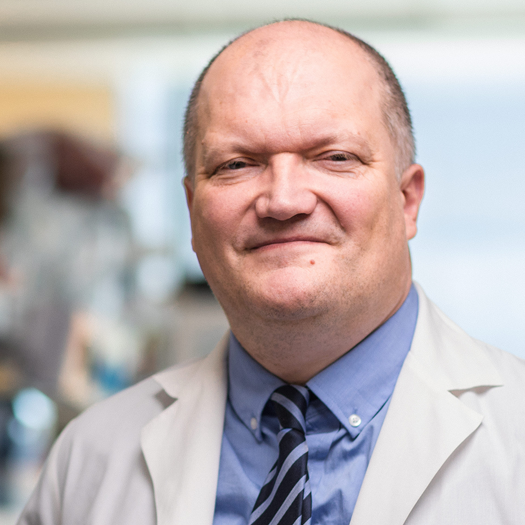 Eric Vilain合作论文数Center for Genetic Medicine Research, Children's National Medical Center;Institut National de Recherche Biomedicale;The George Washington University School of Medicine and Health Sciences6
 Samuel P. Strom合作论文数Department of Pathology and Laboratory Medicine, David Geffen School of Medicine, University of California Los Angeles6
 Azra Hadi Ligon合作论文数Division of Clinical Cytogenetics, Brigham and Women's Hospital;Department of Pathology, Harvard Medical School6
 Fowzan S. Alkuraya (Fowzan Alkuraya)合作论文数College of Medicine, Alfaisal University;Department of Translational Genomics, King Faisal Specialist Hospital and Research Center6Anne W. Higgins合作论文数6Bradley J. Quade合作论文数Division of Women ' s and Perinatal Pathology and Clinical Cytogenetics Laboratory;Brigham and Women ' s Hospital;Department of Pathology;Medical School, Harvard;Brigham and Women ' s Hospital, Harvard Medical School6Michael E. Talkowski合作论文数5Jonathan A Bernstein合作论文数5P. Nagesh Rao合作论文数5
 Sherif Rezk合作论文数5
 Micheil Innes合作论文数Department of Medical Genetics, University of Calgary5
 Eno Celeste C合作论文数Molecular, Cellular and Biomedical Sciences, University of New Hampshire5
 Harrison Brand合作论文数Molecular Neurogenetics Unit and;Center for Human Genetic Research, Massachusetts General Hospital;Departments of Neurology and;Center for Human Genetic Research, Massachusetts General Hospital5
 Anne Slavotinek合作论文数Sherr Lab, University of California, San Francisco5
 Virginia E Kimonis合作论文数Division of Genetics and Genomic Medicine, Pediatrics School of Medicine, University of California, Irvine5
 Hyung-Goo Kim合作论文数Brigham and Women’s Hospital and Harvard Medical School, Massachusetts General Hospital and Harvard Medical School5diana j donovan合作论文数4Jane Peredo合作论文数4David J Harris合作论文数4Małgorzata J M Nowaczyk合作论文数4Anthony Sisk合作论文数Department of Pathology and Laboratory Medicine, University of California, Los Angeles4
 Elizabeth Bhoj合作论文数Department of Genetics, Perelman School of Medicine, University of Pennsylvania4
 Xiaohui Zhao (赵晓辉)合作论文数Xinqiao Hospital, Army Medical University4
 Hodge Jennelle C合作论文数Dept Lab Med & Pathol, Mayo Clin4
 Ghayda M Mirzaa合作论文数4Dubbs Holly合作论文数4Harr Margaret H合作论文数4Biniwale Reshma合作论文数4
 Hakon Hakonarson合作论文数Center for Applied Genomics, The Joseph Stokes Jr. Research Institute, The Children's Hospital of Philadelphia;Pediatrics Department, Perelman School of Medicine, University of Pennsylvania4
 Glen S. Van Arsdell合作论文数Children's Heart Center, University of California Los Angeles4Omar A. Abdul-Rahman合作论文数Department of Pediatrics, University of Mississippi Medical Center4Heather Ferguson合作论文数4
 Daynna J. Wolff合作论文数Department of Pathology and Laboratory Medicine, College of Medicine, Medical University of South Carolina4
 Yanli Fan合作论文数4Tim DeSmet合作论文数3Kerry K Brown合作论文数3




