The human beta-coronavirus strain, OC43, provides a useful model for testing the antiviral activity of various agents. We compared the activity of several antiviral drugs against OC43, including remdesivir, chloroquine, interferon (IFN)-β, IFN-λ1, and IFN-λ4, in two distinct cell types: human colorectal carcinoma cell line (HCT-8 cells) and normal human bronchial epithelial (NHBE) cells. We also tested whether these agents mediate additive, synergistic, or antagonistic activity against OC43 infection when used in combination. When used as single agents, remdesivir exhibited stronger antiviral activity than chloroquine, and IFN-β exhibited stronger activity than IFN-λ1 or IFN-λ4 against OC43 in both HCT-8 and NHBE cells. Anakinra (IL-1 inhibitor) and tocilizumab (IL-6 inhibitor) did not mediate any antiviral activity. The combination of IFN-β plus chloroquine or remdesivir resulted in higher synergy scores and higher expression of IFN-stimulated genes than did IFN-β alone. In contrast, the combination of remdesivir plus chloroquine resulted in an antagonistic interaction in NHBE cells. Our findings indicate that the combined use of IFN-β plus remdesivir or chloroquine induces maximal antiviral activity against human coronavirus strain OC43 in primary human respiratory epithelial cells. Furthermore, our experimental OC43 virus infection model provides an excellent method for evaluating the biological activity of antiviral drugs.
Polysorbate excipients are commonly used as surfactants to stabilize therapeutic proteins in formulations. Degradation of polysorbates could lead to particle formation and instability of the drug formulation. We investigated how the fatty acid composition of polysorbate 80 impacts the degradation profile, particle formation, and product stability under stress conditions. Two polysorbate 80-containing therapeutic protein formulations were reformulated with either Polysorbate 80 NF synthesized from a fatty acid mixture that contains mainly oleic acid (≥58%) or a version of polysorbate 80 synthesized with high oleic acid (>98%). Stress conditions, including high temperature and esterase spiking, were applied and changes to both the polysorbate and the therapeutic protein product were investigated for stability, purity, innate immune response and biological activity. The addition of esterase and storage at 37°C led to significant hydrolysis of the polysorbate and increases in sub-visible particle formation for both polysorbates tested. The fatty acid composition of polysorbate 80 did not directly alter the stability profile of either therapeutic protein as measured by size exclusion chromatography, or significantly impact innate immune response or biological activity. However, formulations with Polysorbate 80 NF showed greater propensity for sub-visible particle formation under stress conditions. These results suggest that composition of fatty acids in polysorbate 80 may be a promoter for sub-visible particulate formation under the stress conditions tested but may not impact protein aggregation or biological activity.
PEGylated recombinant human granulocyte colony stimulating factor (pegfilgrastim) is used clinically to accelerate immune reconstitution following chemotherapy and is being pursued for biosimilar development. One challenge to overcome in pegfilgrastim biosimilar development is establishing pharmacokinetic (PK) similarity, which is partly due to the degree of PK variability. We herein report that commercially available G-CSF and PEG ELISA detection kits have different capacities to detect pegfilgrastim aggregates that rapidly form in vitro in physiological conditions. These aggregates can be observed using SDS-PAGE, size-exclusion chromatography, dynamic light scattering, and real-time NMR analysis and are associated with decreased bioactivity as reflected by reduced drug-induced cellular proliferation and STAT3 phosphorylation. Furthermore, individual variability in the stability and detectability of pegfilgrastim in human sera is also observed. Pegfilgrastim levels display marked subject variability in sera from healthy donors incubated at 37 °C. The stability patterns of pegfilgrastim closely match the stability patterns of filgrastim, consistent with a key role for pegfilgrastim’s G-CSF moiety in driving formation of inactive aggregates. Taken together, our results indicate that individual variability and ELISA specificity for inactive aggregates are key factors to consider when designing and interpreting studies involving the measurement of serum pegfilgrastim concentrations.
Respiratory syncytial virus (RSV) infects small foci of respiratory epithelial cells via infected droplets. Infection induces expression of type I and III interferons (IFNs) and proinflammatory cytokines, the balance of which may restrict viral replication and affect disease severity. We explored this balance by infecting two respiratory epithelial cell lines with low doses of recombinant RSV expressing green fluorescent protein (rgRSV). A549 cells were highly permissive, whereas BEAS-2B cells restricted infection to individual cells or small foci. After infection, A549 cells expressed higher levels of IFN-β-, IFN-λ-, and NF-κB-inducible proinflammatory cytokines. In contrast, BEAS-2B cells expressed higher levels of antiviral interferon-stimulated genes, pattern recognition receptors, and other signaling intermediaries constitutively and after infection. Transcriptome analysis revealed that constitutive expression of antiviral and proinflammatory genes predicted responses by each cell line. These two cell lines provide a model for elucidating critical mediators of local control of viral infection in respiratory epithelial cells.IMPORTANCE Airway epithelium is both the primary target of and the first defense against respiratory syncytial virus (RSV). Whether RSV replicates and spreads to adjacent epithelial cells depends on the quality of their innate immune responses. A549 and BEAS-2B are alveolar and bronchial epithelial cell lines, respectively, that are often used to study RSV infection. We show that A549 cells are permissive to RSV infection and express genes characteristic of a proinflammatory response. In contrast, BEAS-2B cells restrict infection and express genes characteristic of an antiviral response associated with expression of type I and III interferons. Transcriptome analysis of constitutive gene expression revealed patterns that may predict the response of each cell line to infection. This study suggests that restrictive and permissive cell lines may provide a model for identifying critical mediators of local control of infection and stresses the importance of the constitutive antiviral state for the response to viral challenge.
Type III IFNs are important mediators of antiviral immunity. IFN-λ4 is a unique type III IFN because it is produced only in individuals who carry a dG allele of a genetic variant rs368234815-dG/TT. Counterintuitively, those individuals who can produce IFN-λ4, an antiviral cytokine, are also less likely to clear hepatitis C virus infection. In this study, we searched for unique functional properties of IFN-λ4 that might explain its negative effect on hepatitis C virus clearance. We used fresh primary human hepatocytes (PHHs) treated with recombinant type III IFNs or infected with Sendai virus to model acute viral infection and subsequently validated our findings in HepG2 cell line models. Endogenous IFN-λ4 protein was detectable only in Sendai virus–infected PHHs from individuals with the dG allele, where it was poorly secreted but highly functional, even at concentrations < 50 pg/ml. IFN-λ4 acted faster than other type III IFNs in inducing antiviral genes, as well as negative regulators of the IFN response, such as USP18 and SOCS1. Transient treatment of PHHs with IFN-λ4, but not IFN-λ3, caused a strong and sustained induction of SOCS1 and refractoriness to further stimulation with IFN-λ3. Our results suggest unique functional properties of IFN-λ4 that can be important in viral clearance and other clinical conditions.
The influenza virus NS1 protein interacts with a wide range of proteins to suppress the host cell immune response and facilitate virus replication. The amino acid sequence of the 2009 pandemic virus NS1 protein differed from sequences of earlier related viruses. The functional impact of these differences has not been fully defined. Therefore, we made mutations to the NS1 protein based on these sequence differences, and assessed the impact of these changes on host cell interferon (IFN) responses. We found that viruses with mutations at position 171 replicated efficiently but did not induce expression of interferon genes as effectively as wild-type viruses in A459 lung epithelial cells. The decreased ability of these NS1 mutant viruses to induce IFN gene and protein expression correlated with decreased activation of STAT1 and lower levels of IFN-stimulated gene (ISG) expression. These findings demonstrate that mutations at position 171 in the NS1 protein result in decreased expression of IFN and ISGs by A549 cells. Consequently, these viruses may be more virulent than the parental strains that do not contain mutations at position 171 in the NS1 protein.
Influenza A viruses pose a constant potential threat to human health. In view of the innate antiviral activity of interferons (IFNs) and their potential use as anti-influenza agents, it is important to know whether viral resistance to these antiviral proteins can arise. To examine the likelihood of emergence of IFN-λ1-resistant H1N1 variants, we serially passaged the A/California/04/09 (H1N1) strain in a human lung epithelial cell line (Calu-3) in the presence of increasing concentrations of recombinant IFN-λ1 protein. To monitor changes associated with adaptation of this virus to growth in Calu-3 cells, we also passaged the wild-type virus in the absence of IFN-λ1. Under IFN-λ1 selective pressure, the parental virus developed two neuraminidase (NA) mutations, S79L and K331N, which significantly reduced NA enzyme activity (↓1.4-fold) and sensitivity to IFN-λ1 (↓˃20-fold), respectively. These changes were not associated with a reduction in viral replication levels. Mutants carrying either K331N alone or S79L and K331N together induced weaker phosphorylation of IFN regulatory factor 3 (IRF3), and, as a consequence, much lower expression of the IFN genes (IFNB1, IFNL1 and IFNL2/3) and proteins (IFN-λ1 and IFN-λ2/3). The lower levels of IFN expression correlated with weaker induction of tyrosine-phosphorylated STAT1 and reduced RIG-I protein levels. Our findings demonstrate that influenza viruses can develop increased resistance to the antiviral activity of type III interferons.
IFN-λ4, a recently discovered type-III interferon (IFN), is genetically regulated, and has clinical significance because of its association with impaired clearance of hepatitis C virus (HCV) infection. Type-III IFNs play a critical role in the innate immune response to viral infections. Although IFN-λ4 induces a similar antiviral profile as other type-III IFNs, its specific contribution to the innate immune response is unclear. Delineating the functional properties of IFN-λ4 that distinguish it from other type-III IFNs may give insights into its unique role during HCV, and other infections. Here we compared antiviral responses induced by all four type-III IFNs in HepG2 cells. We show that IFN-λ4 attained 50% peak antiviral activity by 4 hours while other type-III IFNs were only at 20% of peak activity at this time. The rapid antiviral response induced by IFN-λ4 was followed by a sharp decline to less than 25% of peak activity within 24 hours, while activity of other type-III IFNs were still over 60% from peak. By comparing antiviral activity of IFN-λ4 and IFN-λ3 against Sendai virus infection in primary human hepatocytes, we show that IFN-λ4 had stronger antiviral activity than IFN-λ3 early in the course of infection, but comparable activity at later time points. We also show that despite its poor secretion, IFN-λ4 was significantly more potent than IFN-λ3 in inducible cell lines engineered to express either protein. In summary, we show that IFN-λ4 is more active during the early stages of viral infection compared to other type-III IFNs, which maybe a clinically important window for restricting viral infection in the host, and affecting disease outcome.
Macrophages coexpress both the interleukin (IL)-2Rγ chain (γc) and IL-13Rα1. These receptor chains can heterodimerize with IL-4Rα to form type I or type II IL-4 receptor complexes, respectively. We used macrophages derived from Il2rg and Il13ra1 knockout (KO) mice to evaluate the requirements for these receptor chains for induction of the alternative macrophage activation (AMA) pathway by IL-4 and IL-13. Absence of γc significantly decreased activation of STAT6 by IL-4 but not IL-13. However, although activation of STAT6 by IL-4 was markedly reduced in γc KO macrophages, it was not abolished, indicating that IL-4 can still signal through type II IL-4 receptors via the IL-13Rα1 chain. IL-13 failed to activate STAT6 in macrophages derived from Il13ra1 KO mice; however, these cells remained fully responsive to IL-4. The inability of IL-13 but not IL-4 to signal in Il13ra1-/- macrophages correlated with the inability of IL-13 but not IL-4 to induce expression of genes such as Arg1, Retnla and Ccl11 that are characteristically expressed by alternatively activated macrophages. In addition, IL-13 but not IL-4 failed to induce membrane fusion and giant cell formation by Il13ra1 KO macrophages. These findings demonstrate that the IL-13Rα1 chain is essential for induction of the AMA pathway by IL-13 but not IL-4.
TLR agonists such as LPS and poly(I:C) induce expression of type I IFNs, such as IFN‐α and ‐β, by macrophages. To examine the role of IFN‐β in the induction of ISGs by LPS, we compared the ability of LPS to induce ISGF3 activity and ISG expression in bone marrow–derived macrophages from WT and Ifnb1−/− mice. We found that LPS treatment activated ISGF3 and induced expression of ISGs such as Oas1, Mx1, Ddx58 (RIG‐I), and Ifih1 (MDA5) in WT macrophages, but not in macrophages derived from Ifnb1−/− mice or Ifnar1−/− mice. The inability of LPS to induce activation of ISGF3 and ISG expression in Ifnb1−/− macrophages correlated with the failure of LPS to induce activation of STAT1 and ‐2 in these cells. Consistent with these findings, LPS treatment also failed to induce ISG expression in bone marrow–derived macrophages from Stat2 KO mice. Although activation of ISGF3 and induction of ISG expression by LPS was abrogated in Ifnb1−/− and Ifnar1−/− macrophages, activation of NF‐κB and induction of NF‐κB‐responsive genes, such as Tnf (TNF‐α) and Il1b (IL‐1β), were not affected by deletion of either the IFN‐β or IFN‐αR1 genes. These findings demonstrate that induction of ISGF3 activity and ISG expression by LPS is critically dependent on intermediate production of IFN‐β and autocrine signaling through type I IFN receptors.
Chronic infection with hepatitis C virus (HCV) is a common cause of liver cirrhosis and cancer. We performed RNA sequencing in primary human hepatocytes activated with synthetic double-stranded RNA to mimic HCV infection. Upstream of IFNL3 (IL28B) on chromosome 19q13.13, we discovered a new transiently induced region that harbors a dinucleotide variant ss469415590 (TT or ΔG), which is in high linkage disequilibrium with rs12979860, a genetic marker strongly associated with HCV clearance. ss469415590[ΔG] is a frameshift variant that creates a novel gene, designated IFNL4, encoding the interferon-λ4 protein (IFNL4), which is moderately similar to IFNL3. Compared to rs12979860, ss469415590 is more strongly associated with HCV clearance in individuals of African ancestry, although it provides comparable information in Europeans and Asians. Transient overexpression of IFNL4 in a hepatoma cell line induced STAT1 and STAT2 phosphorylation and the expression of interferon-stimulated genes. Our findings provide new insights into the genetic regulation of HCV clearance and its clinical management.
We have identified a novel human interferon, designated as interferon-λ4 (IFN-λ4) (Prokunina-Olsson et al., Nature Genetics, 2013). Inducible expression of IFNL4 , along with three related genes ( IFNL1 , IFNL2 and IFNL3 ) located on chromosome 19q13.13 and encoding other interferon-λ proteins, IFN-λ1, IFN-λ2 and IFN-λ3, was revealed by RNA-sequencing in primary human hepatocytes treated with polyI:C. In humans, IFN-λ4 is fully genetically controlled – it is produced only by mRNA transcripts with a frameshift dG allele of a genetic variant ss469415590 (TT/dG) within the first exon of the gene, while transcripts with the TT allele can generate only unrelated proteins or protein fragments. We characterized IFN-λ4 as a class-2 cytokine based on its protein homology with IFN-λ3 (29% amino acid identity), the ability to cause STAT1 and STAT2 phosphorylation, activation of an ISRE-Luc reporter, induction of ISGs and antiviral response in HepG2 cells transiently transfected with an IFNL4-expressing construct. IFN-λ4 signaling is decreased by a JAK inhibitor, siRNA-silencing of IFN-λR1, and blocking of IL10R2. However, HepG2 cells did not respond to treatment with purified recombinant IFN-λ4 protein. Protein expression of IFN-λ4 is detectable in cells transfected with an IFNL4-expressing construct but not in the culture supernatants. It remains unclear whether IFN-λ4 is an unusual intracellular interferon, which signals through yet unknown intracellular partners or it is released at low levels through secretion or other mechanisms. Individuals homozygous for the derived human-specific ss469415590-TT allele (90% of Asians, 50% of Europeans and 10% of individuals of African ancestry) are genetically unable to produce IFN-λ4. This pattern suggests strong positive selection for elimination of IFN-λ4, which might be caused by current or historic infectious diseases. Individuals who are unable to produce IFN-λ4 are more likely to clear hepatitis C virus (HCV) either spontaneously or after treatment with IFN-α and ribavirin.
This study compared the ability of IFN-alpha and IFN-lambda to induce signal transduction and gene expression in primary human hepatocytes, PBLs, and monocytes. IFN-alpha drug products are widely used to treat chronic HCV infection; however, IFN-alpha therapy often induces hematologic toxicities as a result of the broad expression of IFNARs on many cell types, including most leukocytes. rIFN-lambda 1 is currently being tested as a potential alternative to IFN-alpha for treating chronic HCV. Although IFN-lambda has been shown to be active on hepatoma cell lines, such as HepG2 and Huh-7, its ability to induce responses in primary human hepatocytes or leukocytes has not been examined. We found that IFN-lambda induces activation of Jak/STAT signaling in mouse and human hepatocytes, and the ability of IFN-lambda to induce STAT activation correlates with induction of numerous ISGs. Although the magnitude of ISG expression induced by IFN-alpha in hepatocytes was generally lower than that induced by IFN-lambda, the repertoire of regulated genes was quite similar. Our findings demonstrate that although IFN-alpha and IFN-lambda signal through distinct receptors, they induce expression of a common set of ISGs in hepatocytes. However, unlike IFN-alpha, IFN-lambda did not induce STAT activation or ISG expression by purified lymphocytes or monocytes. This important functional difference may provide a clinical advantage for IFN-lambda as a treatment for chronic HCV infection, as it is less likely to induce the leukopenias that are often associated with IFN-alpha therapy. J. Leukoc. Biol. 93: 377-385; 2013.
Plasmacytoid dendritic cells (pDC) are rare cells found in peripheral blood and lymphoid tissues. pDC are considered to be “professional” type I IFN-producing cells and produce 10- to 100-fold more IFN-α than other cell types in response to enveloped viruses or synthetic TLR7 and TLR9 agonists. In this study, purified pDC were found to express high levels of IFN-λ receptor mRNA, as well as cell-surface IFN-λ receptor. We have developed intracellular flow cytometry assays using Abs to IFN-λ1/3 or -λ2 to assess the expression of IFN-λ proteins by pDC. We observed that a subset of human pDC expresses only intracellular IFN-α, whereas another subset produces both IFN-α and IFN-λ after stimulation with virus or the TLR9 agonist, CpG A; the cells that coexpressed IFN-α and IFN-λ were the cells with the highest levels of IFN-α expression. Ab cross-linking of CD4 or CD303 molecules on pDC inhibited both HSV-induced IFN-λ and IFN-α production. Like the production of IFN-α, the HSV-induced IFN-λ production in pDC was mediated through TLR9 and independent of virus replication. Exogenous IFN-λ treatment of pDC resulted in increased virus-induced expression of both IFN-α and IFN-λ. In addition, both exogenous IFN-λ and -α inhibited dexamethasone-induced apoptosis of pDC. We conclude that pDC are major producers of IFN-λ1 and -λ2 in response to viral stimulation and also express functional receptors for this cytokine. Thus, IFN-λ can serve as an autocrine signal to strengthen the antiviral response of pDC by increasing IFN-α and IFN-λ production, resulting in prolonged pDC survival.
Summary. Recently, several SNPs in the region of the IL28B (IFN‐λ) gene have been associated with spontaneous clearance of hepatitis C virus (HCV) and enhanced cure rates for IFN‐alfa‐based therapies, suggesting a potential correlation between IFN‐λ and the ability to clear HCV. To understand the mechanism of IFN‐λ’s as compared to IFN‐α’s antiviral activity, we performed a comprehensive analysis of their anti‐HCV effects, whole genome transcriptome profiling with validation, and signalling of IFN‐α and IFN‐λ using J6/JFH‐1 and Huh7.5 cells in vitro. IFN‐λ and IFN‐α exhibited comparable anti‐HCV activity and gene expression profiles in Huh7.5 cells. While the majority of genes induced by IFN‐α and IFN‐λ were similar, IFN‐λ exhibits profound, but delayed kinetics of IFN‐stimulated genes (ISG) induction, while IFN‐α induced more rapid induction of ISGs. Furthermore, the increased induction of ISG expression by IFN‐λ correlated with up‐regulation of IFN‐λ receptor (IL‐28RA) expression and more prolonged activation of the Jak‐STAT signalling pathway. The findings from our comparative analysis of IFN‐α and IFN‐λ in HCV‐infected and noninfected cells support the clinical use of IFN‐λ as a potential alternative to IFN‐α in the treatment of chronic hepatitis C.
IL-26 is classified as a member of the IL-10 cytokine family because it has limited sequence homology to IL-10 and the IL-10-related cytokines. The human IL-26 gene, IL26, is located on chromosome 12q15 between the genes for two other important class-2 cytokines, IFNG (IFN-γ) and IL22 (IL-22). IL-26 is often co-expressed with IL-22 by activated T cells, especially Th17 cells. It signals through a heterodimeric receptor complex composed of the IL-20R1 and IL-10R2 chains. IL-26 receptors are primarily expressed on non-hematopoietic cell types, particularly epithelial cells. Signaling through IL-26 receptor complexes results in the activation of STAT1 and STAT3 with subsequent induction of IL-26-responsive genes. The biological functions of IL-26 have only begun to be defined.
When type III interferon (IFN-λ; also known as interleukin-28 [IL-28] and IL-29) was discovered in 2003, its antiviral function was expected to be analogous to that of type I IFNs (IFN-α and IFN-β) via the induction of IFN-stimulated genes (ISGs). Although IFN-λ stimulates expression of antiviral ISGs preferentially in cells of epithelial origin, recent studies have defined additional antiviral mechanisms in other cell types and tissues. Viral infection models using mice lacking IFN-λ signaling and SNP associations with human disease have expanded our understanding of the contribution of IFN-λ to the antiviral response at anatomic barriers and the immune response beyond these barriers. In this review, we highlight recent insights into IFN-λ functions, including its ability to restrict virus spread into the brain and to clear chronic viral infections in the gastrointestinal tract. We also discuss how IFN-λ modulates innate and adaptive immunity, autoimmunity, and tumor progression and its possible therapeutic applications in human disease.
 Raymond P. Donnelly合作论文数Department of Microbiology and Immunology, Temple University School of Medicine36harold dickensheets合作论文数14Sergei V. Kotenko合作论文数6
 Ana M Gamero合作论文数5Anita Lewis-Antes合作论文数4
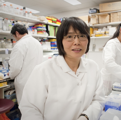 Shioko Kimura合作论文数Laboratory of Molecular Carcinogenesis, Center for Cancer Research, National Cancer Institute4
 Howard A. Young合作论文数Cancer Innovation Laboratory, National Cancer Institute, National Institutes of Health4Harold Dickensheets合作论文数4
 Ludmila Prokunina-Olsson合作论文数Laboratory of Translational Genomics, National Cancer Institute, National Institutes of Health4Atsushi Yamada合作论文数3Shubha Anantha合作论文数3Natalia A. Ilyushina合作论文数3Anthony J. Scarzello合作论文数3Achara Srisodsai合作论文数3L Dumoutier合作论文数2Vitaliy V. Baurin合作论文数2Prabhakar Gupta合作论文数2rachel shepard合作论文数2
 Wei Tang合作论文数Bio-X Life Science Research Center, Shanghai Jiao Tong University, 1954 Huashan Road, Shanghai 200030, PR China2Romero-Weaver Ana L合作论文数2
 Brian Muchmore合作论文数Laboratory of Translational Genomics, Division of Cancer Epidemiology and Genetics, and2Luyang Liu合作论文数2
 Reed S. Shabman合作论文数Office of Data Science and Emerging Technologies, National Institute of Allergy and Infectious Diseases, National Institutes of Health2Abushahba Walid合作论文数2
 Stefanie N. Vogel合作论文数School of Medicine, University of Maryland2
 Bin Gao合作论文数Laboratory of Liver Diseases, Division of Intramural Clinical and Biological Research, National Institute on Alcohol Abuse and Alcoholism, National Institutes of Health2Hillyer Philippa合作论文数2Lasfar Ahmed合作论文数2
 Jean-Christophe Renauld合作论文数Institut de Duve, Faculty of Medicine and Dentistry, Université catholique de Louvain2
 Barbara Rehermann合作论文数National Institute of Diabetes and Digestive and Kidney Diseases, National Institutes of Health2Brand Nathan合作论文数2Singh Inderjit合作论文数2Kenneth R. Reuhl合作论文数2
 Stephen Maher合作论文数Institute of Molecular Medicine, Trinity College Dublin and St. James’s Hospital2Elizabeth S. Raveche合作论文数2
 Zoltan Pos合作论文数National Institutes of Health2Ronald L. Rabin合作论文数2
 S V Smirnov合作论文数Institute Biologie and Biotechnologie, Altai State University2Heiyoung Park合作论文数2Indu Kohaar合作论文数Laboratory of Translational Genomics, National Cancer Institute, National Institutes of Health2Alexander Zdanov合作论文数2
 Thirumalai R Ramalingam合作论文数2
 Mark R. Walter合作论文数Department of Microbiology, University of Alabama at Birmingham2
 Patricia Porter-Gill合作论文数2Thomas R. O'Brien合作论文数2
 Francesco Marincola合作论文数Sonata Therapeutics;Kite Pharma;Refuge Biotechnologies;AbbVie;Sidra Medical and Research Center2Grant Gallagher合作论文数1RT Kenney合作论文数1Ana L. Romero-Weaver合作论文数1




