Mechanistic discoveries mostly rely on animal research or bioengineered human models. However, these platforms often fail to recapitulate human physiology and disease. Here we report a novel approach using adult human hearts, available from the UK NHSBT, explanted exclusively for research and employed to prepare living myocardial slices (LMSs).LMSs are ultrathin (300 μm) sections of living cardiac tissue that can be prepared using a vibratome; LMSs maintain the native structure of the heart and can be kept in culture for weeks/months under physiological electro-mechanical conditions. We assessed whether preparing LMSs from donor human hearts is a viable and practical alternative to other research platforms.From July 2024 to July 2025, we received 203 donor human heart offers. We accepted and used 39 hearts (20 donated after circulatory death, 19 donated after brain death), an almost ten-fold increase compared with previous years. The donors were 62.5 +/− 14.9 years of age (Mean +/− SD). We only accepted hearts from donors without cardiovascular disease or current infections and were within 6 h transport time (transported in saline solution at 4 °C). LMSs were prepared as described by our group previously (Camelliti et al., 2011; Pitoulis et al., 2020; van der Geest et al., 2025; Watson et al., 2019, 2017).We could prepare 30–50 LMSs from a 1 cm3 LV transmural biopsy. LMSs were viable and robust: (force transient amplitude at 2.2 μm sarcomere length: 8.8 +/− 7.2mN/mm2; passive force: 9.4 +/− 6.6mN/mm2 (n = 39)).We also tested if it is possible to delay tissue utilisation, to facilitate high throughput, help with the arrival of tissue out-of-hours, and share it with other centres. There was no statistical difference in both force transient amplitude and passive force between fresh LMS and LMS from biopsies stored in cardioplegia for 5 h at 4 °C (active force fresh: 7.8 +/− 4.8mN/mm2; post-cardioplegia: 26.4+/-24mN/mm2; paired t-test: p = 0.272; passive force fresh: 6.1±1.34mN/mm2; cardioplegia: 13.5±5.4mN/mm2; p = 0.132, n = 3 hearts from 8 fresh and 11 post-cardioplegia LMSs).Adult donor heart tissue is a viable source of LMSs and can lead to valuable research in mechanisms of disease and treatment, as a valid alternative to animal research or bioengineered models.
In search for new treatment options for heart failure patients, human induced pluripotent stem cell-derived (hiPSC-) cardiomyocytes represent the new gold standard for the development of novel cell-replacement therapies in cardiac regenerative medicine. Despite global standardization of differentiation procedures, the resulting hiPSC-cardiomyocytes retain an immature phenotype, revealing a large functional gap toward the characteristics of adult cardiomyocytes. Especially at the level of Ca2+ handling, which is crucial for the contractile activity and modulation of force production, hiPSC-cardiomyocytes reveal deficits that might be causal for the spontaneous contractile activity, one of the hallmarks of immaturity.
BACKGROUND:Promising as a treatment option for life-threatening ventricular arrhythmias, cardiac stereotactic body radiotherapy (cSBRT) has demonstrated early antiarrhythmic effects within days of treatment. The mechanisms underlying the immediate and short-term antiarrhythmic effects are poorly understood. OBJECTIVE:We hypothesize that cSBRT has a direct antiarrhythmic effect on cellular electrophysiology through reprogramming of ion channel and gap junction protein expression. METHODS:After exposure to 20 Gy of x-rays in a single fraction, neonatal rat ventricular cardiomyocytes were analyzed 24 and 96 hours postradiation to determine changes in conduction velocity, beating frequency, calcium transients, and action potential duration in both monolayers and single cells. In addition, the expression of gap junction proteins, ion channels, and calcium handling proteins was evaluated at protein and messenger RNA levels. RESULTS:After irradiation with 20 Gy, neonatal rat ventricular cardiomyocytes exhibited increased beat rate and conduction velocity 24 and 96 hours after treatment. Messenger RNA and protein levels of ion channels were altered, with the most significant changes observed at the 96-hour mark. Upregulation of Cacna1c (Cav1.2), Kcnd3 (Kv4.3), Kcnh2 (Kv11.1), Kcnq1 (Kv7.1), Kcnk2 (K2P2.1), Kcnj2 (Kir2.1), and Gja1 (Cx43) was noted, along with improved gap junctional coupling. Calcium handling was affected, with increased Ryr2 ryanodin-rezeptor 2 and Slc8a1 Na+/Ca2+ exchanger expression and altered properties 96 hours posttreatment. Fibroblast and myofibroblast levels remained unchanged. CONCLUSION:cSBRT modulates the expression of various ion channels, calcium handling proteins, and gap junction proteins. The described alterations in cellular electrophysiology may be the underlying cause of the immediate antiarrhythmic effects observed after cSBRT.
BACKGROUND: Small extracellular vesicles (sEVs) released in the cardiac microenvironment are reported to regulate cardiac remodelling, partially via microRNA transfer. Harvesting sEVs produced exclusively from the myocardium remains challenging and a solid research platform for sEV cardiovascular testing needs to be established. Organotypic living myocardial slices (LMS) allow to mimic cardiac disease and to record electrophysiological responses to biological and pharmacological stimuli. This study aims at understanding how cardiac sEVs obtained from donor and failing human LMS and rat LMS under physiological or heart failure-mimicking conditions impact myocardial function and remodelling. METHODS & RESULTS: Human LMS were obtained from the left ventricle (LV) of human donor non-failing and end-stage failing hearts and cultured at 2.2 μm sarcomere length (SL). Rat LV LMS from healthy Sprague-Dawley rats were cultured at a preload of 2.2 or 2.4 μm SL, to recapitulate physiological load and overload, respectively. Following 48-hours biomimetic culture, sEVs were isolated from the culture media by size exclusion chromatography and characterized for their size, concentration, and expression of exosome markers. LMS from human failing hearts presented impaired contractility (P<0.05 vs donor-LMS), which was improved by application of donor heart-derived sEVs at 15 and 20% stretch. Whilst rat overloaded sEVs did not alter the force production of physiological LMS, physiological sEVs significantly increased the active force and decreased their passive force. In rat LMS, 1x10⁸ physiological EVs/slice restored the contractility of overloaded slices, reduced apoptosis, fibrosis-related gene expression and promoted angiogenesis. microRNAs analysis showed significant upregulation of miR-23a-3p and miR-378a-3p in rat physiological sEVs. Finally, to test whether sEVs have a direct effect on cardiomyocytes, we applied sEVs on cultured induced pluripotent stem cell-derived cardiomyocytes (iPSC-CMs). sEVs did not affect the contractility of iPSC-CM monoculture but increased the contractility of iPSC-CM co-cultured with human microvasculature endothelial cells (MVECs). CONCLUSIONS: Cardiac sEVs isolated from healthy hearts increase the contractility of failing LMS. This effect is associated with, and possibly brought about by, a combination of inhibition of apoptosis, reduction of fibrosis and increased microvascular density, and could involve the transfer of sEV-microRNA into myocardial cells. Our data support the hypothesis that the sEV inotropic action is mediated by endothelial cells. ?### Competing Interest StatementThe authors have declared no competing interest.### Funding StatementWe are grateful to the British Heart Foundation for financial support (FS/19/57/34894).### Author DeclarationsI confirm all relevant ethical guidelines have been followed, and any necessary IRB and/or ethics committee approvals have been obtained.YesThe details of the IRB/oversight body that provided approval or exemption for the research described are given below:UK institutional ethics committee (NRES ethics number for biobank samples: 09/H0504/104 + 5; Biobank approval number: NP001-06-2015 and MED\_CT\_17_079) and Imperial College London. UK Home Office, in accordance with the United Kingdom Animals (Scientific Procedures) Act 1986 and Amendment Regulations 2012; killing procedures were performed in accordance with the established guidelines of the European Directive on the protection of animals used for scientific purposes (2010/63/EU). Human donor hearts were obtained from the NHS Blood and Transplant INOAR program (IRAS project ID: 189069) approved by the NHS Health Research Authority, in accordance with the Governance Arrangements for Research Ethics Committees.I confirm that all necessary patient/participant consent has been obtained and the appropriate institutional forms have been archived, and that any patient/participant/sample identifiers included were not known to anyone (e.g., hospital staff, patients or participants themselves) outside the research group so cannot be used to identify individuals.YesI understand that all clinical trials and any other prospective interventional studies must be registered with an ICMJE-approved registry, such as ClinicalTrials.gov. I confirm that any such study reported in the manuscript has been registered and the trial registration ID is provided (note: if posting a prospective study registered retrospectively, please provide a statement in the trial ID field explaining why the study was not registered in advance).YesI have followed all appropriate research reporting guidelines, such as any relevant EQUATOR Network research reporting checklist(s) and other pertinent material, if applicable.YesAll the data will be available on request
Background: Transverse tubules (t-tubules) form gradually in the developing heart, critically enabling maturation of cardiomyocyte Ca 2+ homeostasis. The membrane bending and scaffolding protein BIN1 (bridging integrator 1) has been implicated in this process. However, it is unclear which of the various reported BIN1 isoforms are involved, and whether BIN1 function is regulated by its putative binding partners MTM1 (myotubularin), a phosphoinositide 3′-phosphatase, and DNM2 (dynamin-2), a GTPase believed to mediate membrane fission. Methods: We investigated the roles of BIN1, MTM1, and DNM2 in t-tubule formation in developing mouse cardiomyocytes, and in gene-modified HL-1 and human-induced pluripotent stem cell-derived cardiomyocytes. T-tubules and proteins of interest were imaged by confocal and Airyscan microscopy, and expression patterns were examined by RT-qPCR and Western blotting. Ca 2+ release was recorded using Fluo-4. Results: We observed that in the postnatal mouse heart, BIN1 localizes along Z-lines from early developmental stages, consistent with roles in initial budding and scaffolding of t-tubules. T-tubule proliferation and organization were linked to a progressive and parallel increase in 4 detected BIN1 isoforms. All isoforms were observed to induce tubulation in cardiomyocytes but produced t-tubules with differing geometries. BIN1-induced tubulations contained the L-type Ca 2+ channel, were colocalized with caveolin-3 and the ryanodine receptor, and effectively triggered Ca 2+ release. BIN1 upregulation during development was paralleled by increasing expression of MTM1. Despite no direct binding between MTM1 and murine cardiac BIN1 isoforms, which lack exon 11, high MTM1 levels were necessary for BIN1-induced tubulation, indicating a central role of phosphoinositide homeostasis. In contrast, the developing heart exhibited declining levels of DNM2. Indeed, we observed that high levels of DNM2 are inhibitory for t-tubule formation, although this protein colocalizes with BIN1 along Z-lines, and binds all 4 isoforms. Conclusions: These findings indicate that BIN1, MTM1, and DNM2 have balanced and collaborative roles in controlling t-tubule growth in cardiomyocytes.
The prospective use of human-induced pluripotent stem cell-derived cardiomyocytes (hiPSC-CM) for cardiac regenerative medicine strongly depends on the electro-mechanical properties of these cells, especially regarding the Ca2+-dependent excitation-contraction (EC) coupling mechanism. Currently, the immature structural and functional features of hiPSC-CM limit the progression towards clinical applications. Here, we show that a specific microarchitecture is essential for functional maturation of hiPSC-CM. Structural remodelling towards a cuboid cell shape and induction of BIN1, a facilitator of membrane invaginations, lead to transverse (t)-tubule-like structures. This transformation brings two Ca2+ channels critical for EC coupling in close proximity, the L-type Ca2+ channel at the sarcolemma and the ryanodine receptor at the sarcoplasmic reticulum. Consequently, the Ca2+-dependent functional interaction of these channels becomes more efficient, leading to improved spatio-temporal synchronisation of Ca2+ transients and higher EC coupling gain. Thus, functional maturation of hiPSC-cardiomyocytes by optimised cell microarchitecture needs to be considered for future cardiac regenerative approaches.
In this study, we report static and perfused models of human myocardial-microvascular interaction. In static culture, we observe distinct regulation of electrophysiology of human induced pluripotent stem cell derivedcardiomyocytes (hiPSC-CMs) in co-culture with human cardiac microvascular endothelial cells (hCMVECs) and human left ventricular fibroblasts (hLVFBs), including modification of beating rate, action potential, calcium handling, and pro-arrhythmic substrate. Within a heart-on-a-chip model, we subject this three-dimensional (3D) co-culture to microfluidic perfusion and vasculogenic growth factors to induce spontaneous assembly of perfusable myocardial microvasculature. Live imaging of red blood cells within myocardial microvasculature reveals pulsatile flow generated by beating hiPSC-CMs. This study therefore demonstrates a functionally vascularized in vitro model of human myocardium with widespread potential applications in basic and translational research.
Until several years ago, for in vitro studies scientists have mostly employed animal-derived cardiomyocytes to investigate the following: (1) the excitation-contraction-coupling (ECC) machinery; (2) The effects of disease-related pathological conditions on the ECC machinery; (3) Cardiomyocyte bioenergetics and metabolism; (4) Developmental changes in the ECC machinery in cardiomyocytes obtained from animals at different ages; (5) Inotropic and chronotropic modulation by autonomic agonists and antagonists; (6) Arrhythmia models and antiarrhythmic efficacies of different drugs; (7) Cardiac toxicity of approved drugs and new chemical entities. Although these experimental models have vastly expanded our knowledge regarding cardiac function under physiological and pathophysiological conditions, in many cases, key ECC characteristics of these animal models differ from those of the human heart. A new thrilling era of cardiac (but not only) research has emerged in the late 90s, early 2000s with the fundamental discovery by Thomson, Itskovitz, and coworkers that pluripotent embryonic stem cells (ESCs) can be derived from the inner cell mass of human blastocytes. These ESCs demonstrated the two fundamental features of stem cells: self-renewal and the ability to differentiate into the three germ layers, endoderm, mesoderm, and ectoderm. This pivotal discovery was received by the scientific community with world-wide enthusiasm, thus embracing the concept of using human ESC-derived cardiomyocytes (ESC-CMs) for cardiac muscle regeneration; for example, replacing dead cardiac tissue following myocardial infarction following coronary artery occlusion. Nonetheless, in addition to major ethical concerns regarding the use of cardiomyocytes generated from (destroyed) human embryos, other drawbacks are the limited supply and the need to use immunosuppression upon ESC-CMs transplantation in experimental animals and human hearts. Nonetheless, the use of human ESC-CMs promptly became the focus of many laboratories worldwide, for both basic research and clinical applications, aiming for example at postmyocardial infarction tissue regeneration. In 2006/7 the scientific community was overwhelmed (again) by the breakthrough discovery of Takahashi and Yamanaka who discovered the four transcription factors critical for cells' pluripotency, thus providing the unique opportunity to reprogram any somatic cell into human-induced pluripotent stem cell (iPSCs). Once human iPSCs are generated, they can be differentiated into almost any cell type, such as cardiomyocytes, neurons, pancreatic cells, and skeletal muscle cells. The ability to generate almost any cell type by reprogramming our own somatic cells (e.g., skin fibroblasts, blood cells, hair keratinocytes) has truly revolutionized the scientific world, with far-reaching implications that are beyond the scope of this chapter. To acquaint the readers with the magical world of iPSCs into cardiomyocytes (iPSC-CMs), this chapter addresses the following topics: (1) Differentiating iPSC-CMs; (2) Functional properties of immature iPSC-CMs; (3) Experimental procedures for promoting maturation of immature iPSC-CMs.
Human induced pluripotent stem cell-derived cardiomyocytes (hiPSC-CMs) enable human cardiac cells to be studied in vitro, although they use glucose as their primary metabolic substrate and do not recapitulate the properties of adult cardiomyocytes. Here, we have explored the interplay between maturation by stimulation of fatty acid oxidation and by culture in 3D. We have investigated substrate metabolism in hiPSC-CMs grown as a monolayer and in 3D, in porous collagen-derived scaffolds and in engineered heart tissue (EHT), by measuring rates of glycolysis and glucose and fatty acid oxidation (FAO), and changes in gene expression and mitochondrial oxygen consumption. FAO was stimulated by activation of peroxisome proliferator-activated receptor alpha (PPARα), using oleate and the agonist WY-14643, which induced an increase in FAO in monolayer hiPSC-CMs. hiPSC-CMs grown in 3D on collagen-derived scaffolds showed reduced glycolysis and increased FAO compared with monolayer cells. Activation of PPARα further increased FAO in cells on collagen/elastin scaffolds but not collagen or collagen/chondroitin-4-sulphate scaffolds. In EHT, FAO was significantly higher than in monolayer cells or those on static scaffolds and could be further increased by culture with oleate and WY-14643. In conclusion, a more mature metabolic phenotype can be induced by culture in 3D and FAO can be incremented by pharmacological stimulation.
Endothelial cells (EC) are active regulators of cardiac function, and bi-directional signalling between EC and cardiomyocytes (CM) is essential for maintenance of cardiac tissue homeostasis. For this reason, cardiac EC show strong promise as therapeutic targets for the prevention and treatment of heart disease. However, due to the lack of physiologically relevant multicellular in vitro platforms, many mechanisms governing the crosstalk between these two cells types are poorly understood. To investigate this relationship, we have developed an induced pluripotent stem cell derived cardiomyocyte (iPSC-CM) and endothelial cell (human cardiac microvascular endothelial cell (HCMEC) and human umbilical vein endothelial cells (HUVEC)) co-culture platform. To enhance physiological relevance of this platform, we have used fibrin hydrogels as a 3D cell culture scaffold to approximate micromechanical tissue-like conditions. We have shown by fixed and live cell immunofluorescence microscopy that this platform supports spontaneous cellular reorganisation, allowing biomimetic physical CM-EC association, thus enabling bidirectional biochemical and mechanical modes of cell signalling. We have observed a significant reduction of iPSC-CM spontaneous beating rate after 1 week 3D co-culture with EC. In electrically paced conditions, optical mapping of a genetically encoded fluorescent Ca2+ reporter, GCaMP6f, revealed significant prolongation of iPSC-CM Ca2+ transient kinetics while in 3D co-culture with EC, via increased time to 50%, 80% and 90% relaxation, and a reduced rate of Ca2+ transient decay. Investigation of iPSC-CM metabolic activity using TMRM mitochondrial inner membrane dye revealed significantly depolarised mitochondrial membrane potential in iPSC-CM in co-culture with EC. These results highlight the importance of EC influence on excitation-contraction coupling, and the necessity of theirs and other non-myocyte inclusion in iPSC derived in vitro models in order to faithfully replicate physiology and further our understanding of cardiac function.
 Nina D Ullrich合作论文数6
 Cesare Terracciano合作论文数Cardiac Electrophysiology National Heart & Lung Institute, Faculty of Medicine, Imperial College London3
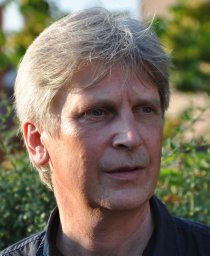 Martin Bastmeyer合作论文数Faculty of Biology;University of Konstanz;Max-Planck-Znstitut fur Entwicklungsbiologie;Faculty of Biology, University of Konstanz3
 Jerome Fourre合作论文数2Barrett Downing合作论文数2Oisín King合作论文数2Kleopatra Rapti合作论文数2M Hecker合作论文数2
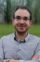 Enrico Lemma合作论文数Karlsruhe Inst Technol KIT, Zool Inst2
 Brian Wang合作论文数StemCyte International Cord Blood Center2
 Anna Randi合作论文数National Heart & Lung Institute, Imperial College London;Faculty of Medicine, Imperial College London2
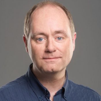 Dirk Grimm合作论文数Grimm-labs, University of Heidelberg;Heidelberg University Hospital2Julia Pfeiffer合作论文数1Marianne Lunde合作论文数1Antoine A F de Vries合作论文数1
 Danielle Regev合作论文数1Warrapong Kit-Anan合作论文数1Anna Bergan-Dahl合作论文数1Laura Nicastro合作论文数1Christine Mages合作论文数1Ilona Sunyovszki合作论文数1
 Colleen Lopez合作论文数1
 Maria da Luz Sousa Fialho合作论文数1Sarah Bertels合作论文数1David B Lipsett合作论文数1CR CARLSON合作论文数1Alaa H. Sayed合作论文数1Sonia Iftekhar合作论文数1Mor Davidor合作论文数1Heike Gampp合作论文数1
 Rita Alonaizan合作论文数1
 Polina Baskin合作论文数Department of Physiology, Biophysics and Systems Biology, Rappaport Faculty of Medicine and Research Institute, Technion, Haifa, Israel.1Katharina Seidensaal合作论文数1
 Ujang Purnama合作论文数1
 Rabia Nazir合作论文数Interdisciplinary Res Ctr Biomed Mat IRCBM, COMSATS Univ Islamabad CUI1Worrapong Kit-Anan合作论文数Natl Heart & Lung Inst, Hammersmith Hosp1Daniel Hachim合作论文数Institute of Biomedical Engineering, Imperial College London1Matthew Kerr合作论文数1Magdalena Laugsch合作论文数Univ Cologne, Ctr Mol Med Cologne CMMC, Cologne, Germany1
 Xin Shen合作论文数K.G. Jebsen Centre for Cardiac Research, University of Oslo1
 Daniela Cruz-Moreira合作论文数ICVS/3B's, PT Government Associate Laboratory1
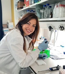 Fabiana Martino合作论文数St Annes Univ Hosp, Int Clin Res Ctr ICRC, CZ-65691 Brno, Czech Republic1
 Norbert Frey合作论文数Univ Med Ctr Schleswig Holstein, Kiel, Germany1
 Costanza Emanueli合作论文数Cardiac Function, National Heart and Lung Institute, Faculty of Medicine, Imperial College London1
 Arne Bruyneel合作论文数Department of Medicine, Stanford School of Medicine1
 Daniël A. Pijnappels合作论文数Department of Cardiology, Leiden University Medical Center1Wasserstrom J Andrew合作论文数1
 Jens Preben Morth合作论文数PUMPKIN Res Ctr, Aarhus Univ1Kholia Sharad合作论文数1




