Epilepsy is a common neurological disorder worldwide. Hydrogen sulfide (H2S) has been found to have anti-seizure effects. However, its mechanism remains to be explored. In the present study, we showed that a novel H2S donor attenuated neuroinflammation by up-regulating ATP-sensitive potassium channel (KATP) expression to reduce seizures. The novel H2S donor significantly reduced the expression of TNF-α and increased the expression of IL-10 in LPS-treated BV2 cells and the hippocampus of pilocarpine-induced epileptic mice. The modulatory effects of the H2S donor on inflammatory cytokines were prevented by glibenclamide, a common KATP channels blocker. The H2S donor promoted the expression of KATP channel subunits SUR2 and Kir6.1 in LPS-treated BV2 cells and the hippocampus of pilocarpine-induced epileptic mice. In addition, the H2S donor reduced the electroencephalography amplitude of hippocampal epileptic waves and reduced seizures in pilocarpine-induced epileptic mice, which were also attenuated by glibenclamide. These results indicated that the novel H2S donor reduced seizures and regulated microglial inflammatory cytokines by activating KATP channels, which may provide a prospective therapeutic strategy for the anti-seizure effects of H2S donor.
Although epilepsy is one of the most common neurologic disorders, there is still a lack of effective therapeutic drugs for it. Recently, we synthesized a novel hydrogen sulfide (H2S) donor, which is found to reduce seizures in animal models effectively. But it remains to be determined for its mechanism. In the present study, we found that the novel H2S donor could reduce pilocarpine-induced seizures in mice. It alleviated the epileptic behavior, the hippocampal electroencephalography (EEG) activity of seizures, and the damage of hippocampal neurons in status epilepticus mice. In addition, the novel H2S donor could reduce microglial inflammatory response. It not only reduced the upregulation of pro-inflammatory markers [inducible nitric oxide synthase (iNOS) and cyclooxygenase 2 (COX2)] in status epilepticus mice, but also increased the levels of microglial anti-inflammatory marker arginase-1 (Arg-1). In lipopolysaccharide-treated microglia BV2 cells, administration of the H2S donor also significantly reduced the lipopolysaccharide-induced upregulation of the expression of the pro-inflammatory markers and increased the expression of the anti-inflammatory markers. Thus, the novel H2S donor regulates microglial inflammatory profile in status epilepticus mice and in vitro. These results suggested that the novel H2S donor can reduce seizures and regulate microglial inflammatory profile, which may be a novel mechanism and potential therapeutic strategy of the H2S donor anti-seizures.
Disrupted follicular development may result in increased follicular atresia, which is a crucial mechanism of various ovarian pathologies. It has been demonstrated that oxidative stress is associated with disrupted follicular development. Catalpol is a natural compound that has been found to possess antioxidative stress. However, the effects of catalpol on oxidative stress-induced disrupted follicular development remain unclear. In the present study, we evaluated the protective effect of catalpol on hydrogen peroxide (H2O2)-induced oxidative damage in granulosa cells (GCs), which play crucial roles in the follicular development. Our results showed that catalpol significantly improved cell viability, reduced reactive oxygen species (ROS) and malondialdehyde (MDA) production, and elevated superoxide dismutase (SOD) and glutathione peroxidase (GSH-Px) activities in H2O2-induced GCs. Catalpol treatment caused significant increase in bcl-2 expression, and decreases in bax and caspase-9 expressions. Compared with the H2O2-induced GCs, caspase-3 activity in catalpol-treated cells was markedly decreased. Furthermore, catalpol caused significant activation of PI3K/Akt/mTOR pathway in GCs in response to H2O2 stimulation. Additionally, inhibition of this pathway reversed the inhibitory effects of catalpol on H2O2-induced oxidative injury and apoptosis in GCs. In conclusion, these findings suggested that catalpol protected GCs from H2O2-induced oxidative injury and apoptosis via activating PI3K/Akt/mTOR signaling pathway. Thus, catalpol might serve as a therapeutic approach for regulating disrupted follicular development.
Despite converging epidemiological evidence for the inverse relationship of regular caffeine consumption and risk of developing Parkinson's disease (PD) with animal studies demonstrating protective effect of caffeine in various neurotoxin models of PD, whether caffeine can protect against mutant α-synuclein (α-Syn) A53T-induced neurotoxicity in intact animals has not been examined. Here, we determined the effect of chronic caffeine treatment using the α-Syn fibril model of PD by intra-striatal injection of preformed A53T α-Syn fibrils. We demonstrated that chronic caffeine treatment blunted a cascade of pathological events leading to α-synucleinopathy, including pSer129α-Syn-rich aggregates, apoptotic neuronal cell death, microglia, and astroglia reactivation. Importantly, chronic caffeine treatment did not affect autophagy processes in the normal striatum, but selectively reversed α-Syn-induced defects in macroautophagy (by enhancing microtubule-associated protein 1 light chain 3, and reducing the receptor protein sequestosome 1, SQSTM1/p62) and chaperone-mediated autophagy (CMA, by enhancing LAMP2A). These findings support that caffeine-a strongly protective environment factor as suggested by epidemiological evidence-may represent a novel pharmacological therapy for PD by targeting autophagy pathway.
BACKGROUND: Working memory (WM) taps into multiple executive processes including encoding, maintenance, and retrieval of information, but the molecular and circuit modulation of these WM processes remains undefined due to the lack of methods to control G protein-coupled receptor signaling with temporal resolution of seconds. METHODS: By coupling optogenetic control of the adenosine A(2A) receptor (A(2A)R) signaling, the Cre-loxP-mediated focal A(2A)R knockdown with a delayed non-match-to-place (DNMTP) task, we investigated the effect of optogenetic activation and focal knockdown of A(2A)Rs in the dorsomedial striatum (n = 8 to 14 per group) and medial prefrontal cortex (n = 16 to 22 per group) on distinct executive processes of spatial WM. We also evaluated the therapeutic effect of the A(2A)R antagonist KW6002 on delayed match-to-sample/place tasks in 6 normal and 6 MPTP-treated cynomolgus monkeys. RESULTS: Optogenetic activation of striatopallidal A(2A)Rs in the dorsomedial striatum selectively at the delay and choice (not sample) phases impaired DNMTP performance. Optogenetic activation of A(2A)Rs in the medial prefrontal cortex selectively at the delay (not sample or choice) phase improved DNMTP performance. The corticostriatal A(2A)R control of spatial WM was specific for a novel but not well-trained DNMTP task. Focal dorsomedial striatum A(2A)R knockdown or KW6002 improved DNMTP performance in mice. Last, KW6002 improved spatial WM in delayed match-to-sample and delayed match-to-place tasks of normal and dopamine-depleted cynomolgus monkeys. CONCLUSIONS: The A(2A)Rs in striatopallidal and medial prefrontal cortex neurons exert distinctive control of WM maintenance and retrieval to achieve cognitive stability and flexibility. The procognitive effect of KW6002 in nonhuman primates provides the preclinical data to translate A(2A)R antagonists for improving cognitive impairments in Parkinson's disease.
 Qiyun Kang合作论文数2
 Ren Xiangpeng合作论文数2Zhongrui Liu合作论文数2Ziting Zhu合作论文数2
 Xingjun Chen合作论文数2Yan He合作论文数2Zheng Wu合作论文数2Wei Guo合作论文数2
 Jiangfan Chen (陈江帆)合作论文数Eye Hospital, Wenzhou Medical University;Boston University School of Medicine2
 Piu Chan合作论文数1Fenfen Ye合作论文数1Zhenhai Zeng合作论文数1Yuwan Lin合作论文数1Zhidong HOU (侯志东)合作论文数1Keming Wu合作论文数1Wenlong Zhang合作论文数1Yanan Luan合作论文数1Yuehua He合作论文数1
 Mingshu Mo合作论文数1Bao-Yi Huang合作论文数1Jin Xue合作论文数1
 Zhang Liping合作论文数1Ying Gao合作论文数1DiSi Deng (邓蒂斯)合作论文数1
 Shuisheng Lei (雷水生)合作论文数The Fifth Affiliated Hospital of Guangzhou Medical University1
 Xiaoqin Zhu (朱晓琴)合作论文数School of Basic Medical Sciences, Guangzhou Medical University1
 Yeke Wu (吴也可)合作论文数Hospital of Chengdu University of Traditional Chinese Medicine1
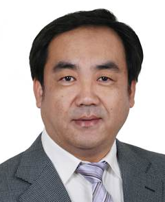 Feng Yue (岳峰)合作论文数School of Biomedical Engineering, Hainan University1
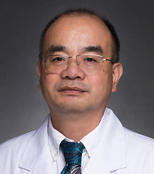 Pingyi Xu (徐评议)合作论文数The First Affiliated Hospital of Guangzhou Medical University;Guangzhou Medical University1H. E. Yan合作论文数1




