Tissue regeneration is not simply a local repair event occurring in isolation from the distant, uninjured parts of the body. Rather, evidence indicates that regeneration is a whole-animal process involving coordinated interactions between different organ systems. Here, we review recent studies that reveal how remote uninjured tissues and organ systems respond to and engage in regeneration. We also discuss the need for toolkits and technological advancements to uncover and dissect organ communication during regeneration.
Mammals have limited capacity for heart regeneration, whereas zebrafish have extraordinary regeneration abilities. During zebrafish heart regeneration, endothelial cells promote cardiomyocyte cell cycle reentry and myocardial repair, but the mechanisms responsible for promoting an injury microenvironment conducive to regeneration remain incompletely defined. Here, we identify the matrix metalloproteinase Mmp14b as an essential regulator of heart regeneration. We identify a TEAD-dependent mmp14b endothelial enhancer induced by heart injury in zebrafish and mice, and we show that the enhancer is required for regeneration, supporting a role for Hippo signaling upstream of mmp14b. Last, we show that MMP-14 function in mice is important for the accumulation of Agrin, an essential regulator of neonatal mouse heart regeneration. These findings reveal mechanisms for extracellular matrix remodeling that promote heart regeneration.
Aging research has shifted from studying phenotypes to studying in-depth mechanisms in recent decades. However, extrapolating cellular and molecular bases of aging from studying traditional model systems to humans has been challenging. The advent of organoids holds promise for overcoming the limitations of monolayer cell culture and bridging the gap between animal models and humans. Here, we mainly discuss recent paradigms for using organoid models in studying organ aging. Pluripotent stem cells–derived organoid provides a promising platform for simulating the pathophysiology of several aging-related diseases, especially neurodegenerative diseases, and adult stem cells organoids derived from different age groups have been applied to detect aging-related functional changes. We also assess the value of organoid model systems in understanding human aging and aging-related diseases, and identify challenges to be addressed in the future, such as the immaturity of organoids, and effective methods of inducing aging.
Regeneration requires the collective effort of multiple organ systems. A recent study of planarian whole-body regeneration finds that Erk kinase activity propagates rapidly across the entire animal through longitudinal muscle cells to coordinate animal-wide wound responses and that this signal propagation is required for regeneration.
Acute trauma stimulates local repair but can also affect structures distant from the injury, for instance through the activity of circulating factors. To study the responses of remote tissues to injury and regeneration, we generated genome-wide profiles from zebrafish brains following experimental cardiac damage. Our findings indicate regulatory mechanisms that distinguish short- and long-range gene expression responses to injury.
Acute trauma stimulates local repair mechanisms but can also impact structures distant from the injury, for example through the activity of circulating factors. To study the responses of remote tissues during tissue regeneration, we profiled transcriptomes of zebrafish brains after experimental cardiac damage. We found that the transcription factor gene cebpd was upregulated remotely in brain ependymal cells as well as kidney tubular cells, in addition to its local induction in epicardial cells. cebpd mutations altered both local and distant cardiac injury responses, altering the cycling of epicardial cells as well as exchange between distant fluid compartments. Genome-wide profiling and transgenesis identified a hormone-responsive enhancer near cebpd that exists in a permissive state, enabling rapid gene expression in heart, brain and kidney after cardiac injury. Deletion of this sequence selectively abolished cebpd induction in remote tissues and disrupted fluid regulation after injury, without affecting its local cardiac expression response. Our findings suggest a model to broaden gene function during regeneration in which enhancer regulatory elements define short- and long-range expression responses to injury.
Adult zebrafish possess an elevated cardiac regenerative capacity as compared with adult mammals. In the past two decades, zebrafish have provided a key model system for studying the cellular and molecular mechanisms of innate heart regeneration. The ease of genetic manipulation in zebrafish has enabled the establishment of a genetic ablation injury model in which over 60% of cardiomyocytes can be depleted, eliciting signs of heart failure. After this severe injury, adult zebrafish efficiently regenerate lost cardiomyocytes and reverse heart failure. In this chapter, we describe the methods for inducing genetic cardiomyocyte ablation in adult zebrafish, assessing cardiomyocyte proliferation, and histologically analyzing regeneration after injury.
A new study establishes genetic tools to ablate tendon progenitor cells in zebrafish larvae, finding that larval tendons display high regenerative capacity. The authors employ this musculoskeletal repair model to explore the source of tendon progenitors by fate mapping and live imaging, as well as underlying molecular stimuli like BMP signaling.
Chromatin regulation is a principal mechanism governing animal development, yet it is unclear to what extent structural changes in chromatin underlie tissue regeneration. Non-mammalian vertebrates such as zebrafish activate cardiomyocyte (CM) division after tissue damage to regenerate lost heart muscle. Here, we generated transgenic zebrafish expressing a biotinylatable H3.3 histone variant in CMs and derived cell-type-specific profiles of histone replacement. We identified an emerging program of putative enhancers that revise H3.3 occupancy during regeneration, overlaid upon a genome-wide reduction of H3.3 from promoters. In transgenic reporter lines, H3.3-enriched elements directed gene expression in subpopulations of CMs. Other elements increased H3.3 enrichment and displayed enhancer activity in settings of injury- and/or Neuregulin1-elicited CM proliferation. Dozens of consensus sequence motifs containing predicted transcription factor binding sites were enriched in genomic regions with regeneration-responsive H3.3 occupancy. Thus, cell-type-specific regulatory programs of tissue regeneration can be revealed by genome-wide H3.3 profiling.
Recently, quantum dots (QDs) have attracted widespread interest in biology and medicine. They are rapidly being used as new tools for both diagnostic and therapeutic purposes. Critical issues for further applications of QDs include the assessment of biocompatibility and biosafety of QDs. Most of previous researches concerning QD cytotoxicity focused on in vitro studies. In the present study, the impairments of acute exposure to well-modified and unmodified QDs (streptavidin-CdSe/ZnS and CdSe QDs, respectively) on synaptic transmission and plasticity were examined in adult rat hippocampal dentate gyrus (DG) area in vivo. The input/output (I/O) functions, paired-pulse ratio (PPR), field excitatory postsynaptic potential (fEPSP) and population spike (PS) amplitude were measured. The results showed that PPR and long-term potentiation (LTP) were all significantly decreased in these two types of QD-exposed rats compared to those in control rats. While the I/O functions and the amplitudes of fEPSP slope and PS amplitude of the baseline were significantly increased under QD exposure. These findings suggest that exposure to QDs, no matter whether they are well modified or not, could impair synaptic transmission and plasticity in the rat DG area in vivo and reveal the potential risks of QD applications in biology and medicine, especially in the toxin-susceptible central nervous system (CNS).
 Kenneth Daniel Poss合作论文数Department of Cell Biology, School of Medicine, Duke University;Department of Medicine, School of Medicine, Duke University;Department of Cell and Regenerative Biology, School of Medicine and Public Health, University of Wisconsin–Madison7
 Adam Shoffner合作论文数2Alexias Safi合作论文数1Roland Wu合作论文数1Nutishia Lee合作论文数1Zhifeng Li合作论文数1Bryan H. Lau合作论文数1Matthew J. Foglia合作论文数Department of Cell Biology, Duke University Medical Center1
 Ivana Zlatanova合作论文数Univ Paris 05, INSERM, UMRS 970, Sorbonne Paris Cite, F-75015 Paris, France1
 Xiao-Xin Chen合作论文数1Lingyun Song合作论文数Duke Univ, Inst Genome Sci & Policy, Durham, NC 27708 USA1Ziyi Wang合作论文数1Yu Luan合作论文数Department of Biochemistry and Molecular Genetics, Feinberg School of Medicine Northwestern University, Chicago, IL, USA.1
 Gregory E. Crawford合作论文数Department of Pediatrics, Duke University School of Medicine1
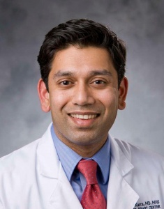 Ravi Karra合作论文数Department of Medicine, Duke University School of Medicine1
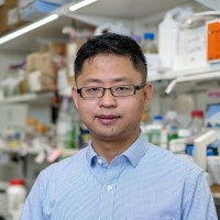 Hongbo Yang合作论文数Department of Biochemistry and Molecular Genetics, Feinberg School of Medicine, Northwestern University1
 Feng Yue合作论文数Department of Biochemistry and Molecular Genetics, Feinberg School of Medicine, Northwestern University1
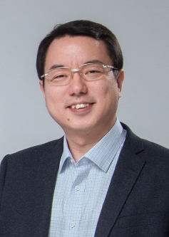 Wei Xiong (熊伟)合作论文数School of Life Sciences, Division of Life Sciences and Medicine, University of Science and Technology of China;Institute of Advanced Technology. University of Science and Technology of China;Key Laboratory of Brain Function Disease, Chinese Academy of Sciences1
 Jianhong Ou合作论文数1
 Zhe Yang合作论文数Dept Biochem & Mol Biol, Wayne State Univ1Kuzu Guray合作论文数Center for Computational Biology and Bioinformatics and College of Engineering, Koc University1
 Ramkumar Nitya合作论文数Sloan-Kettering Institute1Dickson Amy L合作论文数1Gemberling Matthew合作论文数1Black Brian L合作论文数1
 Jing Qu (曲静)合作论文数State Key Laboratory of Stem Cell and Reproductive Biology, Institute of Zoology, Chinese Academy of Sciences;Medical School, University of Chinese Academy of Sciences;University of Chinese Academy of Sciences1
 Cao Jingli合作论文数Department of Cell Biology and Howard Hughes Medical Institute, Duke University Medical Center1
 Celona Barbara合作论文数Cardiovascular Research Institute, University of California1
 Pauline Colombier合作论文数1
 De Simone Alessandro合作论文数School of Life Sciences, Swiss Federal Institute of Technology (EPFL) Lausanne1
 Bo Yang合作论文数1
 Joseph Goldman合作论文数1
 Sinha Tanvi合作论文数Department of Cell, Developmental and Integrative Biology, University of Alabama at Birmingham1
 Chunying Li合作论文数1
 Si Wang (王思)合作论文数Xuanwu Hospital, Capital Medical University1
 Guanghui Liu (刘光慧)合作论文数University of Chinese Academy of Sciences;State Key Laboratory of Membrane Biology, Institute of Zoology, Chinese Academy of Sciences1
 Liang Chen (陈亮)合作论文数Department of Life Sciences and Medicine, University of Science and Technology of China1
 Mingliang Tang (唐明亮)合作论文数School of Life Science and Technology, Southeast University1
 Ming Wang (汪铭)合作论文数School of Life Sciences, University of Science and Technology of China Life Sciences and Medicine1
 Di-Yun Ruan (阮迪云)合作论文数University of Science and Technology of China1
 Weiqi Zhang (张维绮)合作论文数Beijing Institute of Genomics, Chinese Academy of Sciences1
 Michael Tolstorukov合作论文数Dana-Farber Cancer Institute1Xiaoyan Sun (孙晓艳)合作论文数Chinese PLA General Hospital1Shan-Mei Xu合作论文数1
 Guo N. Huang合作论文数1Tairan Xing合作论文数1
 Stefan C. Materna合作论文数Division of Biology, California Institute of Technology1




