Aim There are currently no high-quality studies comparing the static navigation technique with conventional methods of fibre post removal. The aim of this ex vivo study was to compare the effectiveness of fibre post removal between a static navigation technique and a conventional freehand technique using ultrasonics by experienced and inexperienced operators.Methodology Forty-eight extracted single-rooted human premolars were root-filled. A fibre post was cemented in all 48 teeth, which were then divided randomly into the following groups: static navigation group using burs; static navigation-ultrasonic group; and non-guided group using ultrasonic tips. The following parameters were evaluated for both experienced operators and inexperienced operators: reaching the gutta-percha root filling successfully, the time required to remove the entire post, the occurrence of lateral root perforations, and the amount of root dentine removed. The Kolmogorov-Smirnov test was used to examine the normality of the data; the anova test was used to compare the significant differences among groups; and Tukey tests were used for all two-by-two comparisons. The significance level was set at 0.05.Results In the static navigation group, the gutta-percha was reached significantly more frequently than in the non-guided group (p < .05). The static navigation approach required significantly less time than the non-guided approach to reach the gutta-percha (p < .05). The total removal of posts was significantly different between groups (p < .05), but there was no significant difference between experienced and inexperienced operators in the static navigation group (p > .05). More perforations were associated with the non-guided group than with the other two groups. The total mean loss of dentine in the non-guided group in all directions was 0.39 (+/- 0.17) mm, with 0.25 (+/- 0.09) mm for experienced, and 0.42 (+/- 0.16) mm for inexperienced operators.Conclusion When compared to a conventional ultrasonic technique for the removal of fibre posts, the static navigation method using burs resulted in less dentine removal, more rapid access to the gutta-percha root filling, less overall time to remove the posts, and fewer complications. When using static navigation, there was no difference in performance between experienced and inexperienced operators.
Background Regenerative endodontics has evolved in recent years with tissue engineering concepts in particular appearing promising. Endodontic tissue engineering (ETE) describes the various approaches based on the orthograde introduction of scaffolds or biomaterials (with or without cells) into the root canal to achieve pulp tissue regeneration. There are currently no systematic reviews investigating whether ETE is a suitable method for the treatment of endodontic disease in both mature and immature permanent teeth. Objectives The purpose of this systematic review was to determine the effectiveness of ETE in permanent teeth with pulp necrosis in comparison with conventional endodontic treatment. Methods We searched MEDLINE, Embase and the Cochrane Library for published reports as well as Google Scholar for grey literature up to November 2021. Included were studies of patients with permanent immature or mature teeth and pulp necrosis with or without signs of apical periodontitis (P) comparing ETE (I) with calcium hydroxide apexification, apical plug and root canal treatment (C) in terms of tooth survival, pain, tenderness, swelling, need for medication (analgesics and antibiotics), radiographic evidence of reduction in apical lesion size, radiographic evidence of normal periodontal ligament space, function (fracture and restoration longevity), the need for further intervention, adverse effects (including exacerbation, restoration integrity, allergy and discolouration), oral health-related quality of life (OHRQoL), presence of sinus tract and response to sensibility testing (O). An observation period of at least 12 months was mandatory (T) and the number of patients in human experimental studies or longitudinal observational studies had to be at least 20 (10 in each arm) at the end (S). Risk of bias was appraised using the Cochrane risk-of-bias (RoB 2) tool. Two authors independently screened the records, assessed full texts for eligibility and evaluated risk of bias. Heterogeneity of outcomes and limited body of evidence did not allow for meta-analysis. Results Two randomized clinical trials investigating cell transplantation approaches with a total of 76 participants (40 treated immature teeth and 36 treated mature teeth) were included for qualitative analysis. Both studies had moderate concerns in terms of risk of bias. Due to the lack of homogeneity a meta-analysis was not possible. Tooth survival for ETE, root canal treatment and apexification was 100% after 12 months. Teeth treated with ETE showed a higher number of cases with positive pulpal responses to sensitivity tests and with blood perfusion compared with root canal treatment or apexification. Discussion This systematic review highlights that there is limited evidence for ETE approaches. Even though the results of this review suggest a high survival with ETE in mature and immature teeth, there is a moderate risk of bias due to methodological limitations in the included studies, so the overall results should be interpreted with caution. Lack of a robust control group was a common problem during literature screening, and outcomes besides dental survival were reported inconsistently. Future clinical trials need to address methodical as well as assessment concerns and report long-term results. Conclusion The benefits and high survival rates reported for ETE techniques suggest that this procedure might be an alternative to conventional procedures for permanent teeth with pulpal necrosis. However, more appropriate studies are needed to derive clinical recommendations. Registration PROSPERO (CRD42021266350).
BACKGROUND:Autotransplantation is a highly successful technique to replace ankylosed teeth. The authors propose a modified therapeutic approach to guide the autotransplant of an immature maxillary premolar using a tooth-shaped osteotome.CASE DESCRIPTION:A 9-year-old boy reported an avulsion of his maxillary permanent left central incisor with a delayed replantation. An autotransplant of the immature maxillary right first premolar into the position of the affected tooth was planned. A surgical 3-dimensional guiding template and a tooth-shaped osteotome were manufactured to prepare the neo-alveolus (referring to the creation or modification of an alveolus to house the tooth) modification. Although the donor tooth was placed in the recipient socket with the buccal side of the root fully exposed, the transplant outcome was successful.PRACTICAL IMPLICATIONS:The use of 3-dimensionally designed surgical osteotome could improve accuracy and surgical handling of a donor tooth autotransplant, even with substantial bone defects in the recipient site.
Introduction: This research studies and compares the shaping ability of WaveOne Gold (WG; Dentsply Tulsa Dental Specialties, Tulsa, OK), the Reciproc Blue (RB; VDW, Munich, Germany), TRUShape (TS, Dentsply Tulsa Dental Specialties), XP-endo Shaper (XP; FKG, La Chaux-de-Fonds, Switzerland), iRace (IR, FKG), and TruNatomy (TN; Dentsply Sirona, Ballaigues, Switzerland) in the preparation of moderately curved canals and using micro- computed tomographic technology. Methods: Sixty lower molars with 2 mesial canals were randomly distributed into 6 groups of 10 molars and 20 canals per group (n 5 20). Specimens were scanned before and after preparation using the SkyScan 1275 (Bruker microCT, Kontich, Belgium). Group 1 was treated with WG, group 2 with RB, group 3 with TS, group 4 with XP, group 5 with IR, and group 6 with TN. After instrumentation, researchers quantified the changes produced in the canal geometry in terms of surface, volume, structure thickness, surface convexity index, structure model index, percentage of surface touched, and centroids. Wilcoxon and analysis of variance tests were performed to compare the values before and after preparation and the differences between groups. The significance level was established at 5%. Results: There were no significant differences between WG and RB (P > .05) and between TN and XP (P > .05). TN had significant differences with WG, RB, TS, and IR (P , .05). All the files produced similar apical transportation (P > .05). Conclusions: WG and RB and TN and XP had similar shaping effectivity. TS and WG touched the highest percentages of canal surfaces (81% and 73%, respectively) but produced the biggest changes in the canal anatomy. TN and XP better kept the canal anatomy, but TN touched the lowest percentage of canal surface (50%). All the files used were able to clean and to shape moderately curved canals with minimal apical transportation. (J Endod 2021;47:812-819.)
Introduction: The aim of this study was to evaluate and compare the shaping ability of XP-endo Shaper (XP; FKG Dentaire, La Chaux-de-Fonds, Switzerland) and TRUShape (TS; Dentsply/Tulsa Dental Specialties, Tulsa, OK) during the preparation of moderated curved root canals using micro-computed tomographic imaging. Methods: Twenty human maxillary premolars with 2 roots were randomly separated into 2 groups of 10 teeth, which were scanned before and after root canal preparation using the SkyScan 1275 X-ray microtomograph (Bruker micro-CT, Kontich, Belgium) at a nominal resolution of <4 mu m. Premolars and irrigant were maintained at 37 degrees before and during preparation; group 1 was treated using XP and group 2 with TS. After preparation, researchers measured the amount of dentin removed, untreated superficies of canal walls, root canal volume, degree of canal transportation, and centering ability. Values of central tendency and dispersion were calculated using Statgraphics Centurion XV software (StatPoint Technologies, Inc, Warrenton, VA); means and median were compared using the Mann-Whitney-Wilcoxon test. The level of significance was set at 5% (P < .05). Results: No significant statistical differences were observed between the 2 groups in shaping ability, untreated superficies of canal walls, degree of canal transportation, and centering ability (P > .05). Conclusions: Instrumentation of moderately curved root canals using the XP single file and the TS file system were equally effective. XP and TS maintained the original anatomy of the root canals and showed a similar percentage of untreated canal walls, centered ability, and minimal apical transportation.
Aim: The purpose of this study was to determine and evaluate the apical preparation size resulting from different pecking times to the working length (WL) with five different file systems. Materials and methods: Fifty standard simulated endodontic J-shaped blocks were instrumented using ProTaper NEXT (PTN), WaveOne (WO), WaveOne Gold (WOG), OneShape (OS) and the Self- Adjusting File (SAF) (n = 10) with different pecking times (1, 2 and 4) to the WL. For the SAF group, instrumentation was done till WL according to the time, i.e. 1, 3 and 4 min. On completion of each stage, silicone impression material was used to take canal impressions for comparison and evaluation of the apical size preparation, using a stereomicroscope. Two-way analysis of variance was applied to determine differences between groups and pecking times. Results: After four pecking times, a significant increase was observed in the apical diameter of four test groups compared to SAF (P < 0.05), which was not associated with increased apical preparation at all times. Conclusion: A greater apical enlargement occurs with increasing pecking times; however, SAF instrumentation exhibits the minimum changes in the apical preparation after 1, 3 and 4 min. WO, WOG and OS are able to prepare the apical size similar to their tip at a single peck to the WL.
 Francesc Abella合作论文数4Olivieri Juan Gonzalo合作论文数4Maria de las Nieves Perez Morales合作论文数2
 Jose Antonio Gonzalez合作论文数2
 Jaramillo David E合作论文数2Alvaro Zubizarreta-Macho合作论文数1Kjell Laperre合作论文数1
 Gonzalo Gomez Val合作论文数1
 Firas Elmsmari合作论文数Evidence Based Practice Unit, Ajman Univ1
 Tousif Iqbal Nathani合作论文数Dent Fac, Univ Int Catalunya1Helge Knuettel合作论文数1
 Luca Boschini合作论文数Ambulatorio Polispecialist Dottor Boschini Srl1
 Matthias Widbiller合作论文数Department of Conservative Dentistry and Periodontology, University Hospital Regensburg1
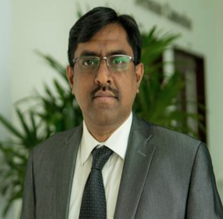 Venkateshbabu Nagendrababu合作论文数Dept Conservat Dent & Endodont, Sri Ramachandra Univ1
 Miguel Roig-Cayon合作论文数1
 Nastaran Meschi合作论文数2 Department of Oral Health Sciences, University Hospitals Leuven1
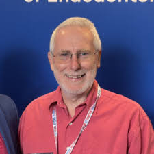 Paul M. H. Dummer合作论文数Cardiff University1
 Phil L Salmon合作论文数1




