BackgroundFatal inflicted head injury in infants requires systematic postmortem evaluation of intracranial, ocular, and cervicomedullary structures. This study summarizes autopsy findings in homicidal blunt head trauma in infants under one year of age and describes an en bloc cervical spine dissection approach intended to improve assessment of the cranio cervical region.MethodsA retrospective review was performed at the Cook County Medical Examiner's Office, Chicago, Illinois, USA, covering 2015 to 2023. Included cases were infants under one year of age with homicide certification and blunt force head injury. Autopsy reports, radiology, neuropathology, and ophthalmology were reviewed, with investigative and child welfare records when available. Injuries were mainly categorized across cranial, spinal, and ocular.ResultsSixty one infant homicides were identified; thirty nine met inclusion criteria. Acute subdural hemorrhage and subarachnoid hemorrhage were each present in 35 of 39 cases (89.7%). Retinal hemorrhages occurred in 34 of 39 (87.2%), with bilateral involvement in 31 (79.5%). Perineural optic nerve subdural hemorrhage was present in 33 (84.6%), bilateral in 32 (82.1%). Skull fractures were identified in 18 (46.2%). Cervical spinal cord injury was documented in 16 (41.1%), most commonly hemorrhagic. Remote extracranial fractures were present in 26 (66.7%), and recent extracranial fractures in 16 (41.1%).ConclusionIntracranial and ocular hemorrhages were frequent in this large medical examiner series, and cervical spinal cord injury was common. The findings support targeted evaluation of the cervical spine in selected infant homicide cases, with en bloc techniques as a useful option when resources and expertise permit.
Personal identification techniques rely on comparing ante-mortem and post-mortem evidence deemed unique to an individual. The Interpol standard of identification establishes fingerprints, DNA, and dental records as primary identification methods. Beyond dentition, odontological analysis can utilize palatal rugae, which are unique anatomical features with high potential for personal identification. However, most forensic approaches to palatal rugae rely on qualitative assessment; a quantitative evaluation of their uniqueness has been largely overlooked. This pilot study preliminarily explores the reliability of a new semi-quantitative method for palatal rugae analysis in forensic identification. Nineteen subjects were recruited, and a dental cast along with a palatal photograph were obtained for each participant. Dental cast images and palatal photographs simulated antemortem and postmortem data, respectively. Six distinct landmarks were identified in both record types, and homologous and heterologous comparisons were performed using image-processing software. Statistically significant differences were evaluated using the Mann-Whitney U test. The results demonstrated statistically significant differences between homologous and heterologous comparisons (p < 0.05) across most software-acquired parameters. These preliminary findings suggest that this new semi-quantitative method has the potential to effectively distinguish matches from mismatches, highlighting its potential utility in personal identification contexts. This pilot study introduces a new semi-quantitative method of palatal rugae analysis. Six different landmarks were identified, enabling homologous and heterologous comparisons via image processing software. This method demonstrates statistically significant differences between homologous and heterologous comparisons across most software parameters. This protocol offers a reliable, alternative approach to personal identification utilizing the palatal rugae.
Hyperactive Delirium with Severe Agitation (HDSA) is characterized by disorientation and aggressive behavior, constituting an acute, life-threatening medical emergency that requires prompt intervention. It is frequently associated with substance use. In prehospital settings, management often involves physical restraint, force, or pharmacological sedation; however, the diagnostic criteria and medico-legal implications of HDSA remain insufficiently defined. We retrospectively analyzed seven fatal cases exhibiting HDSA presentations, as defined by the American College of Emergency Physicians (ACEP), from the archives of two university-based forensic pathology centers. A detailed multidisciplinary review by forensic pathologists and forensic psychiatrists showed that six of seven decedents were males aged 25 to 36 years who experienced sudden cardiorespiratory arrest during episodes of severe agitation. Four deaths were preceded by law enforcement involvement, and five occurred immediately following attempts at manual restraint, implicating restraint-associated asphyxia as a potentially significant factor in mortality. Toxicological analyses detected psychostimulant exposure in five cases. This study represents, to our knowledge, one of the first to employ the updated HDSA terminology to describe fatal cases within a medico-legal forensic psychiatric context.
Background/Objectives: Personal identification is a cornerstone of Forensic Medicine. Historical records indicated that 27 Italian soldiers were executed by Tito’s army in April 1945 and buried in the mass grave of Ossero (Cres Island, Croatia). Methods: The remains, exhumed in 2019 by Croatian authorities and transferred to the Sacrario d’Oltremare (Bari, Italy), underwent radiographic and anthropological analyses. Genetic analysis was later performed on 147 bone samples, leading to the identification of 10 soldiers. Results: Anthropological analyses revealed commingled remains of at least 34 individuals (15–45 years; 161–181 cm), including eight skeletal elements of female sex. Forensic examination confirmed cranial and post-cranial gunshot wounds. Conclusions: Extensive commingling and fragmentation of the remains prevented full reconstruction of individual skeletons, yet comparison with ante-mortem data supported their identification as the aforementioned soldiers. Unexpected findings, including historically undocumented females, were confirmed by DNA analysis. These findings underscore the value of a multidisciplinary approach to optimize recovery and subsequent forensic and genetic investigations.
Bitemark analysis has long been an important field within of forensic odontology, yet it remains controversial due to its inherent subjectivity and the limitations of currently adopted 2D methodologies. The National Institute of Standards and Technology has highlighted the lack of sufficient scientific foundation for conventional bitemark analysis, particularly with respect to bitemarks impressed on human skin, concerning uniqueness, reproducibility, and evidentiary reliability. Rather than addressing the limitations identified for human skin bitemark analysis, this study proposes a three-dimensional quantitative protocol specifically designed for the analysis of bitemarks impressed on a standardized food substrate. Bitemarks were imprinted on standardized Italian cheese (scamorza) by eight adult volunteers. Dental casts and corresponding food items were digitized using a REVOPOINT Blue Light Mini 3D scanner. Digital models were processed with GOM Inspect and analyzed through: surface superimposition with colorimetric deviation mapping, extraction of 2D profiles from 3D data, and computation of three quantitative indicators: three-point angle, distance-perpendicular ratio, and elliptical ratio. A probabilistic model evaluated the likelihood of a match between each bitemark and dental arch. The 3D workflow minimized evidence handling and preserved bite pattern integrity. Colorimetric maps highlighted deviations within 5 mm and 2 mm tolerance ranges, enabling screening and refined matching. Quantitative indicators showed good discriminatory power, with correct matches for all subjects except one. Substrate deformation cases emphasized integrating surface analysis with profile-based indicators to reduce geometric distortion. The combined use of 3D scanning and reverse engineering reduced angular distortion and information loss typical of 2D methods, yielding more consistent and reproducible results within the experimental conditions adopted in this study. This study demonstrates a standardized engineering-based workflow for the quantitative analysis of bitemarks impressed on a stable food substrate. The proposed protocol provides objective and reproducible quantitative parameters for this specific experimental scenario and should not be interpreted as validation or direct support for bitemark analysis performed on human skin, whose biomechanical characteristics and distortion mechanisms differ substantially from those of stable inanimate substrates.
Background/Objectives: Age estimation is of fundamental importance in forensic investigations. When traditional methods based on gross bone morphology or morphometric analysis cannot be applied, forensic experts must rely on multidisciplinary approaches. Histomorphometry has consistently proven to be reliable in cases of highly fragmented or incomplete skeletal remains, particularly in older individuals. Building on the foundational study of Amprino and Bairati, this study evaluated the correlations between bone microstructural features in femoral cross-sections and the age and sex of individuals. Methods: The sample comprised 95 femoral mid-diaphyseal thin sections obtained from autopsy specimens housed at the Institute of Legal Medicine, University of Bari (Italy), representing both male and female individuals aged 18 to 92 years. The numbers and densities of primary, intact secondary, and fragmentary secondary osteons, together with osteon circularity and the mean osteonal area, were measured to investigate age-related variation. Statistical analyses included t-tests, Mann–Whitney tests, Spearman’s rank correlation, and General Linear Models (GLMs). Results: No significant differences in histomorphometric variables were observed between males and females. However, the number of intact secondary osteons and osteon population density increased with age, while the mean osteonal area and osteon circularity decreased with age. Although some variables displayed significant correlations with age, residual analysis indicated a lack of heterogeneity in variance, which limited the development of a robust predictive model. Conclusions: The findings highlight both the potential and the limitations of histomorphometry in forensic age estimation. While certain microstructural variables correlate with age, inter-individual variability reduces predictive accuracy. Further research is needed to refine models that account for biological and biomechanical variability, particularly in older adults.
The rapid advancements in healthcare technologies are reshaping the medical landscape, prompting a reconsideration of clinical methodologies and their ethical foundations. This article explores the need for an updated approach to medical deontology, emphasizing the transition from traditional practices to a hybrid clinical methodology that integrates both human expertise and technological innovations. With the increasing use of Artificial Intelligence, data analytics, and advanced medical tools, healthcare professionals are presented with new ethical and professional challenges. These challenges demand a reevaluation of professional responsibility, highlighting the importance of scientific evidence in decision-making while mitigating the influence of economic and ideological factors. By framing medical practice within a systemic and integrated perspective, this article proposes a model that moves beyond the reductionist and anti-reductionist dualism, fostering a more realistic understanding of healthcare. This new paradigm necessitates the evolution of the Medical Code of Ethics, integrating the concept of “medical intelligence” to address the complexities of data management and its ethical implications. The article ultimately advocates for a dynamic and adaptive approach that aligns medical practice with emerging technologies, ensuring that patient care remains person-centered and ethically grounded in a rapidly changing healthcare environment.
Understanding the motives behind femicides is crucial to design effective prevention strategies and to support women's self-determination, free from threats to their mental and physical integrity. We conducted a retrospective, multicenter forensic study involving 27 Italian Institutes of Legal Medicine, analyzing 1238 female homicides (1950-2023). Cases were classified as femicide or non-femicide female homicide according to the medico-legal definition of femicide as the murder due to the failure to recognize women's right to self-determination. Motives were categorized into 12 groups, and relevant anamnestic and circumstantial data were collected. Of the 1238 cases, 410 were identified as femicides, 395 as non-femicides, and 433 were excluded for insufficient information. Femicides were most frequently driven by jealousy/rejection (n = 185; 45.1 %) and separation/divorce (n = 144; 35.1 %), often in the context of intimate partner violence or inability to accept the end of a relationship. Non-femicides were predominantly associated with the perpetrator's psychiatric disorder and/or drug addiction (n = 126; 31.9 %), violent aggression without gender-related motive (n = 69; 17.5 %), victim's illness (n = 63; 15.9 %), or economic reasons (n = 58; 14.7 %). In the Italian context, most femicides stem from relational dynamics - particularly jealousy, rejection, and separation - whereas non-femicides are more often linked to psychiatric illness, substance abuse, or non-gender-related aggression. Incorporating these findings into clinical risk assessment protocols, especially in emergency and primary care settings, may help identify women at high risk and guide targeted prevention strategies.
Background: Hospital autopsies, once considered fundamental to evidence-based medicine, have declined in many Western countries due to legislative changes, faith-based objections, and inadequate funding in public healthcare. However, the COVID-19 pandemic has revitalized interest in their importance for determining the cause of death and generating public health statistics. This study analyzes the factors contributing to this decline, with a focus on the Italian context and the new medicolegal perspectives arising in the post-pandemic era. Methods: The research was conducted through an analysis of 91 relevant scientific sources, including government documents, policy briefs, and academic studies. Legislative and regulatory factors influencing the reduction in hospital autopsies were examined, along with new technological and medicolegal perspectives that have emerged following the COVID-19 pandemic. Special attention was given to the Italian context and the pandemic’s impact on autopsy practices. Results: The analysis revealed that the perceived “low value” of autopsies often stems from technological advancements, which have led to the view of these procedures as redundant. However, in the post-COVID-19 era, there has been a renewed awareness of the medicolegal value of autopsies, with significant implications for public health statistics and the evolution of forensic medicine. Conclusions: The COVID-19 pandemic highlighted the vital role of autopsies in delivering accurate medical information, prompting renewed attention to these procedures in the post-pandemic era. Moving forward, hospital autopsies will need to balance technological innovations with traditional medicolegal practices, paving the way for new perspectives in forensic medicine and public health data management.
Femicides are grievous, but little is known about risk factors and preventive measures. We present the results of a study conducted across 27 Italian Forensic Institutes. We analyzed 1,238 cases of female homicide and, adopting the definition of femicide as the murder due to the failure to recognize women's right to self-determination, we identified 410 cases as femicides and 395 as non-femicides Current partners were identified as aggressors in a much larger proportion of femicide cases (241 femicides vs. 145 non-femicides; odds ratio (OR) of femicide 2.46, 95 % CI 1.85-3.27), such association being more pronounced for ex-partners (102 vs. 11; OR 11.56, 95 % CI 6.10-21.92). Sharp weapons showed a higher frequency in femicides (168 vs. 140; OR 1.26, 95 % CI 0.95-1.68). Femicides were more often associated with bodies found in vehicles (31 vs. 9; OR 3.50, 95 % CI 1.64-7.45) and outdoor (68 vs. 43; OR 1.62, 95 % CI 1.08-2.45). There was an indication of femicides being more frequently associated with overkilling (87/323 vs. 71/324; OR 1.24, 95 % CI 0.88-1.76) and even more with lesions located in erogenous zones (94/316 vs. 70/325; OR 1.38, 95 % CI: 0.97-1.95). However, the strongest (though statistically imprecise) association emerged for overkilling in erogenous zones (12/398 vs. 6/389; OR 1.95, 95 % CI: 0.72-5.25). The number of lesions showed a nonlinear association with femicide likelihood. These findings offer forensic indicators that could contribute in predicting and potentially prevent femicide occurrence in a Western population such as the Italian one.
The autopsy of the non-fresh cadaver is characterized by inherent difficulties in that it requires the adoption of genetic and histological investigation techniques to the specific case. The case that the authors present in this paper concerns the corpse of a young woman who had already been autopsied, then treated with 20 % neutral buffered formaldehyde, a preserving agent, to enable its intercontinental transport, and then autopsied again. At the judge's request, a second judicial autopsy was performed in order to verify the cause of death and to carry out genetic investigations. As fresh tissue was not available to perform the genetic analysis, three different tissue matrices were taken: retina, liver, and iliopsoas muscle. The biological matrices taken were chosen as they were considered less likely to be in contact with formaldehyde and therefore probably less prone to artifacts caused by the action of formalin on the DNA structure. All three biological matrices were used to obtain a genetic profile. In particular, the genetic data obtained from the retina was found to be better than the genetic profile obtained from the iliopsoas muscle. In turn, the genetic profile obtained from the iliopsoas muscle was found to be better than the genetic data obtained from the liver sample.
Background:Cocaine overdose is a condition in which an increase in blood pressure, heart rate and depth of breath is observed. Cocaine consumption also causes a wide and well-known neuropsychiatric symptomatology that is characterized by incomprehensible behavior, confused and disordered thoughts, and paranoia. Cocaine addiction is a worldwide public health problem, which has somatic, psychological, psychiatric, socio-economic, and judicial complications. Case series:This work shows three cases in which cocaine poisoning and overdose caused a psychotic and extremely violent behavior in the hours leading up to the death of the subjects. Conclusion:The aim of this brief case series is to suggest some diagnostic criteria for the correct definition of this psychosis in order to make an early diagnosis crucial to prevent deaths related to it.
Background: When the body temperature falls below 35 °C, a condition known as hypothermia occurs. This renders it impossible to regulate the body temperature and produce heat. In these cases, death is due to a cessation of vital functions and is triggered by a body temperature below 25 °C. A multidisciplinary approach is typically required when hypothermia is suspected to combine circumstantial evidence, external examination, autopsy, microscopic and biochemical findings, and other data useful to assess the diagnosis of fatal hypothermia. Post-mortem diagnosis of death by hypothermia frequently presents a medico-legal dilemma. Methods and Results: The authors present a case of hypothermia in which site inspection, autopsy, and histological examination simultaneously revealed the forensic and anatomopathological characteristic findings of hypothermia with some peculiar evidence: paradoxical undressing, thanatochronological phenomena, frost erythema, polyvisceral congestion and Wischnewski spots, and subnuclear vacuolization of renal tubular epithelial cells.
INTRODUCTION:In the dynamic universe of new psychoactive substances (NPS), the identification of multiple and chemically diverse compounds remains a challenge for forensic laboratories. Since hair analysis represents a gold-standard to assess the prevalence of NPS, which are commonly detected together with classical drugs of abuse (DoA), our study aimed at developing a wide-screen method to detect and quantify 127 NPS and 15 DoA on hair.MATERIALS AND METHODS:A multi-analyte ultra-high performance liquid chromatography mass spectrometry method for the identification and quantification of 127 NPS (phenethylamines, arylcyclohexylamines, synthetic opioids, tryptamines, synthetic cannabinoids, synthetic cathinones, designer benzodiazepines) and 15 DoA in hair samples was developed. A full validation was performed according to the European medicines Agency (EMA) guidelines, by assessing selectivity, linearity, accuracy, precision, limit of quantification (LOQ), limit of detection (LOD), matrix effect and recovery. As a proof of the applicability, the method was applied to 22 authentic hair samples collected for forensic purposes.RESULTS:Successful validation was achieved, by meeting the required technical parameters, for 137 compounds (122 NPS and 15 DoA), with LOQ set at 4 pg/mg for 129 compounds, at 10 pg/mg for 6 and at 40 pg/mg for 2. The method was not considered validated for 5 NPS, as LLOQ resulted too high for a forensic analysis (80 pg/mg). Among authentic forensic samples, 17 tested positive for DoA, and 10 to NPS, most samples showing positivity for both. Detected NPS were ketamine and norketamine, 5-MMPA, ritalinic acid, methoxyacetyl fentanyl, methylone and RCS-4.CONCLUSION:The present methodology represents an easy, low cost, wide-panel method for the quantification of 122 NPS and 15 DoA, for a total of 137 analytes, in hair samples. The method can be profitably applied by forensic laboratories. Similar multi-analyte methods on the hair matrix might be useful in the future to study the prevalence of NPS and the co-occurrence of NPS-DoA abuse.
Background:Calculating the immersion time at sea of human skeletal remains is a challenge for forensic investigations and the answer to this question would solve many court cases in the shortest possible time. Remains in water create profound structural changes due to countless variables that lead to difficulties in interpretation during investigations. Case report:In this paper, two forensic cases found at sea were analysed, Case A, an extensively skeletonised corpse, and Case B, an isolated, intact foot with soft tissue, both found only 9 km away from the shores of the Tyrrhenian Sea (Southern Italy). A preliminary radiodiagnostic examination was performed for both cases. Subsequently, macroscopic observation of two tattoos found on case A was carried out. Autopsy examination was then carried out with subsequent anthropometric analysis to reconstruct the biological profile of both subjects, (case A) and (case B). Finally, histological and genetic analyses were performed. Conclusions:The examinations carried out made it possible to determine two compatible biological profiles, despite the taphonomic difference, and these examinations were corroborated by the anthropometric and genetic correlation, which led to the solution of a single judicial case. In conclusion, all the investigations carried out made it possible to state that the skeletonised corpse (Case A) and the isolated foot (Case B) belonged to the same individual in life. This result made it possible to identify the victim, thus establishing the profile of an individual who had disappeared in the previous two months due to a shipwreck.
Background: HAIs (Healthcare-Acquired-Infections) have been recently the subject of judgment n. 6386 pronounced on 3rd March 2023 by the Italian Supreme Court. This sentence provided three criteria to determine whether a health facility is responsible for the patient contracting a nosocomial infection, i.e. time criterion, topographical criterion and clinical criterion. Accordingly, the healthcare facility is obliged to prove the fulfillment of a series of preventive hygiene measures specifically detailed by the legislator. Herein, the positive predictive value of these criteria ("juridic criteria") in the identification of professional liability for nosocomial infections was evaluated in comparison with clinical criteria reviewed by Infectious Disease specialists ("Infectious-Disease criteria", i.e. presence of a Multidrug Resistant Organism (MDRO); development of surgical site infection; inadequate antibiotic therapy; inadequate disinfection). Methods: Two retrospective cohorts were compared from the Portal of Telematic Services of the Ministry of Justice; 51 patients were extrapolated from Italian judgments concerning claims for Gram-negative nosocomial infections in the three-year period 2020-2022. On the other side, from the electronic database of University Hospital of Bari we extracted 349 patients affected by Gram-negative infections in the same timespan. Both "juridic" criteria and "Infectious-Disease" criteria were then applied to the full cohort after stratification for cohort of origin and after stratification for nosocomial or non-nosocomial infections. Predictive value of criteria was evaluated through receiver operating characteristic (ROC) curves and area under the curve (AUC). Results: Overall, the incidence of definite nosocomial infections (according to final judgement or clinical records discharge letter) was 84 % in juridic cohort and 46 % in "real-world" series. Data suggested that the presence of all three juridic criteria [ROC AUC = 0.944 (95%CI = 0.924-0.963)] or the four clinical criteria [ROC AUC = 0.948 (95%CI = 0.928-0.969)] predicted well a case of nosocomial infection with professional liability. Moreover, by summarizing both criteria in a single classification system, the generated ROC curve (was the one with the highest AUC [0.9488 (95%CI = 0.928-0.969)]. Accordingly, further tests were performed, evaluating the predictive value of one juridic criterium plus at one of more Infectious-Disease criteria. Interestingly, the ROCs curves demonstrated that the presence of at least 1 juridic criteria plus at least 2 Infectious Disease criteria reached a predictive value comparable to 2 or 3 juridic criteria. Conclusions: The results highlight the efficiency of new criteria laid down in the judgment of the Italian Supreme Court to attribute liability for nosocomial infection despite the disputed distance between juridic and scientific decision-making process. In addition, the use of a combined score combining "juridic" and "Infectious-Disease" criteria provides a high-quality tool to be used by technical consultants to make up for lack of clinical documentation by passing judgments concerning litigation about professional liability in case of nosocomial infections. This sheds light on the possibility to face worldwide judicial inquiries with scientific rigor.
Background:Establishing the cause of death when analysing burnt human remains is limiting due to thermal degradation. The heat generated by high combustion degrades the bone structure, definitively hiding the perimortem trauma in most cases, which is crucial for solving a court case. Case report:In November 2019, a completely burnt corpse was found inside a car set on fire near a location in Reggio Calabria, Italy. The corpse was subsequently subjected to an initial radiodiagnostic examination and an anthropological/medico-legal investigation, in order to confirm the biological profile of the unidentified subject, define the cause of death and assess the presence of perimortal lesions through macroscopic analysis of skull fragments subjected to fleshing. Conclusions:The soft tissue fleshing of the burnt skull fragments allowed the reconstruction of a partial calotte. Macroscopic analysis of the consolidated shell identified in the left fronto-parietal region a clear linear fracture, perimortal in nature, compatible with blunt trauma. Autopsy examination revealed the presence of carbonaceous residues within the larynx and especially the trachea, confirming ante-mortem combustion.The results of the autopsy examination and the anthropological analysis allowed us to state that the net linear fracture, perimortal in nature, caused the subject a complex encephalic trauma, resulting in loss of consciousness and subsequent death due to carbon monoxide inhalation. This result not only confirms the malicious hypothesis, but reveals a deliberate burning of the victim in order to conceal the evidence necessary to solve the forensic case.
The interpretation of tanatochronological data is a fundamental aspect of the medico legal diagnosis, because it allows to trace back the interval of death. Traditionally, the evaluation of the hypostasis plays a relevant role in the interpretation of such information, despite its well-known limits and fallacies. In order to evaluate the degree of hypostatic area discoloration, the methodology currently used is highly subjective and influenced by several variables. The hypostasis pattern in individuals with V-VI phototype is useless because their post-mortem lividity is not estimable due to the color of the skin. This makes much harder to estimate the interval between the death and the detection of the tanatochronological data. This study is aimed at defining a highly accurate procedure to develop an objective method to estimate the hypostasis’ degree of fixation with scientific accuracy on people with darker skin. The technology used is spectrophotometry Antera3D: this device is able to analyse the hypostasis by measuring the mean hemoglobin quantitative level in the skin either before and after a standardized compression, thus obtaining a numerical value that is directly related to the time of death. The method here presented allows analysing the hemoglobin amount in the skin of a dead body, without the influence of the melanin pigment in the definition of the hypostatic area color, therefore enabling us to overcome the objective limits of the direct and empiric estimation of the hypostasis decoloration. By creating a standardized method it’s possible to reduce the operator-dependent error and to introduce a valid and applicable procedure in order to estimate the post-mortem interval.
Background:Aspiration of food or liquids can result in suffocation, evolving in coughing, difficulty breathing and forced exhalation. Asphyxia occurs when the aspirated material occludes the upper airways, either in the proximal or distal tract, resulting in the inability to breathe. The risk of asphyxiation death, is increased if a person makes sudden movements while eating, walks or runs while eating, or even becomes distracted or frightened. It is higher in individuals with neurological diseases, intellectual disability (ID), cognitive impairment, psychiatric pathologies or their pharmacological treatments and people carrying additional physiological impairments, which can cause oral dysfunction and dysphagia. Protective mechanisms may sometimes lack or fail to expel food fragments stuck in the airways, which completely obstruct them. Case series:The authors present some peculiar cases of subjects who died from food bolus choking, namely a case of mozzarella-cheese clogging in a young subject (25 years old) undergoing rehabilitation treatment following a stroke; a 38-year-old man with middle-grade mental retardation died from first airway food bolus (mush of bread) clogging; a 26-year-old subject with epilepsy died from tripe clogging in the course of a seizure; a 38-year-old subject in psychiatric treatment for depressive disorder who died from clogging with octopus tentacles. Conclusion:Food bolus clogging asphyctic deaths generally occur in subjects with psychic/neurological pathologies, resulting in altered deglutition mechanisms or lack of protective reflexes. Foodstuff, especially if large or viscous, obstruct the proximal or distal airways, leading to acute respiratory failure and death. Autopsy is diriment in ascertain the cause of death.
 Antonio De Donno合作论文数39Valeria Santoro合作论文数31Sablone Sara合作论文数27
 Carlo P. Campobasso合作论文数Department of Health Sciences, University of Molise25
 Federica Mele合作论文数19
 Antonella De Donno合作论文数Faculty of Sciences, University of Salento17Enrica Macorano合作论文数13Mariagrazia Calvano合作论文数11V. Santoro合作论文数11Aldo Di Fazio合作论文数10
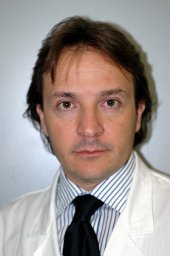 Giancarlo Di Vella合作论文数Department of Internal Medicine and Public Medicine, University of Bari10Alessia Leggio合作论文数9Mirko Leonardelli合作论文数9F Mele合作论文数7M Dragone合作论文数7Stefano Duma合作论文数7M Colonna合作论文数7Alessandro Dell'Erba合作论文数Dipartimento Interdisciplinare Med, Univ Bari Aldo Moro7Piercarlo Lozito合作论文数Department of Legal Medicine, University of Bari7
 Gerardo Cazzato合作论文数Department of Emergency and Organ Transpantations, University of Bari6Roca Roberta合作论文数6John E. Smialek合作论文数6E Macorano合作论文数5G De Gabriele合作论文数5A FRANCHINI合作论文数5Giovanni De Gabriele合作论文数5Matteo Favia合作论文数5R RIZZI合作论文数4D Rizzi合作论文数4Chiara Lauretti合作论文数4Carmelinda Angrisani合作论文数4Innamorato Liliana合作论文数4Lubelli Sergio合作论文数4Maricla Marrone合作论文数4Andrea Marzullo合作论文数Department of Pathological Anatomy and Genetics, University of Bari Policlinico4Giuseppe Strisciullo合作论文数3Bm Altamura合作论文数3Arianna Giorgetti合作论文数Unit Legal Med, Univ Bologna3Pesce Delfino V合作论文数3
 Fulvio Lavecchia合作论文数Dipartimento di Meccanica, Matematica e Management, Politecnico di Bari3
 Stefania Lonero Baldassarra合作论文数3
 Aldo Fiori合作论文数Dipartimento di Ingegneria Civile, Informatica e delle Tecnologie Aeronautiche, Università degli Studi Roma Tre3Fiandaca Chiara合作论文数3
 Ignazio Grattagliano合作论文数Department of Internal and Occupational Medicine (DIMIL),, University of Bari3Eligio Vacca合作论文数3
 Luigi Maria Galantucci合作论文数Dipartimento di Ingegneria Meccanica e Gestionale, Politecnico di Bari3
 Susi Pelotti合作论文数3P Tarzia合作论文数2C Basile合作论文数2




