Genome-wide association studies (GWAS) have identified >1,200 signals associated with type 2 diabetes (T2D), yet identifying functional variants remains challenging because the majority of them lie in noncoding regions of the genome and are in areas of high linkage disequilibrium (LD). While chromatin accessibility QTL (caQTL) and expression QTL (eQTL) analyses are useful for nominating regulatory mechanisms underlying GWAS signals, limitations still exist in pinpointing functional variants within regions of high LD. A complementary approach that has been less frequently applied is to focus on the allele-specific effect on chromatin accessibility at heterozygous single-nucleotide polymorphisms (SNPs), hereafter referred to as "allelic imbalance". We analyzed the allelic imbalance of reads generated from an assay for transposase-accessible chromatin with sequencing (ATAC-seq) across genotyped samples from 490 donors in T2D-relevant tissues: skeletal muscle, liver, pancreatic islets, adipose tissue, and relevant cell types. We identified 119,949 allelically imbalanced SNPs (FDR<0.05) across the genome. The allelic imbalance was often most prominent in one tissue and showed an enrichment overlapping with tissue-specific transcription factor (TF) binding footprints. Focusing on the 8,581 SNPs in previously published 99% credible sets from 338 T2D GWAS signals, we identified 256 imbalanced SNPs across 123 (36.4% of) signals, each showing allelic imbalance in at least one tissue or cell type. Of these, 71 signals contained only a single imbalanced SNP, representing excellent candidate causative variants. As a proof-of-concept, we showed that 23 of the 256 imbalanced SNPs were supported by allelic assays from previous studies. Further, we experimentally validated two imbalanced SNPs as likely functional variants: rs34584161 among a seven-SNP T2D credible set at the RNF6 signal in islets and rs849134 among a 13-SNP credible set at the JAZF1 signal in liver. This study demonstrates the power of integrating ATAC-seq allelic imbalance (ASAI) with GWAS statistical fine-mapping to identify candidate functional regulatory variants from among tightly linked GWAS variants in disease-relevant tissues. While applied here in T2D, this approach represents a widely applicable high-throughput framework for refining the genetic architecture of complex traits.
Type 2 diabetes (T2D) results from the interplay of genetic susceptibility and an unhealthy lifestyle, but their combined effects are not well studied. We examined whether unhealthy modifiable behaviors were associated with similar increases in the risk of incident T2D in individuals with different levels of genetic risk. Among 332,251 UK Biobank participants without diabetes, we constructed a multiancestry genetic risk score (GRS) based on 783 T2D-associated variants, categorized into tertiles. Lifestyle was classified as healthy, intermediate, or unhealthy based on baseline self-reported smoking status, BMI, physical activity level, and diet quality. Cox proportional hazards regression models were used to generate adjusted hazard ratios (HRs) for T2D and associated 95% CIs. During follow-up (median 13.6 years), 13,128 (4.0%) participants developed T2D. GRS (P < 0.001) and lifestyle classification (P < 0.001) were independently associated with increased risk of T2D. Compared with a healthy lifestyle, an unhealthy lifestyle was associated with increased risk in all genetic risk strata, with adjusted HRs ranging from 7.11 to 16.33. High genetic risk and an unhealthy lifestyle were the most significant contributors to T2D development. Individuals at all levels of genetic risk can substantially mitigate their T2D risk through lifestyle modifications. ARTICLE HIGHLIGHTS:Both genetic susceptibility and an unhealthy lifestyle are known to be associated with elevated type 2 diabetes (T2D) risk. However, their combined effects on T2D risk are not well studied. In this large prospective cohort study of more than 332,000 individuals, unhealthy lifestyle factors were associated with risk of incident T2D within and across different levels of genetic risk. These findings suggest individuals at all levels of genetic risk can greatly mitigate their risk of T2D by adhering to a healthy lifestyle.
Proteomics holds great promise for identifying potentially druggable effectors of common diseases, yet its application at population-scale across diverse ancestries, remains challenging. Here, we developed genetic imputation models for 2,594 plasma proteins using proteomic and genetic data from 54,219 UK Biobank participants, validating their performance across multiple ancestry groups and in an independent cohort. Plasma proteomes were then imputed for over 640,000 participants in the UK Biobank and the All of Us Research Program. To assess its aetiological value at population-scale, a further proteome-wide association study of cardiovascular diseases was performed across six genetic ancestries. We identified ∼9000 protein-disease associations across 89 cardiovascular conditions (PheCodes), the majority of which show consistent effects across ancestries and biobanks, with many comprising known targets of drugs either approved or under development. The associations reveal both shared and distinct proteomic signatures across cardiovascular conditions and defined clusters of distinct pathophysiology with shared underlying molecular pathways. Integration of data on tissue specificity and single-cell transcriptomics prioritised liver-derived proteins in circulation as candidate effectors of coronary artery disease, highlighting inter-alpha-trypsin inhibitor heavy chain H4 (ITIH4) as a putative effector. Using a liver-targeted CRISPR gene-editing platform, we show that in vivo disruption of ITIH4 reduces plasma cholesterol and pro-atherogenic lipid species in a preclinical model, consistent with a causal role in cardiovascular disease. Our study enables study of large-scale proteomics in diverse populations, provides a systematic map of protein associations of cardiovascular diseases, and demonstrates the utility of genetically imputed proteomes for target discovery and experimental validation. To facilitate proteomic analyses for the research community, the resultant models and association results have been made freely available through the OmicsPred platform.
The hypothalamus, composed of multiple nuclei, is essential for maintaining the body’s homeostasis. Within the mediobasal hypothalamus, the arcuate nucleus (ARC) contains key neuronal populations, including appetite-suppressing pro-opiomelanocortin (POMC) neurons that regulate energy and glucose balance. Here, we present a chemically defined, scalable method for differentiating human pluripotent stem cells (hPSCs) into hypothalamic neurons enriched for POMC cells, compatible with robotic cell culture platforms for high-throughput use. Neuronal identity was validated by MERFISH single-cell transcriptomics, RNA-Seq, ATAC-Seq, and comparison to human hypothalamus. The method is robust across multiple hPSC lines, showing consistent induction of ventral diencephalon and hypothalamic markers. Derived neurons display metabolic disease-relevant features, including body mass index (BMI)-associated gene enrichment, and ATAC-Seq identifies potential candidate regulatory regions linked to hypothalamic development and metabolic traits. Functional assays reveal neuronal responses to insulin and the GLP-1 receptor agonist Exendin-4, and transcriptional responses to altered glucose conditions. This platform delivers a physiologically relevant model of human hypothalamic neurons that enables deeper mechanistic and therapeutic studies of metabolic disease.
Induced pluripotent stem cells (iPSCs) enabled the generation of diverse cell types; however, certain fundamental biological properties, such as the genetic and epigenetic determinants of proliferation, remain poorly characterized. We quantified proliferation across 602 unique donors with a time-lapse imaging-based growth area under the curve (gAUC) phenotype and correlated gAUC with cell line gene expression and genotype. We identified 3,091 differentially expressed genes and found that rare deleterious variants in WDR54, TMEM250, and C2orf81 were associated with reduced iPSC growth. Notably, WDR54 was differentially expressed with respect to gAUC. Although no common variants were associated, common genetic variation explained 71%-75% of the variance. These results indicate a complex genetic architecture of iPSC growth rates, where rare, large-effect variants in important growth regulators are layered onto a highly polygenic background. These findings can impact the design of pooled iPSC-based studies and disease models, which may be confounded by intrinsic growth differences.
Hutchinson-Gilford progeria syndrome (HGPS) is a premature aging disorder affecting tissues of mesenchymal origin. Most patients harbor a c.1824C>T/p.G608= variant, commonly described as G608G, in exon 11 of LMNA that leads to aberrant splicing and production of the toxic progerin protein. In addition to cardiovascular, dermal, and adipose tissue deterioration, HGPS mouse models also develop progressive bone dysplasia that occurs in patients. Here we characterize the efficacy of in vivo mutation correction with an adenine base editor (ABE) to rescue structural and functional defects in HGPS transgenic murine bone tissue. Treatment of double-copy transgenic osteoblast cultures with a lentiviral-delivered CRISPR-Cas9 ABE achieved nearly 40% gene correction in vitro, resulting in significant reduction of progerin transcripts and protein, in the absence of selective agents. Furthermore, gene correction improved progeroid osteoblasts' capacity to deposit and mineralize extracellular matrix compared to untreated cultures. In vivo, a single intravenous dose of AAV9-delivered ABE corrected the mutation, achieving ~14%, ~22%, ~10% and < 1% correction in bone by six months of age when administered at P3, P14, 1 and 4 months of age, respectively. Partially rescued bone structural and physical parameters were observed in P14-treated mice with concomitant normalization of gene transcriptional programs and intracellular signaling pathways involved in bone remodeling. This work demonstrates in vivo delivery of a locus-specific DNA base editor to bone tissue, delineates the timing of treatment required for maximum efficacy, and suggests that this system might be tailored for application to other monogenic bone disorders.
Abstract Skeletal muscle, a primary site of insulin-mediated glucose uptake, plays a central role in the pathogenesis of type 2 diabetes. It is therefore critical to understand the disease-associated alterations in skeletal muscle and identify the underlying drivers of this dysregulation. Here, we characterize type 2 diabetes associated transcriptional dysregulation using 301 skeletal muscle biopsies from living donors with and without diabetes. Using weighted gene co-expression network analysis, we identify 56 distinct gene modules, which we further characterize using single-nucleus RNA-seq-derived cell type signatures and pathway enrichment analysis. We identify numerous cell type-associated dysregulated pathways in skeletal muscle tissue from individuals with diabetes, including muscle fiber-associated mitochondrial function and mRNA splicing and processing; endothelial vascularization and phospholipase D signaling; and macrophage- and T-cell-associated inflammation. Through analysis of module hub genes and transcription factor regulatory network analysis, we further identify candidate driver genes of this dysregulation including ATP5L , ATF2 , SIRT1, and THRAP3 in muscle fibers; JAM2 and CLEC14A in endothelial cells; and F13A1 and IRF8 in immune cells. Finally, we integrate our co-expression networks with single-nucleus ATAC-seq data to identify proximal and distal genomic regulatory elements and identify context-specific enrichment for type 2 diabetes and related trait GWAS signals in muscle fiber and endothelial modules. Together, our results reveal dysregulation in pathways in muscle tissue from individuals with diabetes, identify candidate drivers, and connect the genomic drivers of this dysregulation across type 2 diabetes and related metabolic traits.
The identification of sex-differential gene regulatory elements is essential for understanding sex-differential patterns of health and disease. We leveraged bulk and single-nucleus RNA sequencing (RNA-seq) and single-nucleus ATAC-seq data from 281 skeletal muscle biopsies to characterize sex differences in gene expression and regulation at the cell-type and whole-tissue levels. We found highly concordant sex-biased expression of over 2,100 genes across the three muscle fiber types and bulk tissue. Gene pathways related to mitochondrial activity and energy metabolism were enriched for male-biased expression, whereas those related to signal transduction and cell differentiation were enriched for female-biased expression. We found widespread sex-biased chromatin accessibility enriched in proximal and distal gene regulatory states; in gene promoters, sex-biased chromatin accessibility was positively associated with sex-biased expression. Long noncoding RNAs (lncRNAs) and microRNAs (miRNAs) also showed extensive sex-biased expression in the fiber-type and bulk data, respectively. Together, these results highlight nuclear and cytoplasmic mechanisms for sex-differential gene regulation in skeletal muscle.
Purpose:To explore the phenotypic spectrum and genetic etiologies of Moebius Syndrome (MBS), a rare neurological disorder defined by congenital, nonprogressive facial weakness and limitations in ocular abduction. Methods:We applied strict diagnostic criteria and conducted clinical phenotyping of 149 individuals with MBS. Subsequently, we performed exome and/or genome sequencing on 67 of these individuals and 117 unaffected family members. Results:All 149 individuals had sporadic MBS, with no recurrence within or across generations. Common co-occurring phenotypes included tongue hypoplasia (81.9%), micrognathia (66.4%), congenital talipes equinovarus (42.3%), major limb anomalies (31.5%), intellectual disability (30.9%), sleep difficulties (22.8%), and Poland anomaly (14.1%). Filtering for rare de novo or autosomal recessive single-nucleotide, insertion/deletion, and structural variants in the sequenced cohort yielded 173 single-nucleotide variant/indels in 113 genes. Although we prioritized 7 candidate genes with de novo variants and 5 with biallelic variants, no compelling recurrently mutated genes were identified. Similarly, we found no convincing variants in 2 putative genes previously implicated in MBS: PLXND1 (HGNC:9107) and REV3L (HGNC:9968). Conclusion:We did not identify a strong or unifying germline genetic etiology for MBS. Future studies may explore alternative causes, including environmental exposures, somatic variants, and/or complex inheritance patterns affecting brainstem and organ embryogenesis.
ObjectiveCongenital facial weakness (CFW) disorders are a heterogeneous group of rare conditions, that present at birth, with reduced facial movement, and mask-like facies. This study utilized a multimodality approach to examine the craniofacial and intraoral phenotypes among CFW disorders: Moebius syndrome (MBS), Hereditary Congenital Facial Palsy (HCFP), β-tubulin isotype 3 syndrome (CFEOM3A-TUBB3), Carey-Fineman-Ziter syndrome (CFZS), and a group of rarer disorders (Other).DesignProspective cohort study.Setting: Dental clinic.Participants: Sixty individuals (sex ratio 1:1, mean age 26.2 ± 17.5 years) with a diagnosis of CFW.Interventions: Deep clinical craniofacial and dental phenotyping, three-dimensional facial surface and cone-beam computed tomography scans, and cephalometric and geometric morphometric analyses.ResultsCFEOM3A-TUBB3, MBS, and CFZS groups had the highest prevalence of craniofacial anomalies; HCFP individuals were least affected. CFEOM3A-TUBB3 had a higher prevalence of short lower face (75%), poor oral hygiene (100%)/decay (75%), and Class II malocclusion (87.5%). Moebius syndrome was associated with lagophthalmos (90.9%), tongue fissures (72.4%), tight/small oral orifice (51.7%), and tongue fasciculations (50%). Carey-Fineman-Ziter syndrome had oblong facial shape (100%), downward lip commissures (100%), and abnormal hearing (60%). Moderate-severe decay/gingivitis correlated with restricted oral orifice, common among patients with facial animation/sling surgery. Morphologically, the CFW cohort had a relatively small craniofacial centroid size, anthropometric measurements, and distinct craniofacial shapes for each subtype.ConclusionsCongenital facial weakness can result in abnormal craniofacial development in addition to the loss of facial movement. Multimodality phenotypic characterization of CFW disorders elucidated key clinical findings and distinct craniofacial shape segregation among the different groups.
Hepatitis C virus (HCV) is predominantly transmitted through parenteral exposures to infectious blood or body fluids. In 2019, approximately 58 million people worldwide were infected with HCV, and 290,000 deaths occurred due to hepatitis C-related conditions, despite hepatitis C being curable. There are substantial barriers to elimination, including the lack of widespread point-of-care diagnostics, cost of treatment, stigma associated with hepatitis C, and challenges in reaching marginalized populations, such as people who inject drugs. The World Health Organization (WHO) has set goals to eliminate hepatitis C by 2030. Several countries, including Australia, Egypt, Georgia, and Rwanda, have made remarkable progress toward hepatitis C elimination. In the United States, the Biden-Harris administration recently issued a plan for the national elimination of hepatitis C. Global progress has been uneven, however, and will need to accelerate considerably to reach the WHO's 2030 goals. Nevertheless, the global elimination of hepatitis C is within reach and should remain a high public health priority.
RNA modifications are critical regulators of gene expression and cellular processes; however, the epitranscriptome is less well studied than the epigenome. Here, we studied transcriptome-wide changes in RNA modifications and expression levels in two human pancreatic beta-cell lines, EndoC-BH1 and EndoC-BH3, after one hour of glucose stimulation. Using direct RNA nanopore sequencing (dRNA-seq), we measured N6-methyladenosine (m6A), 5-methylcytosine (m5C), inosine, and pseudouridine concurrently across the transcriptome. We developed a differential RNA modification method and identified 1,697 differentially modified sites (DMSs) across all modifications. These DMSs were largely independent of changes in gene expression levels and enriched in transcripts for type 2 diabetes (T2D) genes. Our study demonstrates how dRNA-seq can be used to detect and quantify RNA modification changes in response to cellular stimuli at the single-nucleotide level and provides new insights into RNA-mediated mechanisms that may contribute to normal beta-cell response and potential dysfunction in T2D.
Identifying genetic variants that regulate gene expression can help uncover mechanisms underlying complex traits. We performed a meta-analysis of skeletal muscle expression quantitative trait locus (eQTL) using data from 1,002 individuals from two studies. A stepwise analysis identified 18,818 conditionally distinct signals for 12,283 genes, and 35% of these genes contained two or more signals. Colocalization of these eQTL signals with 26 muscular and cardiometabolic trait genome-wide association studies (GWASs) identified 2,252 GWAS-eQTL colocalizations that nominated 1,342 candidate genes. Notably, 22% of the GWAS-eQTL colocalizations involved non-primary eQTL signals. Additionally, 37% of the colocalized GWAS-eQTL signals corresponded to the closest protein-coding gene, while 44% were located >50 kb from the transcription start site of the nominated gene. To assess tissue specificity for a heterogeneous trait, we compared colocalizations with type 2 diabetes (T2D) signals across muscle, adipose, liver, and islet eQTLs; we identified 551 candidate genes for 309 T2D signals representing 36% of T2D signals tested and over 100 more than were detected with any one tissue alone. We then functionally validated the allelic regulatory effect of an eQTL variant for INHBB linked to T2D in both muscle and adipose tissue. Together, these results further demonstrate the value of skeletal muscle eQTLs in elucidating mechanisms underlying complex traits.
Understanding the spatial distribution of gene expression in the pancreas is essential for establishing the molecular basis of pancreatic function in healthy and disease contexts. Recent platforms offer a robust method for quantifying gene expression within a spatial context. Here, we report spatial transcriptomic profiling from pancreas samples obtained from three donors with type 2 diabetes (T2D) and three donors with normal glucose tolerance (NGT). Our analysis identified a major technical challenge: substantial transcript bleed of highly abundant genes (e.g., INS and GCG) into adjacent tissue regions. We demonstrate that this bleed can be computationally corrected using probabilistic models. Our analysis highlights the importance of incorporating bleed-correction techniques in the preprocessing of spatial transcriptomic profiling data. In summary, this study provides a dataset, methods, and resources to investigate the spatial regulation of gene expression in normal and T2D-affected human pancreas.
Characterization of DNA binding sites for specific proteins is of fundamental importance in molecular biology. It is commonly addressed experimentally by chromatin immunoprecipitation and sequencing (ChIP-seq) of bulk samples (103-107 cells). We have developed an alternative method that uses a Chromatin Antibody-mediated Methylating Protein (ChAMP) composed of a GpC methyltransferase fused to protein G. By tethering ChAMP to a primary antibody directed against the DNA-binding protein of interest, and selectively switching on its enzymatic activity in situ, we generated distinct and identifiable methylation patterns adjacent to the protein binding sites. This method is compatible with methods of single-cell methylation-detection and single molecule methylation identification. Indeed, as every binding event generates multiple nearby methylations, we were able to confidently detect protein binding in long single molecules.
Traditional chemical screens have focused on a single assay per screen, making them labor intensive and costly. Here, we combined a chemical screen with single-cell RNA sequencing (scRNA-seq) to perform Chemical Perturb-seq (ChemPerturb-seq), enabling a systematic analysis of the molecular changes of human beta cells upon individual small molecule treatments. Using this platform, we performed an in vivo barcoded screen and discovered a small molecule cocktail, including beta-lipotropin 61-91, insulin growth factor-1, and prostaglandin E2, with which preconditioning human beta cells and primary islets significantly enhanced function and survival when transplanted subcutaneously to female, but not to male, mice. We identified two additional molecules, serotonin and histamine, that promote islet function when transplanted subcutaneously to male mice using ChemPerturb-seq. Such small molecule cocktails could be applied to improve the current FDA-approved islet transplantation procedure. Finally, we developed an artificial intelligence (AI)-powered website, ChemPerturbDB, which provides user-friendly open access analysis of the extensive ChemPerturb-seq dataset.
Genome-wide association studies (GWASs) have identified over 100 signals associated with type 1 diabetes (T1D). However, it has been challenging to translate any given T1D GWAS signal into mechanistic insights, such as causal variants, their target genes, and the specific cell types involved. Here, we present a comprehensive multi-omic integrative analysis of single-cell/nucleus resolution profiles of gene expression and chromatin accessibility in human pancreatic islets under baseline and T1D-stimulating conditions. We nominate effector cell types for all T1D GWAS signals and the regulatory elements and genes for three independent T1D signals acting through β cells at the DLK1/MEG3, RASGRP1, and TOX loci. Subsequently, we validated the functional impact of these genes and regulatory regions using isogenic human embryonic stem cells (hESCs). We found that loss of RASGRP1 or DLK1, as well as disruption of their corresponding regulatory regions, led to increased β cell apoptosis. Furthermore, β cells derived from isogenic hESCs carrying the T1D risk allele of rs3783355 associated with DLK1 showed elevated β cell death. Through additional RNA sequencing (RNA-seq) and assay for transposase-accessible chromatin using sequencing (ATAC-seq) analyses, we identified five genes upregulated in both RASGRP1-/- and DLK1-/- β-like cells, four of which are near T1D GWAS signals. This integrative approach combining single-cell multi-omics, GWASs, and isogenic human pluripotent stem cell (hPSC)-derived β-like cells illuminates cell type context, genes, single nucleotide polymorphisms (SNPs), and regulatory elements underlying T1D-associated signals, providing insights into the biological functions and molecular mechanisms involved.
Cell type-specific chromatin accessibility QTL (caQTL) mapping is a promising approach to understand genetic control of chromatin landscapes and identify regulatory mechanisms underlying GWAS associations. However, current caQTL studies lack resolution and do not distinguish nucleosome-free regions (NFR) from positioned nucleosomes. Here, we leverage statistical modeling of fragment position and length to decompose ATAC-seq profiles into NFRs and phased nucleosomes. With single nucleus (sn)ATAC-seq from 281 human muscle biopsies, we map cell type-specific genetic effects on NFRs (76,027 nfrQTLs) and nucleosome occupancy (24,623 nucQTLs) across skeletal muscle cell types. Colocalization and causal inference between nucQTLs and nearby nfrQTLs and show that nfrQTLs are substantially more likely to causally influence nucQTLs and phase adjacent nucleosomes, indicating a causal relationship in shaping chromatin profiles. Hundreds of nfrQTLs colocalize with GWAS signals for muscle-related traits, including grip strength, atrial fibrillation, and fasting insulin, and the majority of colocalizing signals mapped to credible sets overlapping the corresponding nfrPeak. This approach adds mechanistic insights for how variants underlying caQTLs and GWAS signals exert their cis regulatory effects by initially modifying NFR accessibility and subsequently shaping broader chromatin landscapes, nucleosome positioning, gene expression, and ultimately higher-level traits and disease.
Complete characterization of the genetic effects on gene expression is needed to elucidate tissue biology and the etiology of complex traits. Here, we analyzed 2,344 subcutaneous adipose tissue samples and identified 34K conditionally distinct expression quantitative trait locus (eQTL) signals in 18K genes. Over half of eQTL genes exhibited at least two eQTL signals. Compared to primary signals, non-primary signals had lower effect sizes, lower minor allele frequencies, and less promoter enrichment; they corresponded to genes with higher heritability and higher tolerance for loss of function. Colocalization of eQTL with conditionally distinct genome-wide association study signals for 28 cardiometabolic traits identified 3,605 eQTL signals for 1,861 genes. Inclusion of non-primary eQTL signals increased colocalized signals by 46%. Among 30 genes with ≥2 pairs of colocalized signals, 21 showed a mediating gene dosage effect on the trait. Thus, expanded eQTL identification reveals more mechanisms underlying complex traits and improves understanding of the complexity of gene expression regulation.
Hutchison-Gilford progeria syndrome (HGPS) is a rare genetic disease caused by a mutation in LMNA, the gene encoding A-type lamins, leading to premature aging with severely reduced life span. HGPS is characterized by growth deficiency, subcutaneous fat and muscle issues, wrinkled skin, alopecia, and atherosclerosis. Patients also develop a bone phenotype with reduced bone mineral density, osteolysis and striking demineralization of long bones. To further clarify the tissue modifications in HGPS, we characterized bone mineralization in the LmnaG609G/G609G progeria mouse model. Femurs from 8-week-old mice and humeri from 15-week-old mice were analyzed using quantitative backscattered electron imaging to assess bone mineralization density distribution, osteocyte lacunae sections and structural bone histomorphometry. Tissue sections were stained with Giemsa and Goldner trichrome for histologic evaluation. Bone tissue from Lmna+/+ and LmnaG609G/G609G mice had similar mineral content at 3 different bone sites with specific tissue ages. The osteocyte lacunae features were not statistically different, but more empty lacunae were found in LmnaG609G/G609G at both animal ages. Bone histomorphometry and histology demonstrated decreased bone volume per tissue volume in primary (8W: -23%, p=0.001; 15W: -38%, p=0.002) and secondary spongiosa (8W: -36%, p=0.001; 15W: -49 %, ns), as well as growth plate dysplasia with thinner unmineralized resting and proliferative zones in the LmnaG609G/G609G mice versus controls (8W: -18%, p=0.006; 15W: -25%, p=0.001). Overall, the LmnaG609G/G609G mouse develops chondrodysplasia with reduced trabecular bone volume. Mineral content findings at several tissue sites and ages suggest that bone dysplasia results from impaired bone formation with normal bone turnover.
 Michael Boehnke合作论文数Department of Biostatistics, School of Public Health, University of Michigan;Center for Statistical Genetics, University of Michigan189
 Karen L. Mohlke合作论文数Department of Genetics, School of Medicine, University of North Carolina142
 Markku Laakso合作论文数Institute of Clinical Medicine, School of Medicine, Faculty of Health Sciences, University of Eastern Finland122
 Jaakko Tuomilehto合作论文数Department of Public Health, University of Helsinki;Finnish Institute for Health and Welfare115
 Michael R. Erdos合作论文数Genetics and Molecular Biology Branch, National Human Genome Research Institute109Anne Jackson合作论文数Department of Biostatistics, School of Public Health, University of Michigan106Lori L. Bonnycastle合作论文数105
 Johanna Kuusisto合作论文数Institute of Clinical Medicine, School of Medicine, Faculty of Health Sciences, University of Eastern Finland87Heather M Stringham合作论文数School of Public Health, University of Michigan82
 Laura Scott合作论文数Department of Biostatistics, School of Public Health, University of Michigan79
 Richard Bergman合作论文数Department of Biomedical Sciences, Cedars-Sinai;Cedars-Sinai Medical Center74
 Mark McCarthy合作论文数Genentech73
 Nick Wareham合作论文数MRC Epidemiology Unit, School of Clinical Medicine, University of Cambridge72Amy J Swift合作论文数67
 Claudia Langenberg合作论文数Precision Healthcare University Research Institute, Queen Mary University of London;MRC Epidemiology Unit, School of Clinical Medicine, University of Cambridge67
 Ruth Loos合作论文数Faculty of Health and Medical Sciences, Novo Nordisk Foundation Center for Basic Metabolic Research, University of Copenhagen;Charles Bronfman Institute for Personalized Medicine, Icahn School of Medicine at Mount Sinai66
 Goncalo Abecasis合作论文数Regeneron65
 Cecilia Lindgren合作论文数St Anne’s College, University of Oxford;Big Data Institute, Li Ka Shing Centre for Health Information and Discovery, University of Oxford62
 Jian'an Luan合作论文数MRC Epidemiology Unit, University of Cambridge61
 Andrew Morris合作论文数Institute of Population Health, Faculty of Health & Life Sciences, University of Liverpool61
 Erik Ingelsson合作论文数Wave Life Sciences58
 Cornelia van Duijn合作论文数Nuffield Department of Population Health, Medical Sciences Division, University of Oxford;St Cross College, University of Oxford57
 Veikko Salomaa合作论文数Finnish Institute for Health and Welfare57
 Christian Gieger合作论文数Deutsches Zentrum für Infektionsforschung;Helmholtz Zentrum München - Deutsches Forschungszentrum für Gesundheit und Umwelt55
 Colin Palmer合作论文数School of Medicine, University of Dundee55
 Tim Spector合作论文数Department of Twin Research & Genetic Epidemiology, School of Life Course & Population Sciences, Faculty of Life Sciences & Medicine, King's College London55
 Leif Groop合作论文数Lund University;Institute of Molecular Medicine Finland54
 Timo Lakka合作论文数Institute of Biomedicine, School of Medicine, Faculty of Health Sciences, University of Eastern Finland52Ines Barroso合作论文数52
 Richard M Watanabe合作论文数Keck School of Medicine, University of Southern California52
 Igor Rudan合作论文数Centre for Global Health, Usher Institute, College of Medicine and Veterinary Medicine, The University of Edinburgh;Chinese Academy of Sciences51
 Caroline Hayward合作论文数MRC Human Genetics Unit, Institute of Genetics and Cancer, Deanery of Molecular, Genetic and Population Health Sciences, College of Medicine & Veterinary Medicine, The University of Edinburgh51
 Peter Schwarz合作论文数Department of Clinical Medicine, Faculty of Health and Medical Sciences, University of Copenhagen50Gudmar Thorleifsson合作论文数49
 Reedik Mägi合作论文数Institute of Genomics, University of Tartu;Estonian Genome Centre, University of Tartu;Faculty of Science and Technology, University of Tartu49Alena Stančáková合作论文数Univ Eastern Finland, Dept Med, Kuopio, Finland48
 Unnur Þorsteinsdóttir合作论文数Faculty of Medicine, University of Iceland;School of Health Science, University of Iceland;Decode Genetics48
 Timothy Frayling合作论文数Department of Genetic Medicine and Development, Faculty of Medicine, University of Geneva48
 Harald Grallert合作论文数Institute of Epidemiology II, Helmholtz Zentrum München Deutsches Forschungszentrum für Gesundheit und Umwelt48
 Andres Metspalu合作论文数University of Tartu47
 Jinghua Zhao (赵京华)合作论文数Department of Public Health and Primary Care, School of Clinical Medicine, University of Cambridge;Cardiovascular Epidemiology Unit;Cambridge Cardiovascular46
 Tonu Esko合作论文数Institute of Genomics, University of Tartu;Estonian Business Angels Network;Specialist VC;Genomill;Humanity Inc;Buildit Accelerator;BIOAGE Labs46
 Panos (Panagiotis) Deloukas合作论文数The William Harvey Research Institute, Faculty of Medicine and Dentistry, Queen Mary University of London45
 Eric Boerwinkle合作论文数Department of Epidemiology, Human Genetics and Environmental Sciences, School of Public Health, The University of Texas Health Science Center at Houston;Baylor College of Medicine Human Genome Sequencing Center45
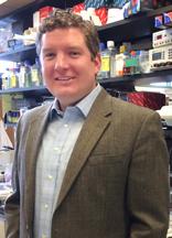 Stephen CJ Parker合作论文数Medical School, University of Michigan44
 Kari Stefansson合作论文数deCODE genetics44
 Anubha Mahajan合作论文数Human Genetics Department, Genentech44
 Jim Wilson合作论文数Usher Institute, Edinburgh Medical School, The University of Edinburgh;Institute of Genetics and Cancer, The University of Edinburgh43
 Daniel I. Chasman合作论文数Brigham and Women’s Hospital43




