Primary liver cancer is the third leading cause of cancer-related deaths, and its incidence and mortality are increasing worldwide. Hepatocellular carcinoma (HCC) accounts for 80% of primary liver cancer cases. Glypican-3 (GPC3) is a heparan sulfate proteoglycan that histopathologically defines HCC and represents an attractive tumor selective marker for radiopharmaceutical imaging and therapy for this disease. Single-domain antibodies are a promising scaffold for imaging because of their favorable pharmacokinetic properties, good tumor penetration, and renal clearance. Although conventional lysine-directed bioconjugation can be used to yield conjugates for radiolabeling fulllength antibodies, this stochastic approach risks negatively affecting target binding of the smaller single-domain antibodies. To address this challenge, site-specific approaches have been explored. Here, we used conventional and sortase-based site-specific conjugation methods to engineer GPC3-specific human single-domain antibody (HN3) PET probes. Methods: Bifunctional deferoxamine (DFO) isothiocyanate was used to synthesize native HN3 (nHN3)-DFO. Site-specifically modified HN3 (ssHN3)-DFO was engineered using sortase-mediated conjugation of triglycine-DFO chelator and HN3 containing an LPETG C-terminal tag. Both conjugates were radiolabeled with 89Zr, and their binding affinity in vitro and target engagement of GPC3-positive (GPC3+) tumors in vivo were determined. Results: Both 89Zr-ssHN3 and 89Zr-nHN3 displayed nanomolar affinity for GPC3 in vitro. Biodistribution and PET/CT image analysis in mice bearing isogenic A431 and A431GPC3+ xenografts, as well as in HepG2 liver cancer xenografts, showed that both conjugates specifically identify GPC3+ tumors. 89Zr-ssHN3 exhibited more favorable biodistribution and pharmacokinetic properties, including higher tumor uptake and lower liver accumulation. Comparative PET/CT studies on mice imaged with both 18F-FDG and 89Zr-ssHN3 showed more consistent tumor accumulation for the single-domain antibody conjugate, further establishing its potential for PET imaging. Conclusion: 89Zr-ssHN3 showed clear advantages in tumor uptake and tumor-to-liver signal ratio over the conventionally modified 89Zr-nHN3 in xenograft models. Our results establish the potential of HN3-based single-domain antibody probes for GPC3directed PET imaging of liver cancers.
Neutralizing antibodies targeting the SARS-CoV-2 spike protein have shown a great preventative/therapeutic potential. Here, we report a rapid and efficient strategy for the development and design of SARS-CoV-2 neutralizing humanized nanobody constructs with sub-nanomolar affinities and nanomolar potencies. CryoEM-based structural analysis of the nanobodies in complex with spike revealed two distinct binding modes. The most potent nanobody, RBD-1-2G(NCATS-BL8125), tolerates the N501Y RBD mutation and remains capable of neutralizing the B.1.1.7 (Alpha) variant. Molecular dynamics simulations provide a structural basis for understanding the neutralization process of nanobodies exclusively focused on the spike-ACE2 interface with and without the N501Y mutation on RBD. A primary human airway air-lung interface (ALI) ex vivo model showed that RBD-1-2G-Fc antibody treatment was effective at reducing viral burden following WA1 and B.1.1.7 SARS-CoV-2 infections. Therefore, this presented strategy will serve as a tool to mitigate the threat of emerging SARS-CoV-2 variants.
Half-sandwich Os-arene complexes exhibit promising anticancer activity, but their photochemistry has hardly been explored. To exploit the photocytotoxicity and photochemistry of Os-arenes, O,O-chelated complexes [Os(η6-p-cymene)(Curc)Cl] (OsCUR-1, Curc = curcumin) and [Os(η6-biphenyl)(Curc)Cl] (OsCUR-2), and N,N-chelated complexes [Os(η6-biphenyl)(dpq)I]PF6 (OsDPQ-2, dpq = pyrazino[2,3-f][1,10]phenanthroline) and [Os(η6-biphenyl)(bpy)I]PF6 (OsBPY-2, bpy = 2,2'-bipyridine), have been investigated. The Os-arene curcumin complexes showed remarkable photocytotoxicity toward a range of cancer cell lines (blue light IC50: 2.6-5.8 μM, photocytotoxicity index PI = 23-34), especially toward cisplatin-resistant cancer cells, but were nontoxic to normal cells. They localized mainly in mitochondria in the dark but translocated to the nucleus upon photoirradiation, generating DNA and mitochondrial damage, which might contribute toward overcoming cisplatin resistance. Mitochondrial damage, apoptosis, ROS generation, DNA damage, angiogenesis inhibition, and colony formation were observed when A549 lung cancer cells were treated with OsCUR-2. The photochemistry of these Os-arene complexes was investigated by a combination of NMR, HPLC-MS, high energy resolution fluorescence detected (HERFD), X-ray adsorption near edge structure (XANES) spectroscopy, total fluorescence yield (TFY) XANES spectra, and theoretical computation. Selective photodissociation of the arene ligand and oxidation of Os(II) to Os(III) occurred under blue light or UVA excitation. This new approach to the design of novel Os-arene complexes as phototherapeutic agents suggests that the novel curcumin complex OsCUR-2, in particular, is a potential candidate for further development as a photosensitizer for anticancer photoactivated chemotherapy (PACT).
With the recent spread of severe acute respiratory syndrome coronavirus ( SARS-CoV-2)_ infecting >16 million people worldwide as of 28 July 2020, causing >650 000 deaths, there is a desperate need for therapeutic agents and vaccines. Building on knowledge of previous outbreaks of SARS-CoV-1 and Middle East respiratory syndrome (MERS), the development of therapeutic antibodies and vaccines against coronavirus disease 2019 (COVID-19) is taking place at an unprecedented speed. Current efforts towards the development of neutralizing antibodies against COVID-19 are summarized. We also highlight the importance of a fruitful antibody development pipeline to combat the potential escape plans of SARS-CoV-2, including somatic mutations and antibody-dependent enhancement (ADE).
The National Center for Advancing Translational Sciences (NCATS) has developed an online open science data portal for its COVID-19 drug repurposing campaign - named OpenData - with the goal of making data across a range of SARS-CoV-2 related assays available in real-time. The assays developed cover a wide spectrum of the SARS-CoV-2 life cycle, including both viral and human (host) targets. In total, over 10,000 compounds are being tested in full concentration-response ranges from across multiple annotated small molecule libraries, including approved drug, repurposing candidates and experimental therapeutics designed to modulate a wide range of cellular targets. The goal is to support research scientists, clinical investigators and public health officials through open data sharing and analysis tools to expedite the development of SARS-CoV-2 interventions, and to prioritize promising compounds and repurposed drugs for further development in treating COVID-19.
Background: Glypican 3 (GPC3), a plasma membrane heparan sulfate proteoglycan, is overexpressed on human hepatocellular carcinoma and may represent a promising biomarker. Several studies have reported peptides that selectively bind to GPC3 and could serve as scaffolds for imaging or therapeutic agents. Materials and Methods: We synthesized variants of two previously published peptides, DHLASLWWGTEL (TJ12P1) and RLNVGGTYFLTTRQ (L5), and evaluated their in vitro binding performance in paired isogenic cell lines, A431(GPC3(-)) and A431-GPC3(+) (G1), as well as the liver cancer cell line HepG2. Using flow cytometry and biolayer interferometry (BLI), we compared the binding of the TJ12P1 and L5 peptide variants to the binding of corresponding scrambled peptides having the same amino acid composition, but in random sequence. Results: While both peptides bound to G1 and HepG2, they also bound to A431. The corresponding scrambled peptides demonstrated greater apparent binding to both G1 and A431 than their specific counterparts. BLI confirmed lack of binding at 0.5-1 mu M for both peptides. Conclusions: We conclude that neither TJ12P1 nor L5 variant demonstrates selectivity for GPC3 at concentrations near the reported K-D, and that the peptides lack potency or are nonspecific, making them inadequate for use as imaging agents.
Hepatocellular carcinoma (HCC) is the second most common cause of cancer-related death in the world. Therapeutic outcomes of HCC remain unsatisfactory, and novel treatments are urgently needed. GPC3 (glypican-3) is an emerging target for HCC, given the findings that 1) GPC3 is highly expressed in more than 70% of HCC; (2) elevated GPC3 expression is linked with poor HCC prognosis; and (3) GPC3-specific therapeutics, including immunotoxin, bispecific antibody and chimeric antigen receptor T cells. have shown promising results. Here, we postulate that GPC3 is a potential target of antibody-drug conjugates (ADCs) for treating liver cancer. To determine the payload for ADCs against liver cancer, we screened three large drug libraries (> 9,000 compounds) against HCC cell lines and found that the most potent drugs are DNA-damaging agents. Duocarmycin SA and pyrrolobenzodiazepine dimer were chosen as the payloads to construct two GPC3-specific ADCs: hYP7-DC and hYP7-PC. Both ADCs showed potency at picomolar concentrations against a panel of GPC3-positive cancer cell lines, but not GPC3 negative cell lines. To improve potency, we investigated the synergetic effect of hYP7-DC with approved drugs. Gemcitabine showed a synergetic effect with hYP7-DC in vitro and in vivo. Furthermore, single treatment of hYP7-PC induced tumor regression in multiple mouse models. Conclusion: We provide an example of an ADC targeting GPC3, suggesting a strategy for liver cancer therapy.
ABSTRACT Currently, four antibody-drug conjugates (ADCs) are approved by the Food and Drug Administration or the European Medicine Agency to treat cancer patients. More than 60 ADCs are in clinical development for cancer therapy. More than 60% of ADCs in clinical trials employ microtubule inhibitors as their payloads. A better understanding of payloads other than microtubule inhibitors, especially DNA-damaging agents, is important for further development of ADCs. In this review, we highlight an emerging trend of using DNA-damaging agents as payloads for ADCs. This review summarizes recent advances in our understanding gained from ongoing clinical studies; it will help to define the utility of DNA-damaging payloads for ADCs as cancer therapeutics. Future directions of the development of ADCs are also discussed, focusing on targeting drug resistance and combination treatment with immunotherapy.
Background Oxidative stress and chronic inflammation can increase cellular levels of reactive oxygen species and lipid peroxidation (LPO) when associated with the pathogenesis of hepatocellular carcinoma (HCC), which can develop following the progression of steatosis, fibrosis and cirrhosis. Using a monoclonal antibody for cyclic γ-hydroxy-1, N2 -propanodeoxyguanosine (γ-OHPdG), a promutagenic DNA adduct formed endogenously by LPO, we examined its formation across liver disease stages to understand it's potential role in HCC development. Methods Formalin-fixed paraffin embedded (FFPE) liver tissue samples from 49 patients representing normal, steatosis, fibrosis, cirrhosis and HCC were stained for γ-OHPdG and 8-hydroxydeoxyguanosine (8-oxo-dG), an oxidative damage biomarker. Quantification of immunohistochemical (IHC) staining was performed using histological scoring of intensity and distribution. Using primary human hepatocytes (HH) and a stellate cell (SC) co-culture, immunocytochemical staining of γ-OHPdG and Nile Red was performed to determine if the formation of γ-OHPdG was consistent between the clinical sample disease stages and the in vitro steatotic and fibrotic conditions. Results γ-OHPdG levels varied significantly between the stages of normal and steatosis, steatosis and fibrosis, and steatosis and cirrhosis (P≤0.005). There was a trend, although not significant, of increased levels of γ-OHPdG in HCC compared to the other groups. A strong correlation was observed (Pearson's, R2 =0.85) between levels of γ-OHPdG and 8-oxo-dG across the disease spectrum. The increase of γ-OHPdG in steatosis and decrease in fibrosis was a pattern confirmed in an in vitro model using primary HH co-cultured with human SCs. Conclusions γ-OHPdG was detected in FFPE liver tissues of patients with different stages of liver disease and in vitro studies, demonstrating that its formation is consistent with LPO in early stages of liver disease and suggesting that it may be a source of mutagenic DNA damage in liver disease progression.
ABSTRACT Background Shark new antigen receptor variable domain (VNAR) antibodies can bind restricted epitopes that may be inaccessible to conventional antibodies. Methods Here, we developed a library construction method based on polymerase chain reaction (PCR)-Extension Assembly and Self-Ligation (named “EASeL”) to construct a large VNAR antibody library with a size of 1.2 × 10¹⁰ from six naïve adult nurse sharks (Ginglymostoma cirratum). Results The next-generation sequencing analysis of 1.19 million full-length VNARs revealed that this library is highly diversified because it covers all four classical VNAR types (Types I–IV) including 11% of classical Type I and 57% of classical Type II. About 30% of the total VNARs could not be categorized as any of the classical types. The high variability of complementarity determining region (CDR) 3 length and cysteine numbers are important for the diversity of VNARs. To validate the use of the shark VNAR library for antibody discovery, we isolated a panel of VNAR phage binders to cancer therapy-related antigens, including glypican-3, human epidermal growth factor receptor 2 (HER2), and programmed cell death-1 (PD1). Additionally, we identified binders to viral antigens that included the Middle East respiratory syndrome (MERS) and severe acute respiratory syndrome (SARS) spike proteins. The isolated shark single-domain antibodies including Type I and Type II VNARs were produced in Escherichia coli and validated for their antigen binding. A Type II VNAR (PE38-B6) has a high affinity (Kd = 10.1 nM) for its antigen. Conclusions The naïve nurse shark VNAR library is a useful source for isolating single-domain antibodies to a wide range of antigens. The EASeL method may be applicable to the construction of other large diversity gene expression libraries.
Hepatocellular carcinoma (HCC) is the second leading cause of cancer-related deaths worldwide. There are two major challenges for HCC, the first being that early detection is generally not applicable, and secondly, it is usually fatal within several months after diagnosis. HCC is an inflammation-induced cancer. It is known that chronic inflammation leads to oxidative/nitrosative stress and lipid peroxidation, generating excess oxidative stress, together with aldehydes which can react with DNA bases to form promutagenic DNA adducts. In this review, the evidence between oxidative stress and liver carcinogenesis is summarized. We focused on the potential of using DNA adducts as oxidative stress biomarkers for liver carcinogenesis.
Hepatocellular carcinoma (HCC) is the third leading cause of cancer–related deaths worldwide, mainly because of its poor prognosis. A valid mechanism‐based prognostic biomarker is urgently needed. γ‐hydroxy‐1,N2‐propanodeoxyguanosine (γ‐OHPdG) is an endogenously formed mutagenic DNA adduct derived from lipid peroxidation. We examined the relationship of γ‐OHPdG with hepatocarcinogenesis in two animal models and its potential role as a prognostic biomarker for recurrence in HCC patients. Bioassays were conducted in xeroderma pigmentosum group A knockout mice and diethylnitrosamine‐injected mice, both prone to HCC development. γ‐OHPdG levels in the livers of these animals were determined. The effects of antioxidant treatments on γ‐OHPdG and hepatocarcinogenesis were examined. Using two independent sets of HCC specimens from patients, we examined the relationship between γ‐OHPdG and survival or recurrence‐free survival. γ‐OHPdG levels in liver DNA showed an age‐dependent increase and consistently correlated with HCC development in all three animal models. Theaphenon E treatment significantly decreased γ‐OHPdG levels in the liver DNA of xeroderma pigmentosum group A knockout mice and remarkably reduced HCC incidence in these mice to 14% from 100% in the controls. It also effectively inhibited HCC development in the diethylnitrosamine‐injected mice. Using clinical samples from two groups of patients, our study revealed that higher levels of γ‐OHPdG are strongly associated with low survival (P < 0.0001) and low recurrence‐free survival (P = 0.007). Conclusion: These results support γ‐OHPdG as a mechanism‐based, biologically relevant biomarker for predicting the risk of HCC and its recurrence. (Hepatology 2018;67:159‐170).
Abstract In various animal models, n-3 polyunsaturated fatty acids (PUFAs) have demonstrated cancer preventive effects. This protective effect is associated with reduced levels of inflammatory cytokines, such as tumor necrosis factor alpha (TNF-α), cyclooxygenase 2 (COX-2) and prostaglandin E2 (PGE2). On the other hand, an endogenous DNA adduct formed from the peroxidation of ω-3 PUFAs, γ-hydroxy-1, N2-propanodeoxyguanosine (γ-OHPdG), is known to be mutagenic. In light of these observations, we hypothesized that the combination of n-3 PUFAs with an antioxidant to inhibit lipid peroxidation (e.g. α-lipoic acid) will result in an enhanced cancer preventive effect than n-3 PUFAs alone. The chemical carcinogen diethylnitrosamine (DEN) induced HCC model is employed in this study. The DEN-induced HCC model has a histology and genetic signature similar to that of human HCCs, such that it has poor prognosis and recapitulates a dependence on inflammatory signaling. It also reflects the same gender disparity seen in human HCCs. Fat-1 transgenic mice were used because they express a Caenorhabditis elegans desaturase converting n-6 to n-3 PUFAs endogenously without using dietary supplementation. This model underlies the importance of dietary control of PUFAs intake and ratios due to the inability of mammalian cells to generate anti-inflammatory n-3 PUFAs from pro-inflammatory n-6 PUFAs, which are a major component of the standard Western diets. The results showed that antioxidant can efficiently suppress the elevated level of γ-OHPdG in fat-1 mice. The tumor incidence, multiplicity, and size are monitored continuously. This work was supported by the NCI grant: RO1-CA-134892. Y.F. thanks the Prevent Cancer Foundation for his fellowship (Marcia and Frank Carlucci Charitable Foundation Award in Cancer Prevention and Early Detection) Citation Format: Ying Fu, Angela Y. Bai, Marcin Dyba, Jing X. Kang, Fung-Lung Chung. Using antioxidant to enhance liver cancer preventive effect of n-3 PUFAs. [abstract]. In: Proceedings of the 107th Annual Meeting of the American Association for Cancer Research; 2016 Apr 16-20; New Orleans, LA. Philadelphia (PA): AACR; Cancer Res 2016;76(14 Suppl):Abstract nr 4314.
AbstractDinuclear trihydroxido‐bridged osmium–arene complexes are inert and biologically inactive, but we show here that linking dihydroxido‐bridged OsII–arene fragments by a bridging di‐imine to form a metallacycle framework results in strong antiproliferative activity towards cancer cells and distinctive knotting of DNA. The shortened spacer length reduces biological activity and stability in solution towards decomposition to biologically inactive dimers. Significant differences in behavior toward plasmid DNA condensation are correlated with biological activity.
Background: We are developing organo-osmium complex 1 as an alternative to platinum-anticancer drugs in the clinic for the treatment of epithelial ovarian cancer (EOC).1,23,4 Method: We used time series RNA-sequencing to determine differential gene expression in A2780 EOC cells in response to treatment of complex 1. This was complemented with reverse-phase protein microarrays to study cellular levels of key proteins involved in DNA-damage repair, and flow cytometry and high-content imaging to investigate activation of oxidative stress and apoptosis. Results: Complex 1 was screened in 809 cancer cell lines as part of the Sanger Institute’s Cancer Genome Project with promising results. Whole transcriptome sequencing identified three missense mutations in the mitochondrial genome of A2780 cells, in the electron transport chain. Time-series RNA-sequencing suggested that osmium-exposed A2780 cells undergo a metabolic shunt from glycolysis to oxidative phosphorylation, where defective machinery, associated with mutations in complex I, could enhance activity. The MOA of 1 is appears to involve redox-mediation. Conclusion: Transcriptomic and proteomic studies suggest an attack on glycolysis which switches energy production towards OXPHOS in A2780 EOC cells. This pathway may already be stressed by the 3 mutations we detected in CI of the ETC.
Abstract In this study we are investigating the role and mechanism of formation of Lipid-peroxidation (LPO)-derived DNA adduct formation in obesity-related hepatocarcinogenesis. Obesity has been implicated as a risk factor for many types of cancer, particularly hepatocellular carcinoma (HCC). HCC incidence correlates with the increasing prevalence of non-alcoholic fatty liver disease (NAFLD) and non-alcoholic steatohepatitis (NASH) in the US. The mechanisms underlying this increased risk of liver cancer in the obese population are still unclear. Liver tissue damage from fat accumulation in NAFLD and NASH produce inflammation and increased levels of reactive oxygen species (ROS). ROS induce LPO of polyunsaturated fatty acids (PUFAs) in cell membranes leading to the formation of reactive aldehydes, such as acrolein and 4-hydroxy-2-nonenal (HNE), which react with DNA forming the DNA adducts γ-OHPdG and DHH-ϵdA, respectively. γ-OHPdG is derived from ω-6 and ω-3 PUFAs, while DHH-ϵdA is specifically from ω-6 fatty acids. Previous data has indicated that mutational hotspots targeted by γ-OHPdG and DHH-ϵdA may be within key cancer driver genes, such as p53. Research has shown that B-6 mice fed a high fat diet (HFD) develop fatty liver disease and eventually HCC. The livers of B-6 mice fed a HFD showed an increased ω-6/ω-3 PUFAs ratio. We have detected and quantified γ-OHPdG and DHH-ϵdA by LC-MS/MS in livers from six healthy individuals and six NAFLD patients and found that levels of DHH-ϵdA in the DNA of the fatty liver samples were nearly three-fold higher than that in normal liver samples. Preliminary in vitro data using primary human hepatocytes has indicated that treatment with oleic and palmitic acid promote the formation of γ-OHPdG. Similarly, treatment of these cells with the epoxide of HNE, 2,3-epoxy-4-hydroxynonanal (EH), and the omega-6 fatty acid, arachidonic acid, induce the formation of DHH-ϵdA, supporting the proposed mechanism of adduct formation. In an 80-week tumor bioassay using C57Bl/6J mice on a HFD, we have observed through live-animal MRI imaging and immunohistochemistry, an increase in pro-inflammatory white adipose tissue accumulation, infiltration of fat into the liver and an increase in overall body weight compared to a low fat diet control. In addition, in mice fed a HFD combined with the green-tea derived antioxidant Theaphenon E, we observed through MRI, reduction in body weight gain, white adipose tissue, and lipid accumulation in liver. Theaphenon E has the potential to decrease fat accumulation and inflammation within the liver that lead to decreases in LPO-derived adduct formation and consequently, the mutations critical for HCC development. Citation Format: Heidi Coia, Hongyi Guan, Ying Fu, Marcin Dyba, Fung-Lung Chung. Lipid peroxidation-derived DNA adduct formation in obesity-related hepatocarcinogenesis. [abstract]. In: Proceedings of the 107th Annual Meeting of the American Association for Cancer Research; 2016 Apr 16-20; New Orleans, LA. Philadelphia (PA): AACR; Cancer Res 2016;76(14 Suppl):Abstract nr 4051.
The acrolein derived cyclic 1,N(2)-propanodeoxyguanosine adduct (Acr-dG), formed primarily from ω-3 polyunsaturated fatty acids such as docosahexaenoic acid (DHA) under oxidative conditions, while proven to be mutagenic, is potentially involved in DHA-induced apoptosis. The latter may contribute to the chemopreventive effects of DHA. Previous studies have shown that the levels of Acr-dG are correlated with apoptosis induction in HT29 cells treated with DHA. Because Acr-dG is shown to be repaired by the nucleotide excision repair (NER) pathway, to further investigate the role of Acr-dG in apoptosis, in this study, NER-deficient XPA and its isogenic NER-proficient XAN1 cells were treated with DHA. The Acr-dG levels and apoptosis were sharply increased in XPA cells, but not in XAN1 cells when treated with 125μM of DHA. Because DHA can induce formation of various DNA damage, to specifically investigate the role of Acr-dG in apoptosis induction, we treated XPA knockdown HCT116+ch3 cells with acrolein. The levels of both Acr-dG and apoptosis induction increased significantly in the XPA knockdown cells. These results clearly demonstrate that NER deficiency induces higher levels of Acr-dG in cells treated with DHA or acrolein and sensitizes cells to undergo apoptosis in a correlative manner. Collectively, these results support that Acr-dG, a ubiquitously formed mutagenic oxidative DNA adduct, plays a role in DHA-induced apoptosis and suggest that it could serve as a biomarker for the cancer preventive effects of DHA.
Deutliche Unterschiede bei der Wechselwirkung mit DNA könnten über die antiproliferative Wirkung von OsII-Aren-Metallacyclen entscheiden. In der Zuschrift auf S. 9055 ff. beschreiben P. J. Sadler et al. zwei vierkernige metallorganische OsII-Komplexe mit starken Abweichungen bei der Zytotoxizität für Krebszellen und dem Verhalten gegenüber der DNA-Kondensation. Es besteht eine Korrelation zwischen der Spacerlänge im Metallacyclus, der Beständigkeit in Lösung und der biologischen Aktivität.
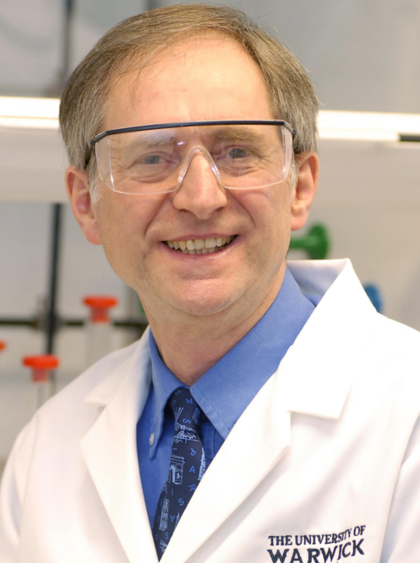 Peter Sadler合作论文数Department of Chemistry, University of Warwick16
 Fung-Lung Chung合作论文数15
 Marcin Dyba合作论文数11
 Abraha Habtemariam合作论文数11Guy J. Clarkson合作论文数8Jishen Pan合作论文数6
 Ana Pizarro合作论文数Department of Chemistry, University of Warwick6Monika Aggarwal合作论文数Dept Oncol, Georgetown Univ5Coia Heidi合作论文数5Hearn Jessica M合作论文数5
 MinHua Chen (陈敏华)合作论文数Beijing Cancer Hospital5María J. Romero合作论文数5
 Isolda Romero-Canelón合作论文数Institute of Clinical Sciences, College of Medical and Dental Sciences, University of Birmingham5
 Mitchell Ho合作论文数Laboratory of Molecular Biology, Center for Cancer Research, National Cancer Institute5
 Deborah Berry合作论文数AstraZeneca5Bhaskar Kallakury合作论文数5
 Luca Salassa合作论文数Department of Chemistry, University of Warwick5
 Elizabeth Sinclair合作论文数Lombardi Comprehensive Cancer Center, Georgetown University4
 Wei Yang (杨薇)合作论文数北京肿瘤医院4
 Wei Wu (吴薇)合作论文数Department of Ultrasound, Key Laboratory of Carcinogenesis and Translational Research (Ministry of Education), Peking University Cancer Hospital and Institute4
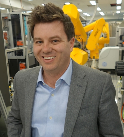 Matthew D. Hall合作论文数Early Translation Branch, Division of Preclinical Innovation, National Center for Advancing Translational Sciences, National Institutes of Health4Steven D. Shnyder合作论文数The Institute of Cancer Therapeutics, University of Bradford4Shana Silverstein合作论文数Lombardi Comprehens Canc Ctr, Georgetown Univ3Jessica Hong合作论文数3
 Alex Renn合作论文数NCATS, Natl Ctr Adv Translat Sci3
 Alison Rodger合作论文数School of Natural Sciences, Macquarie University/MQ Photonics Research Centre, Macquarie University3
 Anton Simeonov合作论文数National Center for Advancing Translational Sciences, National Institutes of Health, U.S. Department of Health & Human Services3AiwuRuth He合作论文数3
 Kun Yan (严昆)合作论文数Beijing Cancer Hospital3Hu Xin合作论文数3Xuan Zhuoli合作论文数3Cheng Xi合作论文数3
 Baocai Xing (邢宝才)合作论文数Beijing Cancer Hospital3
 Giovanni Costantini合作论文数Department of Chemistry, Faculty of Science, Engineering and Medicine, The University of Warwick3
 Sabine H. Van Rijt合作论文数Department of Chemistry, University of Warwick3Ivan Prokeš合作论文数3Patricia A Cooper合作论文数3Angela Y. Bai合作论文数2Karen Creswell合作论文数2Nicholas T. Gutsche合作论文数2Quinlin Hanson合作论文数2M Idalia Cruz合作论文数2marc ferreralegre合作论文数2
 Catherine Chen合作论文数Department of Electrical Engineering and Computer Sciences, University of California, Berkeley2Zina Itkin合作论文数2Manisha Pradhan合作论文数2Bryan D Fleming合作论文数2
 Alison F Munro合作论文数2Qin Wang合作论文数2




