Microautophagy (MI-autophagy) is an umbrella term for intracellular degradative pathways that entail the invagination or protrusion of the limiting membranes of endolysosomal compartments, that is, late endosomes and mammalian lysosomes or yeast and plant vacuoles, followed by pinching-off of the membrane into the lumen of the organelle. During these processes, the material specifically and nonspecifically targeted for degradation is sequestered within the invaginating or protuberating membrane. In contrast to macroautophagy, the molecular mechanisms underlying MI-autophagy are largely unknown due to their diversity and complexity in location, regulation and molecular machinery requirements. Here, we review recent progress in the field of MI-autophagy, describing the molecular basis and functions of the MI-autophagic pathways reported to date in eukaryotic cells, from yeast to mammalian and plant cells.
Atg9-Atg2-Atg18 complexes are essential for the biogenesis of the autophagosome as they mediate the elongation of the phagophore, the precursor structure of autophagosomes. This event occurs by the transfer of lipids through a membrane contact site (MCS) between the phagophore and the endoplasmic reticulum exit sites (ERES). The bridge-like lipid-transfer protein (BLTP) Atg2 interacts with the Atg9 and phosphatidylinositol-3-phosphate (PtdIns3P) on the phagophore and acts as a tether to establish this MCS. While not essential to form the phagophore-ERES MCS, Atg18 plays a crucial role in the phagophore elongation by stimulating Atg2 lipid transfer activity, based on in vitro experiments. To understand the molecular basis of this regulation, we recently solved the structure of the yeast Atg2-Atg18 complex using cryo-electron microscopy (cryo-EM) and identified the critical region in Atg2 required for the Atg2-Atg18 complex formation. Importantly, we applied structure-function analyses to unveil the molecular mechanism behind the Atg18-mediated stimulation of Atg2. We showed that Atg18 binding to Atg2 induces a structural repositioning of the hydrophobic cavity of Atg2 toward the membrane, which allows efficient transfer of lipids from the endoplasmic reticulum to the phagophore. Here, we summarize our recent work and extend our discussion on the molecular regulation of the lipid transfer activity, highlighting open questions concerning the function of the Atg9-Atg2-Atg18 module in the phagophore-ERES MCS.Abbreviations: ATG, autophagy related; BLTP, bridge-like lipid-transfer protein; cryo-EM, cryo-electron microscopy; ER, endoplasmic reticulum; ERES, ER exit sites; MCS, membrane contact site; PAS, phagophore assembly site; PtdIns3P, phosphatidylinositol-3-phosphate; TRAPPIII, transport protein particle III.
The SARS-CoV-2 main protease (3CLpro) is a well-validated target for structure-guided inhibitor discovery. Here, we report α-aminomethyl tetrazole inhibitors accessed via the Ugi tetrazole multicomponent reaction (UT-4CR), enabling rapid exploration of non-classical chemical space. Initial design and modeling suggested a binding mode analogous to Ugi-derived (U-4CR) 3CLpro inhibitors, with heteroaromatic substituents engaging the S1 pocket. However, crystallographic analysis revealed an unexpected binding orientation in which the tetrazole core itself occupies the S1 pocket and forms the key interaction with His163, while the modeled substituents are solvent-exposed. This revised binding mode rationalizes the observed structure-activity relationships. Installation of an electrophilic warhead yielded covalent inhibitors with sub-micromolar enzymatic potency, and lead compound 2a displayed modest antiviral activity in infected cells. These results highlight UT-4CR-derived tetrazoles as a platform for probing the 3CLpro binding space and underscore the importance of early crystallographic validation.
In contrast to the ubiquitin (Ub)-proteasome-system, which only degrades individual proteins, macroautophagy can eliminate protein complexes or aggregates, organelles and even pathogens. Terms such as mitophagy, aggrephagy, lysophagy and xenophagy have been coined based on the targeted substrate. In Ub-dependent selective macroautophagy, cargo selectivity is specified by E3 Ub ligases that append Ub chains that in turn are recognized by selective autophagy receptors (SARs), driving sequestration into autophagosomes. While several Ub-dependent SARs have been identified and characterized, the E3 Ub ligases that ultimately decide target fate remain poorly studied. In this review, we summarize what is known about the E3 Ub ligases involved in selective macroautophagy, with a particular emphasis on the degradation of mitochondria, protein aggregates, lysosomes and pathogens. A better characterization of these enzymes could improve therapeutic strategies for targeted degradation in acute and chronic diseases. A subset of selective autophagy receptors recognizes ubiquitin modifications on their targets. This review provides a comprehensive overview on E3 ubiquitin ligases involved in selective macroautophagy pathways, including mitophagy, aggrephagy, and xenophagy.
BECN1 (beclin 1) is a member of the nucleation complex and considered crucial for induction of macroautophagy/autophagy, leading to the formation and ultimate degradation of autophagosomes. We found that in human B lymphoblastoid cell lines (LCLs) deficient of BECN1 (BECN1-KO), autophagosome formation was intact and autophagic flux could be induced upon nutrient starvation or MTOR inhibition. Remarkably, autophagosomal cargo differed significantly between BECN1-KO and control (CTRL) LCLs, revealing a preferred formation of autophagosomes at the endoplasmic reticulum (ER) and not at endosomes/lysosomes in BECN1-KO LCLs. Endosomal TLR3 (toll like receptor 3) was less frequently incorporated within autophagosomes in BECN1-KO LCLs. Additionally, several proteins of the ER-resident peptide loading complex for MHC class I antigen presentation were found enriched in autophagosomes from BECN1-KO LCLs, resulting in a diminished detection of BECN1-KO LCLs by T cells. Hence, BECN1 seems to be dispensable for autophagosome formation but rather contributes to cargo selection of phagophores and immunosurveillance.Abbreviations: ATG: autophagy-related; BECN1: beclin 1; CTRL: control; LCL: Epstein Barr virus transformed lymphoblastoid cell line; MHC: major histocompatibility complex; PLC: peptide-loading complex; TLR: toll like receptor.
During macroautophagy, the de novo formation of the autophagosome at a membrane contact site (MCS) with the endoplasmic reticulum requires directional lipid flux for the growth of the initial phagophore before its sealing into an autophagosome and subsequent fusion with the lysosome/vacuole. It remains unclear, however, how the formation of this specialized MCS and the directionality of the lipid flux are controlled. Here, we present the structure of the key lipid transfer protein Atg2 from yeast solved together with its Atg18 binding partner, a phosphatidylinositol-3-phosphate (PtdIns3P) effector, using cryo-electron microscopy. We reveal a new interface in Atg2 that, together with PtdIns3P, is required for Atg18 recruitment and lipid transfer activity. Furthermore, we visualize lipid densities along the internal hydrophobic cavity of Atg2, providing structural evidence that Atg2 cavity is filled with lipids throughout the entire length, even when Atg2 is cytosolic. Finally, molecular dynamics simulations show that the complex generates membrane curvature, efficiently positioning the lipid channel of Atg2 towards the membrane to promote lipid transfer into the elongating phagophore.
The clinical outcome of SARS-CoV-2 infection spans from asymptomatic viral elimination to lethal COVID-19 pneumonia, which is due to type I interferon (IFN) deficiency in at least 15-20% of cases. We report two unrelated male patients with critical COVID-19 who are heterozygous for rare deleterious variants in RB1CC1, encoding the autophagy-related FIP200 protein. Airway epithelial cells genetically deprived of FIP200 or cell lines expressing the RB1CC1/FIP200 patient variants exhibit elevated SARS-CoV-2 replication and impaired autophagic flux. The antiviral function of FIP200 is independent of canonical autophagy and type I IFN, but involves the selective autophagy receptor NDP52. We identify a non-canonical function of FIP200 in a novel lysosomal degradation pathway, in which SARS-CoV-2 virions are targeted to single-membrane compartments for degradation of viral RNA in LC3B-positive acidified vesicles. This pathway is impaired in FIP200-deficient cells and in cells expressing FIP200 patient haplotypes. Collectively, we describe a cell-autonomous anti-SARS-CoV-2 restriction pathway, dependent on FIP200 and NDP52, and independent of canonical autophagy and type I IFN, which can underlie critical COVID-19 pneumonia.
The de novo generation of membrane contact sites (MCSs) between the phagophore and the endoplasmic reticulum exit sites (ERES) is important for the acquisition of the lipids necessary for phagophore elongation and autophagosome formation during autophagy. However, it is currently unclear how these MCSs are established. Here, we show that the TRAPPIII complex, the guanine nucleotide exchange factor of the Rab GTPase Ypt1, localizes to and regulates the formation of the MCS between the phagophore and the ERES. In particular, TRAPPIII and the lipid transfer protein Atg2 appear equally essential for the association of the phagophore with the ERES, TRAPPIII activation and Ypt1 activation onto the phagophore. Ypt1 redistributes over the entire surface of the phagophore and promotes its elongation through both stimulation of the local biosynthesis of phosphatidylinositol-3-phosphate and recruitment of the downstream effectors Atg18 and Atg21. Our data suggest that de novo generation of the phagophore-ER MCSs and subsequent Ypt1 activation initiates phagophore elongation.
Macroautophagy mediates the degradation of long-lived proteins and organelles via the de novo formation of double-membrane autophagosomes that sequester cytoplasm and deliver it to the vacuole/lysosome; however, relatively little is known about autophagosome biogenesis. Atg8, a phosphatidylethanolamine-conjugated protein, was previously proposed to function in autophagosome membrane expansion, based on the observation that it mediates liposome tethering and hemifusion in vitro. We show here that with physiological concentrations of phosphatidylethanolamine, Atg8 does not act as a fusogen. Rather, we provide evidence for the involvement of exocytic Q/t-SNAREs in autophagosome formation, acting in the recruitment of key autophagy components to the site of autophagosome formation, and in regulating the organization of Atg9 into tubulovesicular clusters. Additionally, we found that the endosomal Q/t-SNARE Tlg2 and the R/v-SNAREs Sec22 and Ykt6 interact with Sso1-Sec9, and are required for normal Atg9 transport. Thus, multiple SNARE-mediated fusion events are likely to be involved in autophagosome biogenesis.
NF-κB is central for activation of immune responses. Cytosolic DNA activates the cGAS–STING pathway to induce type I interferons (IFNs) and signaling through NF-κB, thus instigating host defenses and pathological inflammation. However, the mechanism underlying STING-induced NF-κB activation is unknown. Here we report that STING activates NF-κB in a delayed manner, following exit from the Golgi to endolysosomal compartments. Activation of NF-κB is dependent on the IFN-inducing transcription factor IRF3 but is independent of type I IFN signaling. This activation pattern is evolutionarily conserved in tetrapods. Mechanistically, the monomer IRF3 is recruited to STING pS358, with delayed kinetics relative to IRF3 recruitment to STING pS366, which promotes type I IFN responses. IRF3 engagement with STING pS358 induces trafficking to late endolysosomal compartments, supporting recruitment of TRAF6 and activation of NF-κB. We identify a TRAF6 binding motif in IRF3 that facilitates recruitment of TRAF6. This work defines a signaling surface on STING and a function for IRF3 as an adaptor in immune signaling. These findings indicate that STING signaling to NF-κB is enabled only within a short time window between exit from the Golgi and lysosomal degradation, possibly limiting inflammation under homeostatic and danger-sensing conditions. Here the authors show how the DNA-sensing cGAS–STING pathway activates NF-κB and inflammatory gene expression with delayed kinetics via post-Golgi endolysosomal signaling.
MAP1LC3/LC3 (microtubule associated protein 1 light chain 3) proteins have long been thought to carry out their cellular and organismal functions, including macroautophagy/autophagy, exclusively in their lipidated form, also referred to as Atg8ylation. They are anchored mainly to the phosphatidylethanolamine present in membranes through the action of two ubiquitin-like conjugation systems. Our recent work, however, uncovered a role of non-lipidated LC3s during influenza A virus (IAV) infection. We revealed that LC3s, together with the centrosomal scaffold protein PCNT (pericentrin), form a dynein adaptor complex that facilitates IAV uncoating at late endosomes (LEs). We also showed that co-opting the LC3s-PCNT complex is an alternative strategy to aggresome processing machinery (APM) hijacking via HDAC6, allowing IAV to exploit the force generated by dynein-dependent motors for virion uncoating and genome delivery in the host cytoplasm. Notably, the function of LC3s in IAV uncoating does not require their Atg8ylation or the core autophagy machinery, and PCNT's role is independent from its centrosomal localization. These findings redefine LC3s as multifunctional adaptor proteins and reveal how viruses can co-opt centrosome assembly machinery components for host invasion.Abbreviation: AKAP9/AKAP450- A-kinase anchoring protein 9; APM- aggresome processing machinery; IAV- influenza A virus; LC3s-I- non-lipidated LC3s; Les- late endosomes; MAP1LC3/LC3s-microtubule associated protein 1 light chain 3 proteins; MT-microtubule; NEU- neuraminidase; PCNT-pericentrin; TNPO1-transportin 1; vRNP-viral ribonucleoprotein.
Recently, rapid progress in the field of microautophagy (MI-autophagy) revealed the existence of multiple subtypes that differ in both intracellular membrane dynamics and molecular mechanisms. As a result, a single umbrella term "microautophagy" has become too vague, even creating some confusion among researchers both within and outside the field. We herein describe different subtypes of MI-autophagic processes and propose a systematic approach for naming them more accurately.Abbreviation: ATG, autophagy related; e-MI, endosomal microautophagy; ER, endoplasmic reticulum; ESCRT, endosomal sorting complex required for transport; EV, extracellular vesicle; HSPA8/HSC70, heat shock protein family A (Hsp70) member 8; ILVs, intralumenal vesicles; l-MI, lysosomal microautophagy; MAP1LC3/LC3, microtubule associated protein 1 light chain 3; MCOLN1, mucolipin TRP cation channel 1; microautophagy, MI-autophagy; MVBs, multivesicular bodies; SQSTM1, sequestosome 1; v-MI, vacuolar microautophagy.
Canonical autophagy is an intracellular pathway that degrades and recycles cellular components. A key step of this pathway is the formation of double-membraned organelles, known as autophagosomes, an emblematic feature of macroautophagy. For convenience, the formation of autophagosomes can be categorized into sequential steps, initiation (X), expansion (Y) and closure (Z). ATG9A is an integral membrane protein known for its role in the X and Y steps. whereby it organizes phagophore membrane assembly and its growth. Here, we report a previously unappreciated function of mammalian ATG9A in directing the last step Z. In particular, ATG9A partners with the key ESCRT-III component CHMP2A through IQGAP1 to facilitate autophagosome closure. Thus, ATG9A orchestrates all stages of autophagosome membrane biogenesis, from phagophore initiation to its closure. This makes ATG9A a unique ATG factor that works as a central hub in autophagosome biogenesis.Abbreviation: ATG9A autophagy related 9A; CCCP carbonyl cyanide m-chlorophenylhydrazone; Co-IP co-immunoprecipitation; ESCRT endosomal sorting complexes required for transport; EBSS Earle's balanced salt solution; ER endoplasmic reticulum; HCM high-content microscopy; HT HaloTag; LC-MS/MS liquid chromatography-tandem mass spectrometry; KO knockout; MPL membrane permeant ligand; MIL membrane impermeant ligand; Mtb Mycobacterium tuberculosis; SolVit sealing of organellar limiting membranes in vitro; TMR tetramethylrhodamine; WT wild type
Influenza A virus (IAV) enters host cells via endocytosis, and fusion of the viral particles (VPs) at endosomes releases the viral ribonucleoproteins (vRNPs) into the cytoplasm. This uncoating step that is vital for IAV infection remains to be fully understood. The aggresome processing machinery (APM) plays a relevant but not essential role in this. Here, we reveal a mechanism in which light chain 3 proteins (LC3s) and pericentrin (PCNT) form an adaptor complex that is required for vRNPs binding to the dynein 1 and IAV uncoating at endosomes. This function of LC3s and PCNT is independent from their established role in autophagy and centrosome assembly, respectively. LC3s or PCNT depletion severely impairs IAV cytoplasm entry and infection, which can be further inhibited by additional silencing of histone deacetylase 6, an APM component. Collectively, our results show that IAV has adopted two redundant strategies to hijack the dynein biomolecular motors and facilitate VP uncoating.
Monitoring the delivery of single proteins and protein complexes to the vacuole by autophagy or other processes in yeast Saccharomyces cerevisiae mainly relies on western blot or fluorescence microscopy analyses using endogenous tagging of the protein of interest with GFP. However, these approaches are semi-quantitative and next to impossible with proteins of low abundancy because of the insensitive nature of the methods. Here, we describe the creation of a new PCR-based integration cassette to endogenously tag specific proteins with the truncated version of the vacuolar phosphatase Pho8. The vacuolar activation of Pho8 allows the quantitative measurement of vacuolar delivery using a colorimetric enzymatic assay. This approach has the advantages of a more quantitative interpretation of data and relies on the appearance of a signal rather than its disappearance. As a proof-of-principle, we examined the vacuolar delivery of known cargoes of bulk autophagy and endocytosis. This new system will be of great value to the whole community working within the field of autophagy and other transport pathways to the vacuole.
Perturbations in protein quality control lead to the accumulation of misfolded proteins and protein aggregates, which can compromise health and lifespan. One key mechanism eliminating protein aggregates is aggrephagy, a selective type of autophagy. Here we reveal that fragmentation is required before autophagic clearance of various types of amorphous aggregates. This fragmentation requires both the 19S proteasomal regulatory particle and the DNAJB6-HSP70-HSP110 chaperone module. These two players are also essential for aggregate compaction that leads to the clustering of the selective autophagy receptors, which initiates the autophagic removal of the aggregates. We also found that the same players delay the formation of disease-associated huntingtin inclusions. This study assigns a novel function to the 19S regulatory particle and the DNAJB6-HSP70-HSP110 module, and uncovers that aggrephagy entails a piecemeal process, with relevance for proteinopathies.
De novo generation of membrane contact sites (MCSs) between the nascent phagophore and the endoplasmic reticulum (ER), particularly the ER exit sites (ERES), are crucial for autophagy as they provide the lipids necessary for the phagophore expansion into an autophagosome. Our recent study provides insights into the mechanism involved in the formation of phagophore-ERES MCSs and uncovers how this event synchronizes the factors involved in phagophore expansion. We revealed that the TRAPPIII complex, the guanine nucleotide exchange factor of the Rab GTPase Ypt1, and the lipid transfer protein Atg2 participate in the phagophore-ERES association. We also show that establishment of phagophore-ERES MCSs leads to TRAPPIII activation and subsequent Ypt1 recruitment onto the phagophore. The presence of active Ypt1 on the growing phagophore enhances local biosynthesis of phosphatidylinositol-3-phosphate (PtdIns3P), triggering the recruitment of the PtdIns3P-effectors Atg18 and Atg21, which play a central role in phagophore expansion. These findings suggest that generation of phagophore-ERES MCSs is one of the signals initiating phagophore expansion.Abbreviations: Atg, autophagy related; ER, endoplasmic reticulum; ERES, ER exit sites; GEF, guanine nucleotide exchange factor; MCS, membrane contact site; PAS, phagophore assembly site; PtdIns3P, phosphatidylinositol-3-phosphate; PtdIns3K, phosphatidylinositol 3-kinase; SNARE, soluble NSF attachment protein receptor; TOR, Target of Rapamycin; WIPI, WD-repeat domain, phosphoinositide interacting.
Canonical autophagy captures within specialized double-membrane organelles, termed autophagosomes, an array of cytoplasmic components destined for lysosomal degradation. An autophagosome is completed when the growing phagophore undergoes ESCRT-dependent membrane closure, a prerequisite for its subsequent fusion with endolysosomal organelles and degradation of the sequestered cargo. ATG9A, a key integral membrane protein of the autophagy pathway, is best known for its role in the formation and expansion of phagophores. Here, we report a hitherto unappreciated function of mammalian ATG9A in directing autophagosome closure. ATG9A partners with IQGAP1 and key ESCRT-III component CHMP2A to facilitate this final stage in autophagosome formation. Thus, ATG9A is a central hub governing all major aspects of autophagosome membrane biogenesis, from phagophore formation to its closure, and is a unique ATG factor with progressive functionalities affecting the physiological outputs of autophagy.
Viruses adapt and modulate cellular pathways to allow their replication in host cells. The catabolic pathway of macroautophagy, for simplicity referred to as autophagy, is no exception. In this review, we discuss anti-viral functions of both autophagy and select components of the autophagy machinery, and how viruses have evaded them. Some viruses use the membrane remodeling ability of the autophagy machinery to build their replication compartments in the cytosol or efficiently egress from cells in a non-lytic fashion. Some of the autophagy machinery components and their remodeled membranes can even be found in viral particles as envelopes or single membranes around virus packages that protect them during spreading and transmission. Therefore, studies on autophagy regulation by viral infections can reveal functions of the autophagy machinery beyond lysosomal degradation of cytosolic constituents. Furthermore, they can also pinpoint molecular interactions with which the autophagy machinery can most efficiently be manipulated, and this may be relevant to develop effective disease treatments based on autophagy modulation.
Pantothenate is a key vitamin for the intracellular biosynthesis of the essential molecule coenzyme A (CoA). Pantothenate can be biosynthesized or is taken up by cells via plasma membrane transporters. In the cell, pantothenate, ATP, and cysteine are required to synthesize CoA via five enzymatic steps. This canonical CoA biosynthesis route is well-studied in various organisms. Alternative routes that begin with the uptake of pantetheine (PanSH) or 4'-phosphopantetheine (PPanSH) as initial CoA precursors also exist. These alternative routes are vital for numerous unicellular organisms and are of interest for treating human diseases caused by defects in the canonical CoA biosynthesis pathway. In contrast to the uptake mechanisms for pantothenate, the cellular uptake mechanisms for PanSH and/or PPanSH are unresolved. Through a combination of in vivo experiments, yeast genetics, and the use of chemically traceable compounds, we uncovered a non-canonical CoA biosynthesis pathway. We demonstrate that extracellularly, PanSH and PPanSH form mixed disulfides with glutathione, followed by uptake via the oligopeptide transporter Opt1. Once PanSH or PPanSH are imported, they are converted into CoA. Via this route, several proteins essential for the canonical pantothenate-cysteine-dependent CoA biosynthesis pathway become dispensable. Additionally, we show that yeast strains cultured on PanSH or PPanSH have a growth advantage under conditions of decreased cysteine biosynthesis. The identified non-canonical CoA biosynthesis route provides a framework to treat CoA-linked diseases and to manipulate the growth of pathogenic or beneficial organisms that grow on PanSH or PPanSH.
 Muriel Mari合作论文数University Medical Centre Utrecht;Department of Cell Biology and Institute of Biomembranes Heidelberglaan;Department of Cell Biology and Institute of Biomembranes, University Medical Centre Utrecht95
 Daniel Klionsky合作论文数Life Sciences Institute, University of Michigan;Department of Molecular, Cell & Developmental Biology, College of Literature, Science, and the Arts, University of Michigan37
 Christian Ungermann合作论文数29
 Mauthe Mario合作论文数21Janice Griffith合作论文数19
 Cornelis A. M. De Haan合作论文数Virol Div, Univ Utrecht17Claudine Kraft合作论文数Research Institute of Molecular Pathology17
 Sharon A. Tooze合作论文数London Research Institute;The Francis Crick Institute13Yingying Cong合作论文数12
 Anne Gjøen Simonsen合作论文数Institute of Clinical Medicine, Faculty of Medicine, University of Oslo;Institute for Cancer Research, Oslo University Hospital;Department of Molecular Medicine, University of Oslo11
 Vojo Deretic合作论文数Department of Molecular Genetics and Microbiology, University of New Mexico School of Medicine11Kriegenburg Franziska合作论文数10
 Pauline Verlhac合作论文数10
 Patrice Codogno合作论文数French Institute for Health and Medical Research;Institut Necker-Enfants Malades10
 Ruben Gomez-Sanchez合作论文数E.U. Enfermería y Terapia Ocupacional, Universidad de Extremadura10
 EL Eskelinen合作论文数Institute of Biomedicine, Faculty of Medicine, University of Turku10
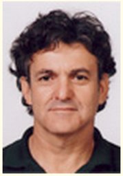 Elazar Zvulun合作论文数Dept Biol Chem, Weizmann Inst Sci9Proikas-Cezanne Tassula合作论文数9
 Jana Sanchez-Wandelmer合作论文数9
 Patricia Boya合作论文数University of Fribourg9Maurizio Molinari合作论文数9Nilima Dinesh Kumar合作论文数8Ralph Hardenberg合作论文数8
 Dengjel Jörn合作论文数University of Freiburg8
 Andreas Conzelmann合作论文数8
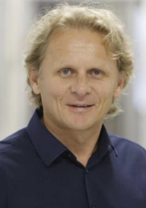 Ivan Đikić合作论文数Institute of Biochemistry II, Goethe-Universität Frankfurt am Main;Vivlion GmbH;Max Planck Institute of Biophysics;Frankfurt Cancer Institute8
 Iryna Monastyrska合作论文数8Lee Allers合作论文数7
 Arlt Henning合作论文数Department of Biology/Chemistry, University of Osnabrück7Ulasli Mustafa合作论文数Faculty of Medicine, University of Gaziantep7
 Terje Johansen合作论文数Department of Medical Biology, Autophagy Research Group, Faculty of Health Science, UiT The Arctic University of Norway7Francesco Cecconi合作论文数7Eduardo Cebollero合作论文数7Aniek van der Vaart合作论文数6Ester Rieter合作论文数6
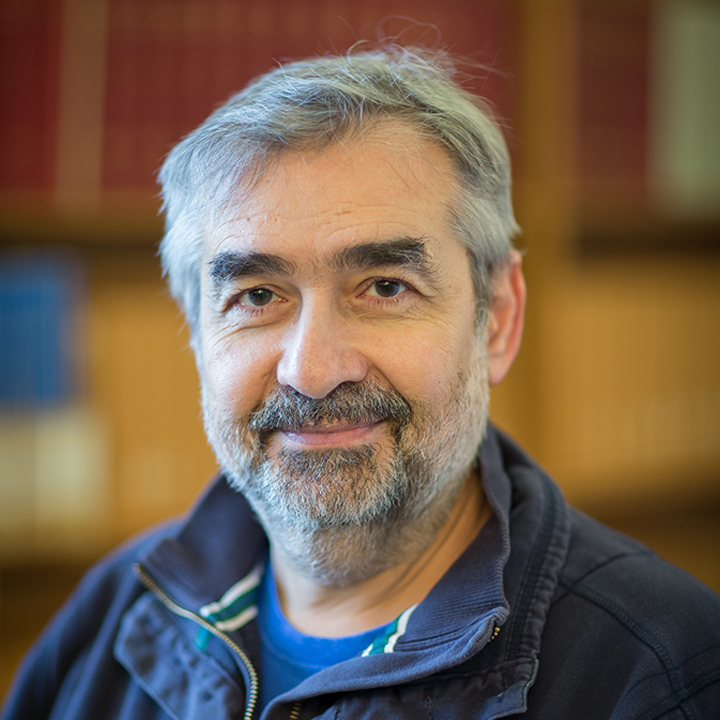 Nicholas Ktistakis合作论文数Babraham Institute6
 Christian Behrends合作论文数Munich Cluster for Systems Neurology, Medical Faculty, Ludwig-Maximilians-Universität
MünchenGermany 6
Christian Behrends合作论文数Munich Cluster for Systems Neurology, Medical Faculty, Ludwig-Maximilians-Universität
MünchenGermany 6 Susana Abreu合作论文数6
 T Yoshimori (吉森保)合作论文数Division of Health Sciences, Graduate School of Medicine, Osaka University6
 Guido Kroemer (吉多·科罗默)合作论文数Faculty of Medicine, University of Paris Cité6Peter J. M. Rottier合作论文数6
 Ana Maria Cuervo合作论文数Department of Developmental & Molecular Biology, Albert Einstein College of Medicine;Department of Medicine, Albert Einstein College of Medicine;Institute for Aging Research, Albert Einstein College of Medicine6
 Christian Münz合作论文数Institute of Experimental Immunology, University of Zurich;Section of Virology, Department of Infectious Disease, Faculty of Medicine, Imperial College London6
 Jayanta Debnath合作论文数Helen Diller Family Comprehensive Cancer Center, University of California San Francisco;Department of Pathology, University of California San Francisco6
 Rodrigo Guimaraes合作论文数5
 Jieqiong Gao合作论文数5
 Maria Isabel Colombo合作论文数5
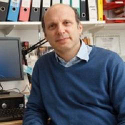 David Rubinsztein合作论文数Department of Medical Genetics, Cambridge Institute for Medical Research, University of Cambridge;UK Dementia Research Institute, University of Cambridge5
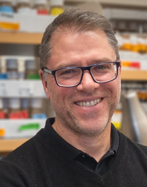 Eric H. Baehrecke合作论文数Department of Molecular, Cell and Cancer Biology, University of Massachusetts Medical School5




