Aim Pathogen inactivation (PI) is a globally recognized method for enhancing the safety of platelet concentrates and plasma. This publication examines the necessity and potential benefits of implementing PI for platelet concentrates in Germany. Method The analysis includes data on the current blood supply, infection risks associated with platelet concentrates, and existing safety standards, also in international comparison. Results The results show that PI improves safety by inactivating known and unknown pathogens, extends shelf life, expands the donor pool, and reduces the number of discarded platelet concentrates. This stabilizes supply and reduces costs. Conclusion The integration of PI into the German hemotherapy guideline and accelerated approval processes are recommended.
Zusammenfassung Zielsetzung Die Pathogeninaktivierung (PI) ist ein weltweit anerkanntes Verfahren zur Verbesserung der Sicherheit von Thrombozytenkonzentraten und Plasma. Diese Arbeit untersucht die Notwendigkeit und potenziellen Vorteile der Implementierung der PI für Thrombozytenkonzentrate in Deutschland. Methodik Analysiert werden sowohl Daten zur aktuellen Blutversorgung als auch zu Infektionsrisiken von Thrombozytenkonzentraten sowie bestehender Sicherheitsstandards auch im interationalen Vergleich. Ergebnisse Die Ergebnisse zeigen, dass PI die Sicherheit durch die Inaktivierung bekannter und unbekannter Pathogene verbessert, die Haltbarkeit verlängert, den Spenderkreis erweitert und die Anzahl verworfener Thrombozytenkonzentrate reduziert. Dies stabilisiert die Versorgung und senkt Kosten. Schlussfolgerung Eine Integration der PI in die deutschen Hämotherapie-Richtlinien und beschleunigte Zulassungsverfahren werden empfohlen.
BackgroundElevated serum ferritin with/without HFE variants in asymptomatic persons leads frequently to referral for blood donation. Hemochromatosis (p.C282Y/p.C282Y) only requires treatment. We evaluated safety and feasibility of iron removal in healthy persons with elevated ferritin and HFE variants using blood donation procedures.Materials and methodsThirty subjects with ferritin >200 ng/mL (women) or >300 ng/mL (men) with p.C282Y/p.C282Y, p.C282Y/p.H63D or p.H63D/p.H63D were randomized to weekly phlebotomy (removal of 450 mL whole blood) or erythrapheresis (removal of 360 mL red blood cells) every 14 days. The ferritin target was <100 ng/mL. A full blood count and ferritin were measured at each visit. Hemoglobin (Hb) ≥140 g/L was required at inclusion. If Hb dropped to <120 g/L (women) or <130 g/L (men), procedures were postponed (7 or 14 days). Primary endpoint was the number of procedures needed to the ferritin target; secondary objectives were duration of treatment and compliance. The treatment effect was tested with Poisson regression; number of procedures and treatment duration were compared between study arms with the Kruskal–Wallis test.ResultsTwenty-five of 30 participants were men (83%); mean age was 47 years (SD 10.5), mean BMI 26.6 kg/m2 (SD 3.6); 17 had p.C282Y/p.C282Y, nine p.C282Y/p.H63D, four p.H63D/p.H63D. Median baseline Hb was 150 g/L (IQR 144, 1,559), median ferritin 504 ng/mL (IQR 406,620). Twenty-seven subjects completed the study. Treatment arm (p < 0.001) and HFE variant (p = 0.007) influenced the primary endpoint significantly. To ferritin levels <100 ng/mL, a median number of 7.5 (IQR 6.2, 9.8) phlebotomies and 4.0 (IQR 3.0, 5.8) erythraphereses (p = 0.001) was needed during a median of 66.5 days (IQR 49,103) and 78.5 days (IQR 46139), respectively (p = 0.448). Low Hb was the principal reason for protocol violation; anemia occurred in 13 participants (48%). Immediate complications were infrequent; fatigue was reported after 25% of phlebotomies and 45% of erythraphereses. Thirty-five procedures were postponed because of low Hb and 15 for non-medical reasons. The median interval was 7.0 (IQR 7.7) and 14.0 (IQR 14, 20) days between phlebotomies and erythraphereses, respectively.ConclusionBlood donation procedures remove iron effectively in HC, but frequent treatments cause Hb decrease and fatigue that can impair feasibility.
Extracellular vesicles (EVs) accumulate during packed red blood cell (PRBC) storage. To date, the involvement of EVs in transfusion-related immunomodulation (TRIM) has not been prospectively evaluated in intensive care unit (ICU) patients. This was a prospective subanalysis of a recent observational feasibility study in postoperative ICU patients after: (1) open aortic surgery (Aorta), (2) bilateral lung transplantation (LuTx), and (3) other types of surgery (Comparison). Patient plasma was collected three times each before and after leukoreduced PRBC transfusion at 30-min intervals. The total number of EVs and EVs derived from erythrocytes (EryEVs), total platelets (total PEVs), activated platelets, granulocytes (GEVs), monocytes, and myeloid cells in PRBC samples and patient plasma were analyzed by flow cytometry. Statistical analysis was performed by Spearman’s correlation test, linear mixed models and pairwise comparisons by Wilcoxon matched-pairs test. Twenty-three patients (Aorta n = 5, LuTx n = 9, Comparison n = 9) were included in the final analysis. All EV subgroups analyzed were detectable in all PRBCs samples (n = 23), but concentrations did not correlate with storage time. Moreover, all EVs analyzed were detectable in all plasma samples (n = 138), and EV counts were consistent before transfusion. Concentrations of total EVs, EryEVs, total PEVs, and GEVs increased after transfusion compared with baseline in the entire cohort but not in specific study groups. Furthermore, the change in plasma EV counts (total EVs and EryEVs) after transfusion correlated with PRBC storage time in the entire cohort. Extracellular vesicles were detectable in all PRBC and plasma samples. Individual EV subtypes increased after transfusion in the entire cohort, and in part correlated with storage duration. Future clinical studies to investigate the role of EVs in TRIM are warranted and should anticipate a larger sample size. Trial registration: Clinicaltrials.gov: NCT03782623.
Introduction: Eicosanoids are bioactive lipids present in packed red blood cells (PRBCs), and might play a role in transfusion-related immunomodulation (TRIM). We tested the feasibility of analyzing eicosanoid profiles in PRBC supernatant and in plasma samples of postoperative intensive care unit (ICU) patients transfused with one unit of PRBCs. Methods: We conducted a prospective, observational feasibility study enrolling postoperative ICU patients: 1) patients treated with acetylsalicylic acid following abdominal aortic surgery (Aorta); 2) patients on immunosuppressants after bilateral lung transplantation (LuTx); and 3) patients undergoing other types of major surgery (Comparison). Abundances of arachidonic acid (AA) and seven pre-defined eicosanoids were assessed by liquid chromatography and tandem mass spectrometry. PRBC supernatant was sampled directly from the unit immediately prior to transfusion. Spearman's correlations between eicosanoid abundance in PRBCs and storage duration were assessed. Patient plasma was collected at 30-min intervals: Three times each before and after transfusion. To investigate temporal changes in eicosanoid abundances, we fitted linear mixed models. Results: Of 128 patients screened, 21 were included in the final analysis (Aorta n = 4, LuTx n = 8, Comparison n = 9). In total, 21 PRBC and 125 plasma samples were analyzed. Except for 20-hydroxyeicosatetraenoic acid (HETE), all analyzed eicosanoids were detectable in PRBCs, and their abundance positively correlated with storage duration of PRBCs. While 5-HETE, 12-HETE/8-HETE, 15-HETE, 20-HETE, and AA were detectable in virtually all plasma samples, 9-HETE and 11-HETE were detectable in only 57% and 23% of plasma samples, respectively. Conclusions: Recruitment of ICU patients into this transfusion study was challenging but feasible. Eicosanoid abundances increased in PRBC supernatants during storage. In plasma of ICU patients, eicosanoid abundances were ubiquitously detectable and showed limited fluctuations over time prior to transfusion. Taken together, larger clinical studies seem warranted and feasible to further investigate the role of PRBC-derived eicosanoids in TRIM.
BackgroundSerological tests are widely used in various medical disciplines for diagnostic and monitoring purposes. Unfortunately, the sensitivity and specificity of test systems are often poor, leaving room for false-positive and false-negative results. However, conventional methods were used to increase specificity and decrease sensitivity and vice versa. Using SARS-CoV-2 serology as an example, we propose here a novel testing strategy: the ‘sensitivity improved two-test’ or ‘SIT²’ algorithm.MethodsSIT² involves confirmatory retesting of samples with results falling in a predefined retesting zone of an initial screening test, with adjusted cut-offs to increase sensitivity. We verified and compared the performance of SIT² to single tests and orthogonal testing (OTA) in an Austrian cohort (1117 negative, 64 post-COVID-positive samples) and validated the algorithm in an independent British cohort (976 negatives and 536 positives).ResultsThe specificity of SIT² was superior to single tests and non-inferior to OTA. The sensitivity was maintained or even improved using SIT² when compared with single tests or OTA. SIT² allowed correct identification of infected individuals even when a live virus neutralisation assay could not detect antibodies. Compared with single testing or OTA, SIT² significantly reduced total test errors to 0.46% (0.24–0.65) or 1.60% (0.94–2.38) at both 5% or 20% seroprevalence.ConclusionFor SARS-CoV-2 serology, SIT² proved to be the best diagnostic choice at both 5% and 20% seroprevalence in all tested scenarios. It is an easy to apply algorithm and can potentially be helpful for the serology of other infectious diseases.
We analysed iron biomarkers and their relationships in 30 subjects with HFE mutations and moderate hyperferritinaemia undergoing iron removal at our blood donation centre. Body mass index (BMI) and liver enzymes were assessed. Serum iron (SI), ferritin, transferrin saturation (TSAT), hepcidin and non-transferrin bound iron (NTBI) were measured serially. Seventeen subjects had p.C282Y/p.C282Y, nine p.C282Y/p.H63D, four p.H63D/p.H63D. Median age (p = 0.582), BMI (p = 0.500) and ferritin (p = 0.089) were comparable. At baseline, 12/17 p.C282Y/p.C282Y and 2/9 p.C282Y/p.H63D had measurable NTBI (p = 0.003). The p. C282Y/p.C282Y had higher TSAT (p < 0.001), lower hepcidin (p = 0.031) and hepcidin/ferritin ratio (p = 0.073). After treatment, iron indices were similar among groups, except TSAT (higher in p.C282Y/p.C282Y; p = 0.06). Strong relationships were observed between ferritin and TSAT (R = 0.71), NTBI and TSAT (R = 0.61), NTBI and SI (R = 0.54) in p.C282Y/p.C282Y. Hepcidin correlated weakly with ferritin in p.C282Y/p.C282Y (R = 0.37) but strongly in p.C282Y/p.H63D (R = 0.66) and p.H63D/p.H63D (R = 0.72), while relationships with TSAT were weak (R = 0.27), moderate (R = 0.55) and strong (R = 0.61), respectively. Low penetrance p.C282Y/p.C282Y phenotype displays hepcidin dysregulation and biochemical risk for iron toxicity.
Pathogen inactivation techniques for blood products have been implemented to optimize clinically safe blood components supply. The INTERCEPT system uses amotosalen together with ultraviolet light wavelength A (UVA) irradiation. Irradiation-induced inactivation of nucleic acids may actually be accompanied by modifications of chemically reactive polyunsaturated fatty acids known to be important mediators of platelet functions. Thus, here, we investigated eicosanoids and the related fatty acids released upon treatment and during storage of platelet concentrates for 7 days, complemented by the analysis of functional and metabolic consequences of these treatments. Metabolic and functional issues like glucose consumption, lactate formation, platelet aggregation, and clot firmness hardly differed between the two treatment groups. In contrast to gamma irradiation, here, we demonstrated that INTERCEPT treatment immediately caused new formation of trans-arachidonic acid isoforms, while 11-hydroxyeicosatetraenoic acid (11-HETE) and 15-HETE were increased and two hydroperoxyoctadecadienoic acid (HpODE) isoforms decreased. During further storage, these alterations remained stable, while the release of 12-lipoxygenase (12-LOX) products such as 12-HETE and 12-hydroxyeicosapentaenoic acid (12-HEPE) was further attenuated. In vitro synthesis of trans-arachidonic acid isoforms suggested that thiol radicals formed by UVA treatment may be responsible for the INTERCEPT-specific effects observed in platelet concentrates. It is reasonable to assume that UVA-induced molecules may have specific biological effects which need to be further investigated.
In severely ill patients undergoing urgent heart transplant (HTX), immunosuppression carries high risks of infection, malignancy, and death. Low-dose immunosuppressive protocols have higher rejection rates. We combined extracorporeal photopheresis (ECP), an established therapy for acute rejection, with reduced-intensity immunosuppression. Twenty-eight high-risk patients (13 with high risk of infection due to infection at the time of transplant, 7 bridging to transplant via extracorporeal membrane oxygenation, 8 with high risk of malignancy) were treated, without induction therapy. Prophylactic ECP for 6 months (24 procedures) was initiated immediately postoperatively. Immunosuppression consisted of low-dose tacrolimus (8-10 ng/ml, months 1-6; 5-8 ng/ml, >6 months) with delayed start; mycophenolate mofetil (MMF); and low maintenance steroid with delayed start (POD 7) and tapering in the first year. One-year survival was 88.5%. Three patients died from infection (POD 12, 51, 351), and one from recurrence of cancer (POD 400). Incidence of severe infection was 17.9% (n = 5, respiratory tract). Within the first year, antibody-mediated rejection was detected in one patient (3.6%) and acute cellular rejection in four (14.3%). ECP with reduced-intensity immunosuppression is safe and effective in avoiding allograft rejection in HTX recipients with risk of severe infection or cancer recurrence.
Background: Antibody tests are essential tools to investigate humoral immunity following SARS-CoV-2 infection or vaccination. While first-generation antibody tests have primarily provided qualitative results, accurate seroprevalence studies and tracking of antibody levels over time require highly specific, sensitive and quantitative test setups. Methods: We have developed two quantitative, easy-to-implement SARS-CoV-2 antibody tests, based on the spike receptor binding domain and the nucleocapsid protein. Comprehensive evaluation of antigens from several biotechnological platforms enabled the identification of superior antigen designs for reliable serodiagnostic. Cut-off modelling based on unprecedented large and heterogeneous multicentric validation cohorts allowed us to define optimal thresholds for the tests’ broad applications in different aspects of clinical use, such as seroprevalence studies and convalescent plasma donor qualification. Findings: Both developed serotests individually performed similarly-well as fully-automated CE-marked test systems. Our described sensitivity-improved orthogonal test approach assures highest specificity (99.8%); thereby enabling robust serodiagnosis in low-prevalence settings with simple test formats. The inclusion of a calibrator permits accurate quantitative monitoring of antibody concentrations in samples collected at different time points during the acute and convalescent phase of COVID-19 and disclosed antibody level thresholds that correlate well with robust neutralization of authentic SARS-CoV-2 virus. Interpretation: We demonstrate that antigen source and purity strongly impact serotest performance. Comprehensive biotechnology-assisted selection of antigens and in-depth characterisation of the assays allowed us to overcome limitations of simple ELISA-based antibody test formats based on chromometric reporters, to yield comparable assay performance as fully-automated platforms. Funding: WWTF, Project No. COV20–016; BOKU, LBI/LBG
Therapeutic apheresis (TA) is prescribed to patients that suffer from a severe progressive disease that is not sufficiently treated by conventional medications. A way to gain more knowledge about this treatment is usually by the local analysis of data. However, the use of large quality assessment registries enables analyses of even rare findings. Here, we report some of the recent data from the World Apheresis Association (WAA) registry. Data from >104,000 procedures were documented, and TA was performed on >15,000 patients. The main indication for TA was the collection of autologous stem cells (45% of patients) as part of therapy for therapy. Collection of stem cells from donors for allogeneic transplantation was performed in 11% of patients. Patients with indications such as neurological diseases underwent plasma exchange (28%). Extracorporeal photochemotherapy, lipid apheresis, and antibody removal were other indications. Side effects recorded in the registry have decreased significantly over the years, with approximately only 10/10,000 procedures being interrupted for medical reasons.CONCLUSION:Collection of data from TA procedures within a multinational and multicenter concept facilitates the improvement of treatment by enabling the analysis of and feedback on indications, procedures, effects, and side effects.
Antibody tests are essential tools to investigate humoral immunity following SARS-CoV-2 infection. While first-generation antibody tests have primarily provided qualitative results with low specificity, accurate seroprevalence studies and tracking of antibody levels over time require highly specific, sensitive and quantitative test setups. Here, we describe two quantitative ELISA antibody tests based on the SARS-CoV-2 spike receptor-binding domain and the nucleocapsid protein. Comparative expression in bacterial, insect, mammalian and plant-based platforms enabled the identification of new antigen designs with superior quality and high suitability as diagnostic reagents. Both tests scored excellently in clinical validations with multi-centric specificity and sensitivity cohorts and showed unprecedented correlation with SARS-CoV-2 neutralization titers. Orthogonal testing increased assay specificity to 99.8%, thereby enabling robust serodiagnosis in low-prevalence settings. The inclusion of a calibrator permits accurate quantitative monitoring of antibody concentrations in samples collected at different time points during the acute and convalescent phase of COVID-19.
Background and aimsNon-O blood type (BT) is a risk factor for thromboses, which has been attributed to its effects on von Willebrand factor (VWF)/factor VIII (FVIII) levels. Although high VWF/FVIII may be risk factors for portal vein thrombosis (PVT) in patients with advanced chronic liver disease (ACLD), the impact of BT on PVT is unknown. We aimed to assess (I) whether non-O-BT is a risk factor for PVT and (II) whether non-O-BT impacts VWF/factor VIII in patients with ACLD. MethodsRetrospective analysis comprising two cohorts: (I) "US" including all adult liver transplantations in the US in the MELD era and (II) "Vienna" comprising patients with a hepatic venous pressure gradient (HVPG) >= 6 mmHg. Results(I) The "US cohort" included 84 947 patients (non-O: 55.43%). The prevalence of PVT at the time of listing (4.37% vs 4.56%; P = .1762) and at liver transplantation (9.56% vs 9.33%; P = .2546) was similar in patients with O- and non-O-BT. (II) 411 patients were included in the "Vienna cohort" (non-O: 64%). Mean HVPG was 18(9) mmHg and 90% had an HVPG >= 10 mmHg. Patients with non-O-BT had slightly increased VWF levels (318(164)% vs 309(176)%; P = .048; increase of 23.8%-23.9% in adjusted analyses), but this difference was driven by patients with less advanced disease. However, non-O-BT explained only 1% of the variation in VWF and had no effect on FVIII. ConclusionsAlthough non-O-BT impacts VWF in patients with early stage ACLD, its contribution to VWF variation is considerably smaller than in the general population. Moreover, non-O-BT had no impact on FVIII. These findings may explain the absence of an association between non-O-BT and PVT in patients with advanced cirrhosis.
BACKGROUND:Pathogen inactivation (PI) techniques for platelet concentrates (PCs) are one of the latest innovations to improve blood safety and reduce the risk of transfusion-transmitted infections (TTIs). An impaired function and in vivo recovery of platelets as well as an increased PC demand are concerns regarding these techniques. The intent of this study was to evaluate the hemostatic effect of PCs treated with the Intercept™ System by thromboelastometry (TEM) and to assess the clinical validity of its results in comparison to post-transfusion increase (PTI) and corrected count increment (CCI). STUDY-DESIGN AND METHODS:This prospective-observational study included 47 patients (m:f = 25:22; median age: 54 years [21-70]) of our Bone Marrow Transplantation unit with hemato-oncological malignancies transfused with Intercept™-treated PCs. Serial TEM measurements were performed just before and 1 hour after PC transfusion and were analyzed for their correlation with PTI and CCI as well as for clinical variables. RESULTS:The majority of our patients had received a hematopoietic stem cell transplantation (HSCT) (n = 41; 87%). In median 9 (1-50) PCs were transfused. Serial TEM, PTI, and CCI measurements were available for 150 transfusion episodes. The median platelet dose transfused was 2.65 × 10¹¹ /unit (1.8-6). The median CCI was 9.250 (0-28.000). We observed a significant improvement in TEM parameters (p < 0.05) after transfusion of PI PCs, which did not mandatory correlate with the 1-hour PTI and CCI. CONCLUSION:Serial TEM measurements indicate the hemostatic effect of Intercept™-treated PCs. The 1-hour PTI and CCI may not appropriately reflect the in vivo function of platelets after PI PC transfusion.
Background SARS-CoV-2 antibody tests have undergone a remarkable improvement in performance. However, due to the low seroprevalence in several areas, very high demands are made on their specificity. Furthermore, the low antibody-response in some individuals requires high test sensitivity to avoid underestimating true seroprevalence. Optimization of testing has been reported through lowering manufacturer cut-offs to improve SARS-CoV-2 assay sensitivity or by combining two tests to improve specificity at the cost of sensitivity. However, these strategies have thus far been used in isolation of each other. Methods To increase sensitivity, cut-offs of three commercially available SARS-CoV-2 automated assays (Roche, Abbott, and DiaSorin) were reduced according to published values in a pre-pandemic specificity cohort (n=1117) and a SARS-CoV-2 positive cohort (n=64). All three testing systems were combined in an orthogonal approach with a confirmatory test, which was one of the remaining automated assays or one of two commercial ELISAs directed against the spike protein receptor binding-domain (RBD) or the nucleocapsid antigen (NP). Results The modified orthogonal test strategy resulted in an improved specificity of at least 99.8%, often even 100%, in all 12 tested combinations with no significant decline in sensitivity. In our cohort, regardless of whether the assays were used for screening or confirmation, combining Roche and Abbott delivered the best overall performance (+~10% sensitivity compared to the single tests and 100% specificity). Conclusion Here we propose a novel orthogonal assay strategy that approaches 100% specificity while maintaining or even significantly improving the screening test's sensitivity.
Abstract Objective To identify a pharmacological compound targeting macrophages, the most affected immune cells in inflammatory X‐linked adrenoleukodystrophy (cerebral X‐ALD) caused by ABCD1 mutations and involved in the success of hematopoietic stem cell transplantation and gene therapy. Methods A comparative database analysis elucidated the epigenetic repressing mechanism of the related ABCD2 gene in macrophages and identified the histone deacetylase (HDAC) inhibitor Vorinostat as a compound to induce ABCD2 in these cells to compensate for ABCD1 deficiency. In these cells, we investigated ABCD2 and pro‐inflammatory gene expression, restoration of defective peroxisomal β‐oxidation activity, accumulation of very long‐chain fatty acids (VLCFAs) and their differentiation status. We investigated ABCD2 and pro‐inflammatory gene expression, restoration of defective peroxisomal ß‐oxidation activity, accumulation of very long‐chain fatty acids (VLCFA) and differentiation status. Three advanced cerebral X‐ALD patients received Vorinostat and CSF and MRI diagnostics was carried out in one patient after 80 days of treatment. Results Vorinostat improved the metabolic defects in X‐ALD macrophages by stimulating ABCD2 expression, peroxisomal ß‐oxidation, and ameliorating VLCFA accumulation. Vorinostat interfered with pro‐inflammatory skewing of X‐ALD macrophages by correcting IL12B expression and further reducing monocyte differentiation. Vorinostat normalized the albumin and immunoglobulin CSF‐serum ratios, but not gadolinium enhancement upon 80 days of treatment. Interpretation The beneficial effects of HDAC inhibitors on macrophages in X‐ALD and the improvement of the blood‐CSF/blood‐brain barrier are encouraging for future investigations. In contrast with Vorinostat, less toxic macrophage‐specific HDAC inhibitors might improve also the clinical state of X‐ALD patients with advanced inflammatory demyelination.
BackgroundThe generation of functional blood vessels remains a key challenge for regenerative medicine. Optimized in vitro culture set-ups mimicking the in vivo perivascular niche environment during tissue repair may provide information about the biological function and contribution of progenitor cells to postnatal vasculogenesis, thereby enhancing their therapeutic potential.AimWe established a fibrin-based xeno-free human 3D in vitro vascular niche model to study the interaction of mesenchymal stromal cells (MSC) with peripheral blood mononuclear cells (PBMC) including circulating progenitor cells in the absence of endothelial cells (EC), and to investigate the contribution of this cross-talk to neo-vessel formation.Materials and MethodsBone marrow-derived MSC were co-cultured with whole PBMC, enriched monocytes (Mo), enriched T cells, and Mo together with T cells, respectively, obtained from leukocyte reduction chambers generated during the process of single-donor platelet apheresis. Cells were embedded in 3D fibrin matrices, using exclusively human-derived culture components without external growth factors. Cytokine secretion was analyzed in supernatants of 3D cultures by cytokine array, vascular endothelial growth factor (VEGF) secretion was quantified by ELISA. Cellular and structural re-arrangements were characterized by immunofluorescence and confocal laser-scanning microscopy of topographically intact 3D fibrin gels.Results3D co-cultures of MSC with PBMC, and enriched Mo together with enriched T cells, respectively, generated, within 2 weeks, complex CD31+/CD34+ vascular structures, surrounded by basement membrane collagen type-IV+ cells and matrix, in association with increased VEGF secretion. PBMC contained CD31+CD34+CD45dimCD14– progenitor-type cells, and EC of neo-vessels were PBMC-derived. Vascular structures showed intraluminal CD45+ cells that underwent apoptosis thereby creating a lumen. Cross-talk of MSC with enriched Mo provided a pro-angiogenic paracrine environment. MSC co-cultured with enriched T cells formed “cell-in-cell” structures generated through internalization of T cells by CD31+CD45dim/– cells. No vascular structures were detected in co-cultures of MSC with either Mo or T cells.ConclusionOur xeno-free 3D in vitro vascular niche model demonstrates that a complex synergistic network of cellular, extracellular and paracrine cross-talk can contribute to de novo vascular development through self-organization via co-operation of immune cells with blood-derived progenitor cells and MSC, and thereby may open a new perspective for advanced vascular tissue engineering in regenerative medicine.
Reliable antibody tests are an essential tool to identify individuals who have developed an adaptive immune response to SARS-CoV-2. However, attempts to maximize the specificity of SARS-CoV-2 antibody tests have come at the cost of sensitivity, exacerbating the total test error with increasing seroprevalence. Here, we present a novel method to maximize specificity while maintaining or even increasing sensitivity: the “Sensitivity Improved Two-Test” or “SIT²” algorithm.SIT² involves confirmatory re-testing of samples with results falling in a predefined retesting-zone of an initial screening test, with adjusted cut-offs to increase sensitivity. We verified and compared the performance of SIT² to single tests and orthogonal testing (OTA) in an Austrian cohort (1,117 negative, 64 post-COVID positive samples) and validated the algorithm in an independent British cohort (976 negatives, 536 positives).The specificity of SIT² was superior to single tests and non-inferior to OTA. The sensitivity was maintained or even improved using SIT² when compared to single tests or OTA. SIT² allowed correct identification of infected individuals even when a live virus neutralization assay could not detect antibodies. Compared to single testing or OTA, SIT² significantly reduced total test errors to 0·46% (0·24-0·65) or 1·60% (0·94-2·38) at both 5% or 20% seroprevalence.SIT² proved to be the best diagnostic choice at both 5% and 20% seroprevalence in all tested scenarios. It is an easy algorithm to apply to different available SARS-CoV-2 antibody testing systems and can potentially be helpful for the serology of other infectious diseases.
 Nina Worel合作论文数Department of Blood Group Serology and Transfusion Medicine, Medical University of Vienna26Simon Panzer合作论文数14Werner Rabitsch合作论文数13M Horvath合作论文数12Gabriela Stiegler合作论文数12Paul Hoecker合作论文数11
 Peter Quehenberger合作论文数10Paul Hocker合作论文数10Peter Kalhs合作论文数9
 Hildegard Greinix合作论文数Medical University of Vienna9Michael B Fischer合作论文数8P Kalhs合作论文数7Michaela Horvath合作论文数7Renz Renate合作论文数6Margit Pavelka合作论文数6Christof Jungbauer合作论文数6
 Andreas S. Buser合作论文数Reg Blood Transfus Serv, Swiss Red Cross6Adolf Ellinger合作论文数6
 Josef Neumuller合作论文数6Margit Mitterbauer合作论文数6Gerjan Fleurke合作论文数5Yvonne Fischer合作论文数5Thijs C van Holten合作论文数5
 Audrey Labrie合作论文数5
 Christian Weidmann合作论文数Fac Hlth Safety & Soc, Hsch Furtwangen5List J合作论文数5
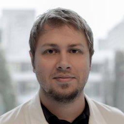 Helmuth Haslacher合作论文数Dept Lab Med, Med Univ Vienna5
 Thomas Volken合作论文数5Rummler Silke合作论文数5von Papen Michael合作论文数5Giussani Barbara合作论文数5Da Prada Luca合作论文数5Gargantini Chiara合作论文数5Giaccherini Cinzia合作论文数5Bedi Harjot合作论文数5
 Thomas Bart合作论文数5
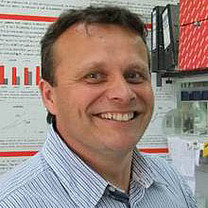 Peter Bugert合作论文数Institute of Transfusion Medicine and Immunology, Heidelberg University, Medical Faculty Mannheim, German Red Cross Blood Service Baden-Wurttemberg – Hessen5
 Bernd Jilma合作论文数Department of Clinical Pharmacology Medical University of Vienna5
 Thomas Perkmann合作论文数Department of Laboratory Medicine, Medical University of Vienna5
 Christoph J. Binder合作论文数Department of Laboratory Medicine, Medical University of Vienna5Marie-Kathrin Breyer合作论文数5
 Petra Jilma-Stohlawetz合作论文数5
 Jasper A Remijn合作论文数5Alfonso Vignoli合作论文数Department of Mathematics;University of Florence;Department of Mathematics, University of Florence5
 Beate Kehrel合作论文数Faculty of Medicine and Health Sciences, University of Nottingham;School of Medicine, University of Nottingham;University Hospital Münster;OxProtect GmbH5Andrea Doescher合作论文数5Guenter Weigel合作论文数5Harald Kluter合作论文数5Martin F. Brodde合作论文数5




