Proliferative growth of the ventricular myocyte (cardiomyocyte) is primarily limited to fetal and early neonatal periods of development. In concert with the neonatal "transition" from proliferative to hypertrophic growth, ventricular remodeling of the nonmyocyte compartment is characterized by increased extracellular matrix synthesis/deposition and capillary angiogenesis. A role for locally generated and bioactive ventricular acidic fibroblast growth factor (aFGF) in these processes is proposed and substantiated by the following: 1) colocalization of aFGF peptide and fibroblast growth factor receptor (fig) transcripts to the developing fetal cardiomyocyte by immunohistochemistry, immunoelectron microscopy, and in situ hybridization, 2) continued localization of aFGF peptide and transcripts to the neonatal/mature cardiomyocyte, and 3) localization offig immunoreactivity and transcripts to specific neonatal ventricular nonmuscle cell types. Specific ventricular cell types at distinct developmental stages appear to be responsive to ventricular myocyte-derived aFGF (myocytes in the fetal heart and nonmyocytes/endothelial cells in the neonatal heart). These data indicate that expression of aFGF and one of its receptors (fig) are most pronounced in the fetal to early neonatal ventricle, the presence of both suggesting an autocrine/paracrine growth regulatory function. As the animal matures, ventricular capillary angiogenesis may be facilitated by "release" of cardiomyocyte-derived fibroblast growth factors into the surrounding extracellular space/matrix functioning as a "paracrine" angiogenic stimuli. Therefore, the results of our study suggest that myocyte-derived aFGF may function to increase the fetal ventricular cardiomyocyte population in absolute number as well as to facilitate the subsequent increase in capillary angiogenesis that occurs during cardiomyocyte maturation and ventricular remodeling. (Circulation Research 1993;72:7-19)
Proliferative growth of the ventricular myocyte (cardiomyocyte) is primarily limited to embryonic, fetal and very early neonatal periods of heart development. In contrast, cardiomyocyte maturation, as evidenced by cellular hypertrophy, is a long-term process that can occupy the bulk of the life-span of the mature organism. As the newborn myocyte undergoes a 'transition' from proliferative to hypertrophic growth, ventricular remodeling of the non-myocyte compartment is characterized by increased extracellular matrix (ECM) formation and coronary capillary angiogenesis. A role for ventricular-derived growth factors (GFs) in these inter-related processes are examined in an animal model of altered heart development produced by neonatal aortic banding. The suprarenal abdominal aorta of five day old rat pups were banded (B), sham operated (S), or untreated (C) and ventricular tissue (left ventricular free wall and septum) obtained at 7-, 14-, and 21-days post-intervention. Using Northern blot RNA hybridizations, expression of growth factors (GFs) and/or GF-receptors (GFR's) temporally associated with heart development were evaluated. Transcript levels for TGF-beta 1, IGF-II, and their associated cell surface receptors were increased in B animals. Concomitant changes in extracellular matrix (ECM) genes (as evaluated by Collagens Type I, III, and IV) were also increased in B animals. In addition, transcript levels for the vascular morphogenesis and remodeling-related protein SPARC (Secreted Protein, Acidic and Rich in Cysteine) was also elevated in the B animals. In several instances, S animals demonstrated changes in steady state transcript levels for genes which may influence myocyte maturation during the postnatal period. This suggests that normal autocrine/paracrine growth regulatory stimuli and responses can be modified (by surgical intervention and/or abdominal aortic banding) and these perturbations in gene expression may be related to previously documented changes in myocyte cell number, vascular composition, and ventricular architecture of the banded, neonatal heart. Future studies using this model will provide an opportunity to evaluate and possibly identify the stimuli and signal transduction machinery that regulate the final phases of myocyte proliferation, stimulate capillary formation and ECM deposition, and orchestrate the transition to hypertrophic growth during heart development.
Recently developed rat heart myocyte cell lines have afforded us the opportunity to evaluate the expression of several transcription factors associated with early cardiac development. These factors include, but are not limited to, Nkx-2.5/Csx, MEF-2C and MLP (Muscle LIM Protein). These factors have been shown to be temporally expressed in pre-cardiac mesenchyme coincident with the earliest stages of heart development. Using the BWEM and CLEM myocyte cell lines as models of the embryonic, committed cardiomyocyte, we have evaluated the basal expression levels of these three genes over multiple passages. Both cell lines express these genes, with MEF-2C being the most abundant based on Northern blot hybridization analyses. Interestingly, as these cells increased their passage number, there was a corresponding increase in their basal expression levels. To evaluate potential ‘downstream’ effectors of these genes, we examined the basal expression levels of two cardiac-specific genes cTNC and MLC-2v. Transcript levels for both of these contractile filament genes were elevated with passage, suggestive of a inductive process mediated by one or all of these three transcription factors. Promoter analysis of MLC-2v expression in the CLEM line shows that this increase is transcriptionally-mediated and the lines retain the necessary regulatory factors to maintain and control the transcription of this gene. Analysis of the dynamics of the regulatory role(s) that these three transcription factors play in cardiac development can now be evaluated in a homogeneous, cell culture system.
TGF-beta affects proliferation, differentiation and maturation of T cells; however, the effect of TGF-beta on thymic stromal cells has not been characterized. To better understand the role of TGF-beta in T cell development, we determined whether TGF-beta is present in the human thymus, and identified stromal cells that express TGF-beta receptors and respond to TGF-beta. We demonstrate that primary cultured human thymic epithelial cells (TEC) express TGF-beta 1, TGF-beta 2 and TGF-beta 3, as well as TGF-beta type I receptor (T beta RI) (ALK-5) and TGF-beta type II receptor (T beta RII) transcripts. In vitro, epidermal growth factor (EGF) increases transcript levels of TGF-beta 1, TGF-beta 3 and T beta RII, suggesting that EGF may modulate TGF-beta responses in TEC; however, TGF-beta 2 and T beta RI transcript levels were not affected. We also detect TGF-beta 3 and T beta RII protein in association with keratin-positive TEC in vitro and in vivo. TEC culture supernatants contain TGF-beta 3 as detected by Western blots and, upon heat and acid activation, display growth inhibitory activity on the CCL-64 cells that is neutralized by anti-TGF-beta mAb treatment. We further demonstrate that TGF-beta 1 increases leukemia inhibitory factor transcript levels in TEC, indicating that TEC express functional TGF-beta receptors. Thus, we have shown in the human thymus that TEC produce TGF-beta 3 and express T beta RI and T beta RII. The data suggest that TGF-beta is present in the human thymus and may indirectly affect T cell development by regulating TEC cytokine production.
Stenotic or occlusive (two different techniques) lesions were surgically induced in the canine common carotid artery. The lesions were evaluated angiographically prior to as well as after laser angiosurgery (LAS). LAS was performed in 30 dogs using a continuous wave (CW) argon ion laser (Innova 20) coupled to a multifiber, quartz-shielded, fiberoptic catheter. Successful removal of lesion tissue was achieved in 89% of the animals with stenoses and in 40% and 82% in each of the two groups of dogs with occlusions. In the latter two groups, perforation of the vessel wall during LAS was encountered in 60% and 18%, respectively. No perforations occurred in the stenotic animals, where long-term follow-up was possible for up to 60 days with a patency rate of 63% and morphological evidence of excellent healing with complete reendothelialization.
In conjunction with our project in laser angiosurgery, our group has demonstrated that laser-induced auto-fluorescence can be a reliable means of distinguishing normal artery from atherosclerotic plaque. Recently a number of groups have become interested in this technique as a means of directing ablation of arterial obstructions in vivo. However, until now, diagnostic spectroscopy has been conducted almost solely on excised cadaveric tissues. As a result, questions still remain about 1) the validity of correlating cadaveric tissue fluorescence spectra with those collected in vivo, and 2) the ability to collect reliable florescence spectra from a subject as dynamic as a living patient.
Sodium butyrate, at millimolar concentrations, seems to mediate or initiate multiple effects on many mammalian cells in culture. Although many transformed cell lines respond to butyrate treatment with acquisition of normal cellular characteristics, the effect of butyrate on a normal cell type, the parenchymal hepatocyte, has not been studied. Serum-free primary cultures of adult rat hepatocytes maintain many adult characteristics, yet after several days in culture a loss of adult characteristics occurs while fetal characteristics are often reexpressed. Therefore, we investigated whether butyrate treatment would improve the morphologic and biochemical characteristics of cultured hepatocytes. Exposure to 5 mM butyrate for 3 d did not affect hepatocyte viability or morphology but retarded the progressive decline in cytochrome P-450 levels and 5′-nucleotidase activity. The spontaneous increase in alkaline phosphatase activity was reduced and the induction of tyrosine aminotransferase was inhibited after 3 d in culture. The fetal liver characteristic, gamma glutamyltranspeptidase, was not affected by butyrate treatment. Results of this study suggest that butyrate represents a nontoxic compound capable of improving the maintenance of cell culture characteristics of adult rat hepatocytes.
We describe in detail the technique of hepatocyte isolation and establishment of primary cell cultures of adult rat hepatocytes. These cultures contain hormonally responsive hepatocytes that retain many adult characteristics under completely serum-free conditions. The cells retain a normal morphology and do not exhibit fetal characteristics during a 4 d culture period.
 Arlan Richardson合作论文数College of Medicine, The University of Oklahoma;Oklahoma City VA Medical Center, The University of Oklahoma3
 Rebecca Richards-Kortum合作论文数Rice 360° Institute for Global Health, Rice University2
 Michael Stephen Feld合作论文数George R. Harrison Spectroscopy Laboratory, Massachusetts Institute of Technology2
 Carter Kittrell合作论文数2Allen M. Samarel合作论文数2Joshua A. Fierer合作论文数2Patricia S. Grutkoski合作论文数2Robert M. Cothren合作论文数2Tatiana Kolubayev合作论文数2N RATLIFF合作论文数2gary b hayes合作论文数1F LOOP合作论文数1Jf Haskell合作论文数1John R Kramer合作论文数1JR KRAMER合作论文数1Scott E. Campbell合作论文数1michael jaye合作论文数1Cook J E合作论文数1Bott-Silverman Corinne合作论文数1Kjellström B T合作论文数1
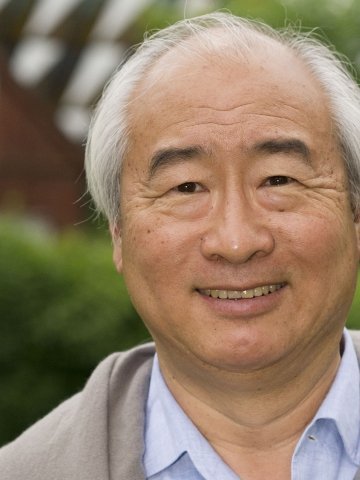 Kenneth Chien (钱肯)合作论文数Department of Cell and Molecular Biology, Karolinska Institutet1
 Richard Harvey合作论文数Development and Stem Cell Laboratory, Victor Chang Cardiac Research Institute;Developmental and Regenerative Biology Division, Victor Chang Cardiac Research Institute;University of New South Wales1Karel Rakusan合作论文数1David G. Penney合作论文数1
 Phong Le合作论文数1Keith D. Boehm合作论文数1Elizabeth J Pielsticker合作论文数1B. Thomas Kjellstrom合作论文数1Robert A. Worrell合作论文数1Maryann Fitzmaurice合作论文数1Jeffrey L. Staecker合作论文数1Richard A. Duff合作论文数1
 Kimberly S Schluns合作论文数1Craig A. Dionne合作论文数1




