Nonspeech (or paraspeech) parameters are widely used in clinical assessment of speech impairment in persons with dysarthria (PWD). Virtually every standard clinical instrument used in dysarthria diagnostics includes nonspeech parameters, often in considerable numbers. While theoretical considerations have challenged the validity of these measures as markers of speech impairment, only a few studies have directly examined their relationship to speech parameters on a broader scale. This study was designed to investigate how nonspeech parameters commonly used in clinical dysarthria assessment relate to speech characteristics of dysarthria in individuals with movement disorders. Maximum syllable repetition rates, accuracies, and rates of isolated and repetitive nonspeech oral-facial movements and maximum phonation times were compared with auditory-perceptual and acoustic speech parameters. Overall, 23 diagnostic parameters were assessed in a sample of 130 patients with movement disorders of six etiologies. Each variable was standardized for its distribution and for age and sex effects in 130 neurotypical speakers. Exploratory Graph Analysis (EGA) and Confirmatory Factor Analysis (CFA) were used to examine the factor structure underlying the diagnostic parameters. In the first analysis, we tested the hypothesis that nonspeech parameters combine with speech parameters within diagnostic dimensions representing domain-general motor control principles. In a second analysis, we tested the more specific hypotheses that diagnostic parameters split along effector (lip vs. tongue) or functional (speed vs. accuracy) rather than task boundaries. Our findings contradict the view that nonspeech parameters currently used in dysarthria diagnostics are congruent with diagnostic measures of speech characteristics in PWD.
Assessment of semantic processing capacities often relies on verbal tasks which are, however, sensitive to impairments at several language processing levels. Especially for persons with aphasia there is a strong need for a tool that measures semantic processing skills independent of verbal abilities. Furthermore, in order to assess a patient's potential for using alternative means of communication in cases of severe aphasia, semantic processing should be assessed in different nonverbal conditions. The Nonverbal Semantics Test (NVST) is a tool that captures semantic processing capacities through three tasks-Semantic Sorting, Drawing, and Pantomime. The main aim of the current study was to investigate the relationship between the NVST and measures of standard neurolinguistic assessment. Fifty-one persons with aphasia caused by left hemisphere brain damage were administered the NVST as well as the Aachen Aphasia Test (AAT). A principal component analysis (PCA) was conducted across all AAT and NVST subtests. The analysis resulted in a two-factor model that captured 69% of the variance of the original data, with all linguistic tasks loading high on one factor and the NVST subtests loading high on the other. These findings suggest that nonverbal tasks assessing semantic processing capacities should be administered alongside standard neurolinguistic aphasia tests.
Neurological patients with apraxia of pantomime provide us with a unique opportunity to study the neural correlates of higher-order motor function. Previous studies using lesion-behaviour mapping methods led to inconsistent anatomical results, reporting various lesion locations to induce this symptom. We hypothesised that the inconsistencies might arise from limitations of mass-univariate lesion-behaviour mapping approaches if our ability to pantomime the use of objects is organised in a brain network. Thus, we investigated apraxia of pantomime by using multivariate lesion behaviour mapping based both on support vector regression and sparse canonical correlations in a sample of 130 left-hemisphere stroke patients. Both multivariate methods identified multiple areas to underlie high-order motor control, including inferior parietal lobule, precentral gyrus, posterior parts of middle temporal cortex, and insula. Further, long association fibres were affected, such as the superior longitudinal fascicle, inferior occipito-frontal fascicle, uncinated fascicle, and superior occipito-frontal fascicle. The findings underline the benefits of multivariate lesion-behaviour mapping in brain networks and provide new insights into the brain networks underlying higher-order motor control.
For over a century, pantomime of tool use has been employed to diagnose limb apraxia, a disorder of motor cognition primarily induced by left brain damage. While research consistently implicates damage to a left frontotemporo-parietal network in limb apraxia, findings are inconsistent regarding the impact of damage to anterior versus posterior nodes within this network on pantomime. Complicating matters is the fact that tool use pantomime can be affected and evaluated at multiple levels. For instance, the production of tool use gestures requires the consideration of semantic characteristics (e.g. how to communicate the action intention) as well as motor features (e.g. forming grip and movement). Together, these factors may contribute substantially to apparent discrepancies in previously reported findings regarding neural correlates of tool use pantomime. In the current study, 67 stroke patients with unilateral left-brain damage performed a classic pantomime task. In order to analyze different error characteristics, we evaluated the proper use of grip and movement for each pantomime. For certain objects, healthy subjects may use body parts as representative for the object, e.g. use of the fingers to indicate scissors blades. To specify the pathological use of body parts as the object (BPO) we only assessed pantomime items that were not prone to this response in healthy participants. We performed modern voxel-based lesion analyses on MRI or CT data to determine associations between brain injury and the frequency of the specific types of pantomime errors. Our results support a model in which anterior and posterior nodes of the left fronto-temporo-parietal network contribute differentially to pantomime of tool use. More precisely, damage in the inferior frontal cortex reaching to the temporal pole is associated with an increased frequency of BPO errors, whereas damage to the inferior parietal lobe is predominantly linked to an increased frequency of movement and/or grip errors. Our work suggests that the validity of attempts to specify the neural correlates of limb apraxia based on tool use pantomime depends on differentiating the specific types of errors committed. We conclude that successful tool use pantomime involves dissociable functions with communicative aspects represented in more anterior (rather ventral) regions and motor-cognitive aspects in more posterior (rather dorsal) nodes of a left fronto-temporoparietal network.
The term apraxia denotes 'higher-order' disturbances of motor control. Apraxia differs from more mundane disorders of motor skill like hemiparesis or ataxia by its bilaterality: unilateral hemisphere lesions cause apraxia of both limbs. Consequently, apraxia can be demonstrated on the limb ipsilateral to the lesioned hemisphere and contrasts with normal motor skill of the same limb in other situations. This definition embraces a wide variety of heterogeneous disturbances, but traditionally diagnosis and research on apraxia concentrate on three domains of human motor actions: imitation of gestures, performance of communicative gestures on command, and skillful use of tools and objects.Disturbances of these domains have traditionally been interpreted as affecting subsequent stages of a unique "praxis system" that is centered on left parietal regions, but empirical evidence does not fit well with this postulate. We discuss each of the manifestations on their own with an emphasis on the laterality of lesions and of the significance of parietal lesions.
Communicative gestures can compensate incomprehensibility of oral speech in severe aphasia, but the brain damage that causes aphasia may also have an impact on the production of gestures. We compared the comprehensibility of gestural communication of persons with severe aphasia and non-aphasic persons and used voxel based lesion symptom mapping (VLSM) to determine lesion sites that are responsible for poor gestural expression in aphasia. On group level, persons with aphasia conveyed more information via gestures than controls indicating a compensatory use of gestures in persons with severe aphasia. However, individual analysis showed a broad range of gestural comprehensibility. VLSM suggested that poor gestural expression was associated with lesions in anterior temporal and inferior frontal regions. We hypothesize that likely functional correlates of these localizations are selection of and flexible changes between communication channels as well as between different types of gestures and between features of actions and objects that are expressed by gestures.
This study examined how 5- and 9-year-old children (N=40) produce pantomimes of object use on verbal request. The task required participants to enact an action with an imagined object. Results showed that with age, children (a) proceeded from body part as object to imaginary object and (b) incorporated into their pantomimes more distinctive features of the object and its use. While the first result replicated previous findings with more items and a more sophisticated coding procedure, the latter result revealed new insights. Children's selection of distinctive features in pantomime was related to their performance on a semantic odd-one-out task that requires the abstraction of specific features without situational support. The discussion focuses on the cognitive requirements of the task and the potential of pantomime being a reflection of children's representational development. We suggest an explanation of the findings within the more general developmental model of Karmiloff-Smith.
AbstractObjectives: Exploring the nature of defective pantomime in apraxia. Methods: Critical review of behavioral associations and dissociations between defective pantomime, imitation of gestures, and real tool use. Analysis of congruencies between crucial lesions for pantomime, imitation, and tool use. Results: There are behavioral double dissociations between pantomime and imitation, and their cerebral substrates show very little overlap. Whereas defective pantomime is bound to temporal and inferior frontal lesions, imitation is mainly affected by parietal lesions. Pantomime usually replicates the motor actions of real use but on scrutiny there are important differences between the movements of real use and of pantomime that cast doubt on the assumption that pantomime is produced by the same motor programs as actual use. A more plausible proposal posits that pantomime is a communicative gesture that uses manual actions for conveying information about objects and their use. The manual actions are constructed by selection and combination of distinctive features of tools and actions. They frequently include replications of characteristic motor actions of real use, but the main criterion for selection and modification of features is the comprehensibility of the gestures rather than the accurate replication of the motor actions of real use. Conclusions: Pantomime of tool use is a communicative gesture rather than a replication of the motor actions of real use. (JINS, 2017, 23, 121–127)
La presence d’une apraxie, notamment apres un accident vasculaire cerebral, est un element limitant l’autonomie de vie au quotidien. Des prises en charge reeducatives sont proposees a la phase aigue et permettent souvent une regression des symptomes. Le propos de cet article est de determiner si l’amelioration constatee en seances peut s’observer au quotidien. A l’aide de trois etudes, nous exposerons les possibilites de generalisation de contenu (c’est a dire que des items non entraines peuvent egalement s’ameliorer apres la prise en charge) et de contexte (l’amelioration est egalement observee en dehors du lieu de la reeducation). En effet, l’enjeu principal de la prise en charge est de rendre service au patient et d’ameliorer son autonomie dans le quotidien.
Apraxia is regularly associated with aphasia, but there is controversy whether their co-occurrence is the expression of a common basic deficit or results from anatomical proximity of their neural substrates. However, neither aphasia nor apraxia is an indivisible entity. Both diagnoses embrace diverse manifestations that may occur more or less independently from each other. Thus, the question whether apraxia is always accompanied by aphasia may lead to conflicting answers depending on which of their manifestations are considered. We used voxel based lesion symptom mapping (VLSM) for exploring communalities between lesion sites associated with aphasia and with apraxia. Linguistic impairment was assessed by the Aachen Aphasia Test (AAT) subtests naming, comprehension, repetition, written language, and Token Test. Apraxia was examined for imitation of meaningless hand and finger postures and for pantomime of tool use. There were two areas of overlap between aphasia and apraxia. Lesions in the anterior temporal lobe interfered with pantomime of tool use and with all linguistic tests. In the left inferior parietal lobe there was a large area where lesions were associated with defective imitation of hand postures and with poor scores on written language and the Token Test. Within this large area there were also two spots in supramarginal and angular gyrus where lesions were also associated with defective pantomime. We speculate that the coincidence of language impairment and defective pantomime after anterior temporal lesions is due to impaired access to semantic memory. The combination of defective imitation of hand postures with poor scores on Token Test and written language is not easily compatible with a crucial role of parietal regions for the conversion of concepts of intended actions into motor commands. It accords better with a role of left inferior parietal lobe regions for the categorical perception of spatial relationships.
Due to the brain damage caused by stroke, apraxic patients suffer from tool use impairment, and sequencing actions during daily tasks (ADL). Patients fail to use tools in a purposeful manner, often adopting an inappropriate speed of the movement and a disrupted movement path (Laimgruber et al., 2005). The core of this symptom lies in the compromised ability to access the appropriate motor program relevant to the task goal (Hermsdorfer et al., 2006). Although many studies have explored kinematic and spatial features of apraxia both in object and non-object related motor tasks, there is a niche in the research to provide a spatiotemporal biomarker for this behaviour. We propose a novel approach based on dynamical systems framework (Bootsma et al., 2004), looking into the temporal and spatial components of movements. Preliminary data shows that this measure has a potential to encapsulate the disrupted motor behaviour in those patients. We put forward a circular-fit based model to quantify deviations from the regular movement pattern. The application of this study is to create a measure of motor behaviour to be implemented in the autonomous assistance system (CogWatch) that could facilitate performance of ADL both in the clinical and
The current concept of a dual loop system of brain organization predicts a domain-general dual-pathway architecture involving dorsal and ventral fiber connections. We investigated if a similar dichotomy of brain network organization applies for pantomime (P) and imitation of meaningless gestures (I). Impairments of these tasks occur after left hemispheric brain lesions causing apraxia. Isolated impairments and double-dissociations point towards an anatomical segregation. Frontal and parietal areas seem to contribute differently. A special role of the inferior frontal gyrus and underlying fiber pathways was suggested recently. Using a combined fMRI/DTI-approach, we compared the fiber pathway architecture of left hemispheric frontal, temporal and parietal network components of pantomime and imitation. Thereby, we separated object effects from pantomime-specific effects. P and I both engage a fronto-temporo-parietal network of cortical areas interconnected by a dorsal fiber system (superior longitudinal fascicle) for direct sensory-motor interactions. The pantomime-specific effect additionally involved the triangular part of the inferior frontal gyrus, the middle temporal gyrus, the inferior parietal cortex and the intraparietal sulcus, interconnected by ventral fibers of the extreme capsule, likely related to higher-order conceptual and semantic operations. We discuss this finding in the context of the dual loop model and recent anatomical concepts.
 Joachim Hermsdörfer合作论文数School of Medicine and Health, Technische Universität München47
 Jennifer Randerath合作论文数Clinical Neuropsychology Research Group;Clinic for Neuropsychology;Clinical Neuropsychology Research Group, Clinic for Neuropsychology23
 Luder - Deecke合作论文数20Ivo Podreka合作论文数19Yong Li合作论文数16
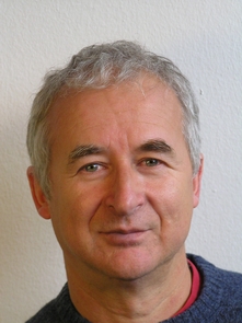 Wolfram Ziegler合作论文数Institute of Phonetics and Speech Processing, Ludwig-Maximilians-University of Munich15Hans-Otto Karnath合作论文数11W Lang合作论文数10W Oder合作论文数10
 Josef Spatt合作论文数Neurol Krankenhaus Rosenhugel9Katharina Hogrefe合作论文数7P Samec合作论文数6A. Wimmer合作论文数6G Lindinger合作论文数5
 Margarete Steiner合作论文数5Frank Uhl合作论文数5Th Reisner合作论文数5Sonja Hagmann合作论文数5B Mamoli合作论文数4J. Hermsdoerfer合作论文数4H Binder合作论文数4E Suess合作论文数4Marie-Luise Brandi合作论文数Max Planck Inst Psychiat, Independent Max Planck Res Grp Social Neurosci, D-80804 Munich, Germany4
 Klaus Willmes合作论文数SignGes Competence Center for Sign Language and Gesture, RWTH Aachen University4
 Nicole Weidinger合作论文数4
 Eduard Auff合作论文数Department of Neurology, Medical University of Vienna4
 Marta Bienkiewicz合作论文数Institute of Movement Science, Technical University of Munich4K HOLL合作论文数3Dal Bianco P合作论文数3A O Ceballos-Baumann合作论文数3
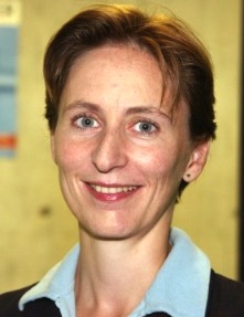 Aude Billard合作论文数Learning algorithms and systems Laboratory, EPFL3
 Henning Boecker合作论文数Department of Radiology, Rheinische Friedrich-Wilhelms-Universität Bonn3Maike Daumüller合作论文数3Peter Wessely合作论文数3
 Leif Johannsen合作论文数Center of Neurology;Clinical Brain Research;University of Tübingen;Center of Neurology, University of Tübingen3Karoline Hartmann合作论文数3
 Christian Sorg合作论文数Faculty of Psychology and Educational Sciences, Ludwig-Maximilians-Universität München;Department of Psychiatry and Neuroradiolgy, Technische Universität München3Wolfgang Lalouschek合作论文数3Bastian Conrad合作论文数3G. Schnaberth合作论文数3
 Georg Kerkhoff合作论文数Department of Clinical Neuropsychology, Saarland University3Biljana Petreska合作论文数3J Hufgard合作论文数2Ralf Glindemann合作论文数2
 Anselm Kornhuber合作论文数2M Lang合作论文数2Becker M合作论文数2R. Blank合作论文数2
 Philipp Gulde合作论文数2




