INTRODUCTION:The prevalence of esophageal webs in the pharyngoesophageal segment (PES) is unknown, in part because webs produce inconsistent symptomatology and can be difficult to diagnose. This study aims to determine the prevalence of PES webs in patients undergoing endoscopic laryngeal surgery. METHODS:This retrospective cohort study included patients undergoing laryngoscopy for pathology unrelated to webs. Evaluation for the presence of a web was performed on standard/routine examination of the PES during direct laryngoscopy. Demographic and clinical factors were analyzed for associations with webs. RESULTS:Of 123 patients included in this study, 42 (34.1%) were found to have a PES web. A total of 22 webs (52.4%) were on the left, 16 (38.1%) were on the right, and 4 (9.5%) were bilateral. There was no difference in age (58.1 vs. 58.7, p = 0.864) or BMI (29.7 vs. 29.8, p = 0.900) between patients with and without PES webs. Webs were significantly more common in patients with a history of irradiation to the head and neck (70% vs. 31%, p = 0.031) with a RR of 2.26 (CI: 1.38-3.69). There was no association of webs with gender, race, history of gastroesophageal reflux disease, or other clinical factors. Only 33.3% of patients with a web had documented symptoms of dysphagia. CONCLUSIONS:PES webs may be more prevalent than what is historically cited in the literature, and webs may be more common in patients with a history of irradiation to the head and neck. LEVEL OF EVIDENCE: 4:
OBJECTIVE:To identify characteristics of patients who have poor improvement in symptoms following surgical management of Zenker Diverticulum (ZD). METHODS:Prospective, multicenter cohort study of all individuals enrolled in the Prospective OUtcomes of Cricopharyngeus Hypertonicity (POUCH) Collaborative who underwent surgical repair of ZD between August 2017 and January 2024. Patient demographics, esophagrams, and the 10-item Eating Assessment Tool (EAT-10) pre- and post-procedure were obtained from a REDCap database. t-tests, Wilcoxon rank sum tests, Chi-square or Fisher's exact tests were used to compare the characteristics. Patients with <50% improvement in their EAT-10 scores were deemed surgical nonresponders (SNRs). Those with ≥50% improvement in their EAT-10 scores were deemed surgical responders (SRs). RESULTS:A total of 184 patients were prospectively followed after undergoing either open or endoscopic surgical management. Twenty-two patients (12%) were deemed SNRs. Preoperative presence of a hiatal hernia was statistically significant characteristic between the SNRs (63.6%) and SRs (32.1%) (p = 0.004). Size of the ZD and history of previous ZD surgery was not a significant characteristic. The length of stay and complication rate were not statistically different between the groups. CONCLUSION:Coexistent esophageal pathology may lead to poor symptomatic improvement following ZD surgery. Preoperative workup of other esophageal disorders is recommended to detect likely SNRs. For SNRs, further esophageal workup may be necessary to evaluate for other esophageal causes related to poor symptomatic improvement following ZD surgery. LEVEL OF EVIDENCE:3 Laryngoscope, 134:4897-4902, 2024.
A variety of surgical treatment options exist for adductor spasmodic dysphonia (ADSD) with selective adductor recurrent laryngeal nerve denervation and reinnervation (SLAD-R) being one of the more popular. We present a case of bilateral vocal fold paralysis (BVFP) for SLAD-R resulting in the need for total laryngectomy. We suggest BVFP is more common than reported and that we all must insure optimal long term follow up of our surgical patients. Laryngoscope, 134:4582-4584, 2024.
Objectives The aim of the study was to identify trends in postoperative management of persons undergoing surgery for Zenker diverticula (ZD) by evaluating length of stay (LOS), diet on discharge, and imaging with or without surgical complication. Methods Prospectively enrolled adult patients with cricopharyngeal muscle dysfunction with diverticula undergoing surgery from August 1, 2017 to February 1, 2023 were included. Data were extracted from a multi‐institutional REDCap database, summarizing means, medians, percentages, and frequencies. Fisher's exact or chi squared analyses were utilized, as appropriate, to compare subsets of data. Descriptive analysis assessed differences in clinical course and the relationship to postoperative management. Results There were 298 patients with a mean (standard deviation) age of 71.8 (11.2) years and 60% male. Endoscopic surgery was performed in 79.5% (237/298) of patients versus 20.5% (61/298) open surgery. Sixty patients (20.1%) received postoperative imaging, with four leaks identified. Complications were identified in 9.4% of cases ( n = 29 complications in 28 patients), more commonly in open surgery. Most (81.2%) patients were discharged within 23 h. About half of patients (49%) were discharged from the hospital on a pureed/liquid diet; 36% had been advanced to a soft diet. In patients without complications, LOS was significantly longer following open cases ( p = 0.002); postoperative diet was not different between open and endoscopic ( p = 0.26). Conclusions Overall, most patients are discharged within 23 h without imaging. However, LOS was affected by surgical approach. Postoperative complications are different in endoscopic versus open surgery. Complications with either approach were associated with prolonged LOS, need for imaging, and diet restriction. Level of Evidence Level III Laryngoscope , 134:2678–2683, 2024
Objective To compare improvement in patient‐reported outcomes (PROM) in persons undergoing endoscopic and open surgical management of Zenker diverticula (ZD). Methodology Prospective, multicenter cohort study of all individuals enrolled in the Prospective OUtcomes of Cricopharyngeus Hypertonicity (POUCH) Collaborative who underwent surgery for ZD. Patient survey, radiography reports, and the 10‐item Eating Assessment Tool (EAT‐10) pre‐ and post‐procedure were abstracted from a REDCap database, which summarized means, medians, percentages, and frequencies of. Outcome based on operative intervention (endoscopic vs. open) was compared using t ‐test, Wilcoxon rank sum test or chi‐square test, as appropriate. Results One hundred and forty‐seven persons were prospectively followed. The mean age (SD) of the cohort was 68.7 (11.0). Overall, 66% of patients reported 100% improvement in EAT‐10; 81% of patients had greater than 75% improvement; and 88% had greater than 50% improvement. Endoscopic was used for n = 109 patients, and open surgical intervention was used for n = 38. The median [interquartile range, IQR] EAT‐10 percent improvement for endoscopic treatment was 93.3% [72, 100], and open was 100% [92.3, 100] ( p = 0.05). The incidence of intraoperative complications was 3.7% for endoscopic and 7.9% for open surgical management. The median [IQR] in follow‐up was 86 and 97.5 days, respectively. Conclusion Both endoscopic and open surgical management of ZD provide significant improvement in patient‐reported outcomes. The data suggest that open diverticulectomy may provide a modest advantage in symptomatic improvement compared to endoscopic management. The data suggest that the postoperative complication rate is higher in the open surgical group. Level of Evidence 3 Laryngoscope , 134:97–102, 2024
Myasthenia gravis (MG) is the most common autoimmune disease affecting the neuromuscular junction (NMJ). Untreated this disease can lead to profound skeletal muscle weakness and respiratory failure.1 Although rare, intraoperative events can precipitate the first clinical presentation of MG, potentially creating diagnostic uncertainty in the postoperative period, and requiring early recognition and management of this condition to prevent the possibility of life-threatening complications in the immediate postoperative period.2 This project received Institutional Review Board approval, Medical College of Georgia, Augusta University # 611858. A 60-year-old white male underwent endoscopic repair of a Zenker's diverticulum under general anesthesia. Medication administration included ampicillin and sulbactam prior to incision, lidocaine, propofol, and remifentanil for induction and maintenance of anesthesia (total intravenous anesthesia), rocuronium, ketoroloc, ondansetron, dexamethasone, and neostigmine and glycopyrrolate for reversal of neuromuscular blockade (NMB). Emergence from anesthesia, return of spontaneous ventilation and extubation was uneventful; however, during recovery, the patient began to complain of new-onset upper and lower extremity weakness. Muscle strength grading using the Medical Research Council Manual Muscle Testing scale revealed upper extremity greater than lower extremity weakness (4− out of 5 vs 4 out of 5). The patient was only able to ambulate with 2-person assistance and did not exhibit sensory deficits or difficulty urinating. The clinical features were suggestive of acute cervical spinal cord injury, or central cord syndrome, raising the concern that this devastating complication had resulted from prolonged neck extension during surgery. Central cord syndrome has previously been recognized as a complication of neck extension during surgical procedures or emergency intubations.3 Fortunately, emergency magnetic resonance imaging and computed tomography imaging were able to rule out spinal cord injury or other traumatic lesions. A focused repeat neurological examination uncovered mild bilateral ptosis, some weakness in eye closure, no diplopia, no dysarthria, and a normal cough. A bedside icepack test was applied to the eyelids for 2 minutes, which was strongly positive for MG and resulted in complete resolution of the ptosis. After additional clinical and laboratory testing, the postoperative diagnosis of new-onset MG was confirmed, and the patient received appropriate treatment at our hospital. In MG autoantibodies damage the nicotinic acetylcholine receptor (AChR) at the postsynaptic membrane and impair neuromuscular transmission. In this case, it is reasonable to assume that exposure to several intraoperative drugs further reduced the efficiency of neuromuscular transmission and unmasked this patient's previously undiagnosed disease. Specifically, the NMJ in MG is exquisitely sensitive to nondepolarizing neuromuscular-blocking agents (eg, rocuronium), different classes of antibiotics (eg, aminoglycosides, ampicillin), possibly local anesthetics (eg, lidocaine), and glucocorticoids.1 Our patient received the above medications during surgery, which are postulated to have various pre- and postsynaptic effects, negatively impacting the availability of acetylcholine and AChR function. In addition, more recently published work has demonstrated that reversal of NMB with neostigmine and glycopyrrolate tends to be incomplete with a reported incidence of residual NMB of up to 63.5% at tracheal extubation and 56.5% at arrival in the postanesthesia care unit.4 Any degree of residual NMB would have contributed to this patient's clinical picture. These considerations highlight that in a surgical patient with known MG, the perioperative pharmacology is complex, requires careful preoperative planning, and aims to preserve the function of the compromised NMJ. Of note, the ice pack test used in this patient increases the availability of acetylcholine in the NMJ through cold inhibition of the acetylcholine-degrading enzyme acetylcholinesterase, which explains the high diagnostic performance of this bedside test for MG.5 This report presents a rare case of new-onset MG diagnosed in the immediate postoperative period after a Zenker's repair. It demonstrates that intraoperative events can unmask MG during the subclinical stage of the disease, lead physicians to other diagnostic considerations because of the general unfamiliarity with this complication in the postoperative period, and challenges surgeons and anesthesiologists to be aware of this potentially life-threatening clinical scenario. Steffen E. Meiler, directed the anesthesia care, diagnosed the patient postoperatively, and drafted the manuscript; Tylin J. Siwemuke, performed the literature search and assisted in writing the manuscript; Gregory N. Postma, directed the surgical care and contributed to the final manuscript. No conflicts of interest to report. None.
Purpose of ReviewThe purpose of this review is to summarize otolaryngology office-based evaluation and management of dysphagia. This review seeks to describe in detail state-of-the-art practice in advanced deglutology practice.Recent FindingsMuch of dysphagia evaluation and management can currently be practiced in the otolaryngology office, from swallowing function assessment with endoscopic and fluoroscopic techniques, to dysphagia interventions such as dilation of the pharyngoesophageal segment under local anesthesia.SummaryAdvanced dysphagia evaluation and management can be performed in the outpatient otolaryngology clinic. However, given the specialized nature of these diagnostic and procedural interventions, laryngology fellowship training with a focus on deglutology and collaboration with a dysphagia speech pathologist will likely become a pre-requisite for the otolaryngologist to run a comprehensive dysphagia clinic.
Objective To assess barium esophagram (BAS) as a diagnostic marker for patients with Killian Jamieson diverticula (KJD). Methods Prospective, multicenter cohort study of individuals enrolled in the Prospective OUtcomes of Cricopharyngeus Hypertonicity (POUCH) Collaborative. Patient demographics, comorbidities, radiographic imaging reports, laryngoscopy findings, patient‐reported outcome measures (PROM), and operative reporting were abstracted from a REDCap database and summarized using means, medians, percentages, frequencies. Paired t‐tests and Wilcoxon Signed Rank test were used to test pre‐ to post‐operative differences in RSI, EAT‐10, and VHI‐10 scores. Diagnostic test evaluation including sensitivity, specificity, positive, and negative predictive value with 95% confidence intervals were calculated comparing BAS findings to operative report. Results A total of 287 persons were enrolled; 13 (4%) patients were identified with confirmed KJD on operative reports. 100% underwent open transcervical excision. BAS has a 46.2% (95% confidence interval [CI]: 23.2, 70.9) sensitivity and 97.8% (95% CI: 95.3, 99.0) specificity in detecting a KJD and 50% (95% CI: 25.4, 74.6) positive predictive value but 97.4% (95%CI: 94.8, 98.7) negative predictive value. Preoperatively, patients reported mean (SD) RSI and EAT‐10 of 19.4 (9) and 8.3 (7.5) accordingly. Postoperatively, patients reported mean (SD) RSI and EAT‐10 as 5.4 (6.2) and 2.3 (3.3). Both changes in RSI and EAT‐10 were statistically significant ( p = 0.008, p = 0.03). Conclusion KJD are rare and represent <5% of hypopharyngeal diverticula undergoing surgical intervention. Open transcervical surgery significantly improves symptoms of dysphagia. BAS has high specificity but low sensitivity in detecting KJD. Level of Evidence 4 Laryngoscope , 133:2110–2115, 2023
Objective To describe demographics and imaging and compare findings and symptoms at presentation in a large cohort of persons with cricopharyngeus muscle dysfunction (CPMD) with and without hypopharyngeal diverticula. Methodology Prospective, multicenter cohort study of all individuals enrolled in the Prospective OUtcomes of Cricopharyngeal Hypertonicity (POUCH) Collaborative. Patient survey, comorbidities, radiography, laryngoscopy findings, and patient‐reported outcome measures (e.g., Eating Assessment Tool [EAT‐10]) data were abstracted from a REDCap database and summarized using means, medians, percentages, and frequencies. Diagnostic categories were compared using analysis of variance. Results A total of 250 persons were included. The mean age (standard deviation [SD]) of the cohort was 69.0 (11.2). Forty‐two percent identified as female. Zenker diverticula (ZD) was diagnosed in 85.2%, 9.2% with CPMD without diverticula, 4.4% with a Killian Jamieson diverticula (KJD), and 1.2% traction‐type diverticula. There were no differences between diagnostic categories in regard to age, gender, and duration of symptoms ( p = 0.25, 0.19, 0.45). The mean (SD) EAT‐10 score for each group was 17.1 (10.1) for ZD, 20.2 (9.3) for CPMD, and 10.3 (9.4) for KJD. Patients with isolated CPMD had significantly greater EAT‐10 scores compared to the other diagnostic groups ( p = 0.03). Conclusion ZD is the most common, followed by CPMD without diverticula, KJD, and traction‐type. Patients with isolated obstructing CPMD may be more symptomatic than persons with ZD or KJD. Level of Evidence 4 Laryngoscope , 133:1349–1355, 2023
ObjectivesTo develop an expert consensus statement on the clinical use of swallowing fluoroscopy in adults that reduces practice variation and identifies opportunities for quality improvement in the care of patients suffering from swallowing impairment.MethodologyA search strategist reviewed data sources (PubMed, Embase, Cochrane, Web of Science, Scopus) to use as evidence for an expert development group to compose statements focusing on areas of controversy regarding swallowing fluoroscopy. Candidate statements underwent two iterations of a modified Delphi protocol to reach consensus.ResultsA total of 2184 publications were identified for title and abstract review with 211 publications meeting the criteria for full text review. Of these, 148 articles were included for review. An additional 116 publications were also included after reviewing the references of the full text publications from the initial search. These 264 references guided the authors to develop 41 candidate statements in various categories. Forty statements encompassing patient selection, fluoroscopic study choice, radiation safety, clinical team dynamics, training requirements, videofluoroscopic swallow study and esophagram techniques, and interpretation of swallowing fluoroscopy met criteria for consensus. One statement on esophagram technique reached near‐consensus.ConclusionsThese 40 statements pertaining to the comprehensive use of swallowing fluoroscopy in adults can guide the development of best practices, improve quality and safety of care, and influence policy in both the outpatient and inpatient settings. The lack of consensus on some aspects of esophagram technique likely reflects gaps in knowledge and clinical practice variation and should be a target for future research. Laryngoscope, 133:255–268, 2023
Laryngopharyngeal reflux (LPR) is a common otolaryngologic diagnosis. Treatment of presumed LPR remains challenging, and limited frameworks exist to guide treatment. Using RAND/University of California, Los Angeles (UCLA) Appropriateness Methods, a modified Delphi approach identified consensus statements to guide LPR treatment. Experts independently and blindly scored proposed statements on importance, scientific acceptability, usability, and feasibility in a four-round iterative process. Accepted measures reached scores with ≥ 80
A 37-year-old incarcerated male with a history of schizophrenia presented to the emergency department with dysphagia after intentional ingestion of a metallic foreign body. Past medical history was remarkable for multiple prior procedures for removal of esophageal and gastrointestinal foreign bodies. The patient was not in distress and tolerating his secretions. Flexible laryngoscopy was unremarkable. Plain radiography was remarkable for a radiopaque metallic foreign body, consistent with the face of a combination lock, located at the thoracic inlet within the proximal esophagus (Figure 1). The patient failed flexible endoscopic removal by gastroenterology and underwent rigid esophagoscopy with removal of the esophageal foreign body in the operating room (Figure 2). Intentional ingestion of foreign bodies is increasing in incidence and is associated with incarceration, psychiatric disorders, and substance abuse. Motivating factors include malingering, psychosis, pica, and personality disorders. Repeat offenders account for a disproportionately high percentage of intentional ingestions, underscoring the psychosocial factors that drive this behavior. Psychiatric treatments specific to intentional ingestion have been poorly studied, though the role of behavioral therapy and pharmacologic intervention with antipsychotics or antidepressants appears to be limited in preventing recurrent foreign body ingestion. Prevention of intentional ingestion in the incarcerated population may need to focus on limiting unsupervised access to potentially ingestible foreign bodies. The most commonly ingested objects in prisoners include pens, eating utensils, razor blades, toothbrushes, and various other metal objects, such as combination locks. Management of ingested foreign bodies is dependent on presenting symptoms, foreign body location, number of ingested objects, and associated complications. Most foreign bodies can be successfully removed endoscopically, with surgery reserved for failed endoscopic removal or in the presence of other indications, such as perforation. Approximately 50% of patients with intentional ingestions present with a foreign body in the stomach, and less than 10% present in the esophagus. Although some advocate for a conservative observational approach specifically for inmates, foreign bodies that are impacted in the esophagus typically warrant removal, and emergent intervention may be indicated with esophageal obstruction or with specific foreign bodies, such as disc batteries or sharp objects. Figure 1. AP radiograph of the neck showing a radiopaque metallic foreign body, consistent with the face of a combination lock, located at the thoracic inlet within the proximal esophagus.
Objective: To present international recommendations regarding the proper evaluation of oropharyngeal dysphagia (OD), both objectively and subjectively (self-evaluation). Methods: Following a thorough review of the literature, 5 experts in the field from 4 different continents answered separately a questionnaire regarding the work-up of OD. Individual answers were presented and discussed during the world ENT conference that was held in Paris in June 2017. This article will present the recommendations issued from that meeting. Results: For the initial objective assessment of OD, it is recommended to perform either a functional endoscopic evaluation of swallowing (FEES) or a videofluoroscopic swallowing study (VFSS). FEES is the more popular investigation given its increased ease of use and accessibility. When evaluating for the presence of aspiration during the objective evaluation of OD, it is recommended to perform either a FEES or a VFSS. In this case, FEES is the favored investigation given its likely increased sensitivity. In order to highlight the presence of oropharyngeal food residue following the deglutition process, it is recommended to perform either a FEES or a VFSS; FEES likely being the more sensitive investigation while VFSS allows a better quantification of the amount of pharyngeal residue. Is it also recommended to objectify the quality of the deglutition process by means of a score during the objective evaluation of OD. Finally, it is recommended to utilize a self-evaluation questionnaire during research studies exploring the deglutition process. (C) 2018 Elsevier Masson SAS. All rights reserved.
Acronyms and abbreviations are frequently used in otorhinolaryngology and other medical specialties. CO2 laser-assisted transoral surgery of the pharynx, the larynx and the upper airway is a family of commonly performed surgical procedures termed transoral laser microsurgery (TLM). The abbreviation TLM can be confusing because of alternative modes of delivery. Classification and definition of the different types of procedures, performed transorally or transnasally, are proposed by the Working Committee for Nomenclature of the European Laryngological Society, emphasizing the type of laser used and the way this laser is transmitted. What is usually called TLM, would more clearly be defined as CO2 laser transoral microsurgery or CO2 TOLMS or CO2 laser transoral surgery only (with a handpiece) would be defined as CO2 TOLS. KTP transnasal flexible laser surgery would be KTP TNFLS. Transoral use of the flexible CO2 wave-guide with a handpiece would be a CO2 TOFLS. One can argue that these clarifications are not necessary and that the abbreviation TLM for transoral laser microsurgery is more than sufficient. But this is not the case. Laser surgery, office-based laser surgery and microsurgery are frequently and erroneously interchanged for one another. These classifications allow for a clear understanding of what was performed and what the results meant.
Abstract Objectives: To validate the ovine model of profound oropharyngeal dysphagia and compare swallowing outcomes of laryngotracheal separation with those of total laryngectomy. Methods: Under real-time fluoroscopy, swallowing trials were conducted using the head and neck of two Dorper cross ewes and one human cadaver, secured in lateral fluoroscopic orientation. Barium trials were administered at baseline, pre- and post-laryngohyoid suspension, following laryngotracheal separation, and following laryngectomy in the ovine model. Results: Mean pre-intervention Penetration Aspiration Scale and National Institutes of Health Swallow Safety Scale scores were 8 ± 0 and 6 ± 0 respectively in sheep and human cadavers, with 100 per cent intra- and inter-species reproducibility. These scores improved to 1 ± 0 and 2 ± 0 post-laryngohyoid suspension (p < 0.01). Aerodigestive tract residue was 18.6 ± 2.4 ml at baseline, 15.4 ± 3.8 ml after laryngotracheal separation and 3.0 ± 0.7 ml after total laryngectomy (p < 0.001). Conclusion: The ovine model displayed perfect intra- and inter- species reliability for the Penetration Aspiration Scale and Swallow Safety Scale. Less aerodigestive tract residue after narrow-field laryngectomy suggests that swallowing outcomes after total laryngectomy are superior to those after laryngotracheal separation.
 Peter C. Belafsky合作论文数Department of Otolaryngology Head and Neck Surgery, University of California, Davis45
 Koufman James A合作论文数25James A. Koufman合作论文数16Bach Kevin K合作论文数12
 Jamie A. Koufman合作论文数Department of Clinical Otolaryngology, New York Medical College;Voice Institute of New York12
 Mark A Fritz合作论文数9
 Paul Weinberger合作论文数Deaconess Clinic;SpheroFill, LLC8Halum Stacey L合作论文数Dept Otolaryngol Head & Neck Surg, Indiana Univ Sch Med7Rebecca J. Howell合作论文数6Christopher Johnson合作论文数6Milan R. Amin合作论文数6S Carter Wright合作论文数5Keith Wilson合作论文数5Ashli O'Rourke合作论文数5
 Yue Ma合作论文数5
 Dale C Ekbom合作论文数Department of Otolaryngology/Head and Neck Surgery, Mayo Clinic Rochester5Kuhn Maggie A合作论文数5
 Elizabeth Guardiani合作论文数5
 Jan L. Kasperbauer合作论文数School of Continuous Professional Development, Mayo Clinic;Mayo Medical School5Meredith E. Tabangin合作论文数5Mekibib Altaye合作论文数Dept. of Radiology, Cincinnati Children's Hospital Medical Centre5Brad W. Desilva合作论文数5
 Laura Matrka合作论文数5Jacqui E. Allen合作论文数5William S Halsey合作论文数4
 Paul E Kwak合作论文数4
 Nikki Johnston合作论文数4Semirra L Bayan合作论文数4Young VyVy N合作论文数4Friedman Aaron D合作论文数4
 Kim Brandon J合作论文数4
 Gregory Robert Dion合作论文数Department of Otolaryngology, San Antonio Military Medical Center4
 Madden Lyndsay L合作论文数University of Pittsburgh, University of Pittsburgh Medical Center4Mayerhoff Ross合作论文数4Marvin P. Fried合作论文数4
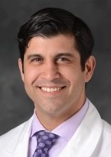 Cyrus R Piraka合作论文数Henry Ford Cancer Institute;Division of Gastroenterology, Department of Internal Medicine, Henry Ford Hospital4
 John-Paul Giliberto合作论文数Division of Cardiovascular Surgery, St. Michael's Hospital4P Blalock合作论文数4Paul E Johnson合作论文数3Shaun A. Wahab合作论文数3Anne Phillips合作论文数3
 Brittany Nicole Krekeler合作论文数Dept Med, Univ Wisconsin Madison3
 Brian Cervenka合作论文数3John H. Krouse合作论文数3
 Jacob T. Cohen合作论文数Sackler Faculty of Medicine, Tel-Aviv University3
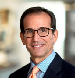 Kenneth W. Altman合作论文数Department of Otolaryngology-Head and Neck Surgery, Mount Sinai School of Medicine3C. Blake Simpson合作论文数Surgery Foundation, Inc.3Michele Morrison合作论文数2Zao M. Yang合作论文数2




