In optical microscopy, the intrinsic trade-off between resolution and depth of field (DOF) prevents the observation of 3D samples, limiting the observable thickness to some micrometers. The usual solution to this problem is to mechanically scan the object in depth. However, this slows down the process and can induce vibrations in soft samples. To overcome these drawbacks, we propose the use of an electrically tunable lens (ETL) based on electrowetting effect. Specifically, we propose an extended-DOF microscope set up in a very compact architecture in which full telecentricity is no longer achieved. On top of that, the axial scanning is carried out by the non-afocal coupling between two M12 lenses and the ETL. The main feature of the proposed extended-DOF microscope is that, despite its compactness, it can capture stacks of images with long depth range, constant magnification and a spatial resolution that equals that of the standard microscope and remains invariant along the depth range. We also show that the proposed idea can be implemented as an add-on attachable to any commercial microscope, which enables the use of all the microscope facilities.
Structured illumination microscopy (SIM) provides accurate 3D images of translucent samples by using non-uniform irradiance patterns in combination with digital post processing. We present here a novel setup, tunable SIM, that provides shorter registration times, higher signal-to-noise ratios, and increased optical sectioning capabilities respect to classical SIM implementations. Full-text article not available; see video presentation
We have designed and implemented an approach for three-dimensional (3D) structured illumination (SI) microscopy (SIM) based on a quasi-monochromatic extended source illuminating a Wollaston prism to improve robustness, light efficiency and flexibility over our previous design. We show through analytical and experimental verification of the presented theoretical framework for our proposed tunable structured illumination microscopy (TSIM) system, that a simple and accurate determination of the axial modulation of the SI pattern is achieved, enabling a realistic characterization of the system's effective optical transfer function (OTF). System performance as a function of the extended source size is investigated with simulations. Results from a comparative performance analysis of the proposed TSIM system and traditional SIM systems show some advantages over the traditional two-wave and three-wave interference SIM systems. We show that by controlling the source size and thereby the axial modulation of the 3D SI pattern, the TSIM scheme offers increased OTF compact support and improved optical sectioning capability, quantified by the integrated intensity, under certain conditions, which may be desirable when imaging optically thick samples. The additional tunability of the 3D SI pattern, provides a unique opportunity for OTF engineering in our TSIM system.
The resolution limit achievable with an optical system is a fundamental piece of information when characterizing its performance, mainly in case of microscopy imaging. Usually this information is given in the form of a distance, often expressed in microns, or in the form of a cutoff spatial frequency, often expressed in line pairs per mm. In modern imaging systems, where the final image is collected by pixelated digital cameras, the resolution limit is determined by the performance of both, the optical systems and the digital sensor. Usually, one of these factors is considered to be prevalent over the other for estimating the spatial resolution, leading to the global performance of the imaging system ruled by either the classical Abbe resolution limit, based on physical diffraction, or by the Nyquist resolution limit, based on the digital sensor features. This estimation fails significantly to predict the global performance of opto-digital imaging systems, like 3D microscopes, where none of the factors is negligible. In that case, which indeed is the most common, neither the Abbe formula nor the Nyquist formula provide by themselves a reliable prediction for the resolution limit. This is a serious drawback since systems designers often use those formulae as design input parameters. Aiming to overcome this lack, a simple mathematical expression obtained by finely articulating the Abbe and Nyquist formulas, to easily predict the spatial resolution limit of opto-digital imaging systems, is proposed here. The derived expression is tested experimentally, and shows to be valid in a broad range of opto-digital combinations.
Fourier lightfield microscopy (FLMic) is a powerful technique to record 3D images of thick dynamic samples. Belonging FLMic to the general class of computational imaging techniques, its efficiency is determined by sev-eral factors, like the optical system, the calibration process, the reconstruction algorithm, or the computation architecture. In the case of FLMic the calibration and the reconstruction algorithm should be fully adapted to the singular features of the technique. To this end, and concerning the reconstruction, we discard the use of experimental PSFs, and propose the use of a synthetic one, which is calculated on the basis of paraxial optics and taking into account the equal influence of diffraction and pixelation. Using this quite simple PSF, performing the adequate calibration and finally implementing the algorithm in GPU, we demonstrate here the possibility of obtaining 3D images with good results in terms of resolution and strong improvement in terms of computation time. In summary, and aiming to accelerate the widespread of FLMic among microscopy users and researchers, we are proposing a fast protocol fully adapted to FLMic and that is very flexible and robust against any slight misalignment or against the change of any optical element.
Using a mathematical approach, this paper presents a generalization of semi-analytical expressions for the point spread function (PSF) of plenoptic cameras. The model is applicable in the standard regime of the scalar diffraction theory while the extension to arbitrary main lens transmission functions generalizes a priori formalism. The accuracy and applicability of the model is well verified against the exact Rayleigh-Sommerfeld diffraction integral and a rigorous proof of convergence for the PSF series expression is made. Since vignetting can never be fully eliminated, it is critical to inspect the image degradation it poses through distortions. For what we believe is the first time, diffractive distortions in the diffraction-limited plenoptic camera are closely examined and demonstrated to exceed those that would otherwise be estimated by a geometrical optics formalism, further justifying the necessity of an approach based on wave optics. Microlenses subject to the edge diffraction effects of the main lens vignetting are shown to translate into radial distortions of increasing severity and instability with defocus. The distortions due to vignetting are found to be typically bound by the radius of the geometrical defocus in the image plane, while objects confined to the depth of field give rise to merely subpixel distortions.
A practical guide for the easy implementation of a Fourier light-field microscope is reported. The Fourier light-field concept applied to microscopy allows the capture in real time of a series of 2D orthographic images of microscopic thick dynamic samples. Such perspective images contain spatial and angular information of the light-field emitted by the sample. A feature of this technology is the tight requirement of a double optical conjugation relationship, and also the requirement of NA matching. For these reasons, the Fourier light-field microscope being a non-complex optical system, a clear protocol on how to set up the optical elements accurately is needed. In this sense, this guide is aimed to simplify the implementation process, with an optical bench and off-the-shelf components. This will help the widespread use of this recent technology.
In this work, the design, building, and testing of the most portable, easy-to-build, robust, handheld, and cost-effective Fourier Lightfield Microscope (FLMic) to date is reported. The FLMic is built by means of a surveillance camera lens and additional off-the-shelf optical elements, resulting in a cost-effective FLMic exhibiting all the regular sought features in lightfield microscopy, such as refocusing and gathering 3D information of samples by means of a single-shot approach. The proposed FLMic features reduced dimensions and light weight, which, combined with its low cost, turn the presented FLMic into a strong candidate for in-field application where 3D imaging capabilities are pursued. The use of cost-effective optical elements has a relatively low impact on the optical performance, regarding the figures dictated by the theory, while its price can be at least 100 times lower than that of a regular FLMic. The system operability is tested in both bright-field and fluorescent modes by imaging a resolution target, a honeybee wing, and a knot of dyed cotton fibers.
Fourier lightfield microscopy is an emerging technique for real-time acquisition of three-dimensional microscopic samples. Here, we present the lightfield eyepiece, an add-on device capable of converting any conventional microscope to a Fourier lightfield microscope.
In this work, a practical guide for the design of a Fourier lightfield microscope is reported. The fundamentals of the Fourier lightfield are presented and condensed on a set of contour plots from which the user can select the design values of the spatial resolution, the field of view, and the depth of field, as function of the specifications of the hardware of the host microscope. This work guides the reader to select the parameters of the infinity-corrected microscope objective, the optical relay lenses, the aperture stop, the microlens array, and the digital camera. A user-friendly graphic calculator is included to ease the design, even to those who are not familiar with the lightfield technology. The guide is aimed to simplify the design process of a Fourier lightfield microscope, which sometimes could be a daunting task, and in this way, to invite the widespread use of this technology. An example of a design and experimental results on imaging different types of samples is also presented.
The use of microlens arrays for lightfield display has the drawback of providing images with strong chromatic aliasing. To overcome this problem, pinhole-type lightfield monitors are proposed. This paper is devoted to evaluating the capability for such lightfield monitors to offer the user a convincing 3D experience with images with enough brightness and continuous aspect. Thus, we have designed a psychophysical test specifically adapted for lightfield monitors, which allowed us to confirm the usability of pinhole-type monitors.
Lightfield microscopy has raised growing interest in the last few years. Its ability to get three-dimensional information about the sample in a single shot makes it suitable for many applications in which time resolution is fundamental. In this paper we present a novel device, which is capable of converting any conventional microscope into a lightfield microscope. Based on the Fourier integral microscope concept, we designed the lightfield microscope eyepiece. This is coupled to the eyepiece port, to let the user exploit all the host microscope's components (objective turret, illumination systems, translation stage, etc.) and get a 3D reconstruction of the sample. After the optical design, a proof-of-concept device was built with off-the-shelf optomechanical components. Here, its optical performances are demonstrated, which show good matching with the theoretical ones. Then, the pictures of different samples taken with the lightfield eyepiece are shown, along with the corresponding reconstructions. We demonstrated the functioning of the lightfield eyepiece and lay the foundation for the development of a commercial device that works with any microscope.
Alejandro Gimeno-Gomez1, Hui Yun1, Cong T.S. Van2, Chrysanthe Preza2,*, Juan C. Barreiro-Hervas1, Genaro Saavedra1,+ 13D Imaging and Display Laboratory, Universitat de València, Burjassot, Spain. 2Computational Imaging Research Laboratory, Dept. of Electrical & Computer Eng., University of Memphis, Memphis, TN, USA. e-mail: *cpreza@memphis.edu; +genaro.saavedra@uv.es
Lightfield (also known as integral or plenoptic) microscopy has been introduced successfully in 3D bioimaging. The strongest point of this technique when compared with conventional 3D microscopy arquitectures is its capability of capturing the 3D information of the sample in a single-shot manner. Despite this advantage, lightfield microscopy currently faces some challenges, like improving the resolution and depth of field of the reconstructed specimens or the development and optimization of specially-adapted reconstruction algorithms. In this contribution we review a new paradigm, namely, the Fourier lightfield microscope (FLMic), that improves the capabilities of the technique and we present recent advances and applications of this new architecture. As an example, we describe a protocol that takes profit from FLMic concept for providing, in a single-shot, 3D dark-field images of volumetric transparent samples.
This Roadmap article on digital holography provides an overview of a vast array of research activities in the field of digital holography. The paper consists of a series of 25 sections from the prominent experts in digital holography presenting various aspects of the field on sensing, 3D imaging and displays, virtual and augmented reality, microscopy, cell identification, tomography, label-free live cell imaging, and other applications. Each section represents the vision of its author to describe the significant progress, potential impact, important developments, and challenging issues in the field of digital holography.
We report a scanning non-confocal fluorescence microscopy scheme that provides images with optical sectioning and with a lateral resolution that surpasses by a factor of two the diffraction resolution limit. This technique is based on the type-1 microscopy concept combined with patterned illumination. The method does not require the application of phase-shifting or post-processing algorithms and provides artifact-free superresolved 3D images. We have validated the theory by means of experimental data.
We propose the combination of squared cubic phase profiles and spherical aberration patterns to further improve the quality of multiphoton microscopy images. The benefit will be analyzed through the use of objective image quality metrics.
Multiphoton (MP) microscopy is mainly limited by specimen-induced aberrations. Among them spherical aberration (SA) is the dominant term at deeper layers within a sample [1]. Adaptive optics has been demonstrated to improve MP imaging performance [1,2]. Wavefront coding techniques (or alternatively, pupil masks) have also been used to manipulate the wavefront and reduce the impact of SA when imaging thick tissues [3]. In particular, a symmetric squared cubic pupil phase mask (known as SQUIBIC) was able to produce a depth-invariant focal spot with noticeable enhanced confocal microscopy images [4]. Here, we propose a wavefront sensor-less approach based on combining SQUBIC masks and SA patterns to further improve the quality of MP images. A spatial light modulator was implemented into the illumination pathway of a custombuild MP microscope to manipulate the incident laser beam wavefront [2]. This was used to generate phase maps with different amounts of SA, and to produce a number of SQUBIC masks attending to its design parameter [4]. Individual or combined SA and SQUIBIC patterns were systematically generated during image recording. A set of image quality metrics was used to evaluate the impact for different nonlinear signals and depth locations within the samples under study. Both negative SA maps and SQUBIC patterns were able to improve MP images. However, results show that particular combinations of both yield improved images, better that those obtained when using those wavefront patterns separately (see Figure 1 as an example). Moreover, those controlled combinations of phase conditions led to an increase in depth-offocus, noticeable better than that obtained just using SA [1], independently of the specimeninduced aberrations.
Plenoptic (also known as lightfield) cameras began to be commercialized at the end of the 2000’s and now many companies are beginning to present lightfield displays. Both devices are based on integral photography technique [1]: by inserting a lens array in front of the sensor, it is possible to register the angular information of the rays proceeding from the scene, which is fundamental for the 3D reconstruction.
DATA ACQUISITION BY MEANS OF A WOLLASTON PRISM Alejandro Gimeno-Gomez, Hui Yun, Cong T.S. Van, Hasti Shabani Chrysanthe Preza, Juan C. Barreiro-Hervas, Genaro Saavedra 3D Imaging and Display Laboratory, Universitat de València, Burjassot, Spain. Dept. of Electrical & Computer Eng., University of Memphis, Memphis, TN, USA. e-mail: cpreza@memphis.edu; genaro.saavedra@uv.es
 Manuel Martínez-Corral合作论文数Department of Optics, University of Valencia;Doitplenoptic171
 Sánchez-Ortiga Emilio合作论文数3D Imaging and Display Laboratory, University of Valencia54
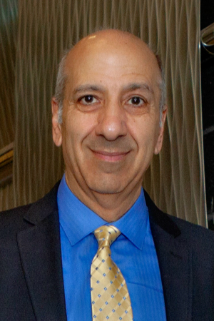 Bahram Javidi合作论文数Multidimensional Optical Sensing and Imaging Systems Laboratory, College of Engineering, University of Connecticut;Electrical & Computer Engineering Department, University of Connecticut54
 Ana Doblas合作论文数46
 Walter D Furlan合作论文数41
 Raúl Martínez Cuenca合作论文数Departamento de Ingeniería Mecánica y Construcción, Universidad Jaime I36
 Hector Navarro合作论文数31
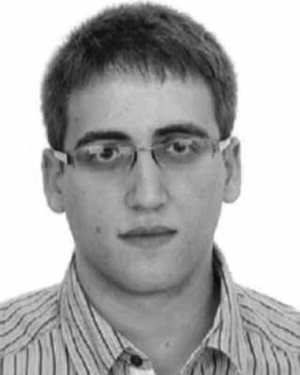 Adrián Dorado合作论文数22
 Anabel Llavador合作论文数21
 Jorge Ivan Garcia Sucerquia合作论文数School of Physics, Universidad Nacional de Colombia Sede Medellin20
 Juan A. Monsoriu合作论文数Departamento de Física Aplicada;Universidad Politécnica de Valencia;Departamento de Física Aplicada, Universidad Politécnica de Valencia19
 Chrysanthe Preza合作论文数Department of Electrical and Computer Engineering, The University of Memphis;Department of Biomedical Engineering, The University of Memphis;Computational Imaging Research Laboratory, The University of Memphis18
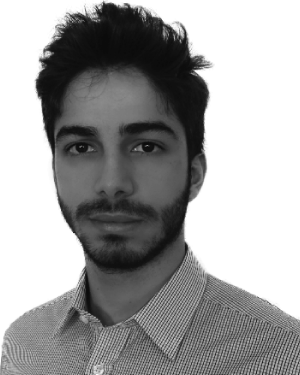 G. Scrofani合作论文数3DID Lab, Univ Valencia18
 Pedro Andrés合作论文数Departamento de Óptica, University of Valencia17
 Seokmin Hong合作论文数IAI Lab., Dongseo Univ.;c16
 Jorge Sola-Pikabea合作论文数16J.C. Barreiro合作论文数12
 Hasti Shabani合作论文数10Amparo Pons合作论文数9a marti合作论文数8
 Nicolo Incardona合作论文数8
 Nurmohammed Patwary合作论文数Dept Elect & Comp Engn, Univ Memphis8
 Isabel Escobar合作论文数8A. Pons合作论文数7Enrique Silvestre合作论文数7C. Ibáñez-López合作论文数7
 Arnau Calatayud合作论文数7J Ojedacastaneda合作论文数6
 Yi-Pai Huang合作论文数Apple Inc.6
 Jesús Lancis合作论文数Departamento de Física, Universitat Jaume I6
 Angel Tolosa Ruiz合作论文数Asociación Industrial de Óptica, Color e Imagen (AIDO), Calle Nicolás Copérnico 7-13, Parque Tecnológico, 46980 Paterna, Valencia, Spain6C. Preza合作论文数5
 Amir Ansari合作论文数Univ Valencia, Dept Opt, E-46100 Burjassot, Spain5
 Sharon V. King合作论文数Department of Electrical and Computer Engineering, University of Colorado5
 Carlos J. Zapata-Rodríguez合作论文数Departamento de Óptica, Universidad de Valencia5Juan Carlos Barreiro Hervás合作论文数4Juan C Barreiro合作论文数4A. Tolosa合作论文数4Maria T. Caballero合作论文数4H. Shabani合作论文数3r mourelle合作论文数3Alejandro Gimeno-Gomez合作论文数3Laura Galdón合作论文数3Cong T. S. Van合作论文数Center for Cancer Research, National Cancer Institute3
 Anca Stefanoiu合作论文数Dept Informat, Tech Univ Munich3Hui Yun合作论文数3
 Myungjin Cho合作论文数School of ICT, Robotics, and Mechanical Engineering, Hankyong National University3
 Joaquin de Nova合作论文数3
 Gonzalo Feijoo合作论文数3




