A. Meents, P. Roedig, I. Vartiainen, R. Duman, S. Panneerselvam, N. Stuebe, O. Lorbeer, M. Warmer, G. Sutton, D. I. Stu art, E. Weckert, C. Da vid, and A. Wag ner Deutsches Elektronen Syn chro tron DESY, Pho ton Sci ence, Notkestraße 86, 22607 Ham burg Paul Scherrer Institut, Villigen PSI, 5232, Swit zer land Di a mond Light Source Ltd., Di a mond House, Harwell Sci ence & In no va tion Cam pus, Didcot, Oxfordshire, OX11 0DE, United King dom Di vi sion of Struc tural Bi ol ogy, Wellcome Trust Cen tre for Hu man Ge net ics, Uni ver sity of Ox ford, Ox ford, OX3 7BN, United King dom alke.meents@cfel.de
At low emittance synchrotron sources it has become possible to perform structure determinations from the measurement of multiple microcrystals which were previously considered too small for diffraction experiments. Conventional mounting techniques do not fulfill the requirements of these new experiments. They significantly contribute to background scattering and it is difficult to locate the crystals, making them incompatible with automated serial crystallography. We have developed a micro-fabricated sample holder from single crystalline silicon with micropores, which carries up to thousands of crystals and significantly reduces the background scattering level. For loading, the suspended microcrystals are pipetted onto the chip and excess mother liquor is subsequently soaked off through the micropores. Crystals larger than the pore size are retained and arrange themselves according to the micropore pattern. Using our chip we were able to collect 1.5 Å high resolution diffraction data from protein microcrystals with sizes of 4 micrometers and smaller.
Single-wavelength anomalous dispersion of sulfur atoms (S-SAD) is an elegant phasing method to determine crystal structures that does not require heavy atom incorporation or selenomethionine derivatization. Nevertheless this technique has been limited by the paucity of the signal at usual X-ray wavelengths, requiring very accurate measurement of the anomalous differences. Here we report the data collection and structure solution of the N-terminal domain of the ectodomain of Hepatitis C virus (HCV) E1, from crystals that diffracted very weakly. By combining the data from 32 crystals it was possible to solve the sulfur substructure and calculate initial maps at 7Å resolution, and after density modification and phase extension, using a higher resolution native dataset, to 3.5Å resolution, model building was achievable. The crystal structure of the N-terminal domain of reveals a complex network of covalently linked intertwined homodimers that do not harbor the expected truncated class II fusion protein fold.
 David Stuart合作论文数Division of Structural Biology, Nuffield Department of Medicine, University of Oxford34
 Jonathan M. Grimes合作论文数Division of Structural Biology;Wellcome Trust Centre for Human Genetics;University of Oxford;Wellcome Trust Centre for Human Genetics, University of Oxford20
 Danny Axford合作论文数14
 Helen M Ginn合作论文数Diamond Light Source, Ltd.13R. Owen合作论文数12
 Kamel El Omari合作论文数The Wellcome Trust Centre for Human Genetics, University of Oxford10
 Dennis H. Bamford合作论文数Department of Biological and Environmental Sciences, Institute of Biotechnology, University of Helsinki10E.J. Mancini合作论文数9
 Peter Mertens合作论文数Faculty of Medicine & Health Sciences, University of Nottingham;School of Veterinary Medicine and Science, University of Nottingham9C. Meier合作论文数7Poranen Minna M合作论文数7
 Denis E Kainov合作论文数Inst Mol Med Finland FIMM, Helsinki 00290, Finland7
 Houssam Attoui合作论文数Laboratoire de Virologie, Etablissement de Transfusion Sanguine Alpes-Provence7
 Roman Tuma合作论文数School of Biological Sciences, University of Missouri-Kansas City7N S Maan合作论文数5
 Simon John Anthony合作论文数Department of Pathology, Microbiology, and Immunology, School of Veterinary Medicine, University of California, Davis5N. Maan合作论文数4A.S. Brewster合作论文数3R. Duman合作论文数3N. Sauter合作论文数3
 Roedig, P.合作论文数3
 Ismo Vartiainen合作论文数3
 Wally Thomas合作论文数School of Biomedical Sciences, Faculty of Medicine, The University of Queensland3
 Karin E Darpel合作论文数3
 Alke Meents合作论文数DESY;Center for Free-Electron Laser Science3Jonathan M. Diprose合作论文数3
 Elizabeth Fry合作论文数Division of Structural Biology, Nuffield Department of Medicine, Oxford University3M.W. Bahar合作论文数2O. Lorbeer合作论文数2Ray Owens合作论文数2E. Weckert合作论文数2M. Warmer合作论文数2S. Panneerselvam合作论文数2R.J. Gildea合作论文数2J. Hattne合作论文数2Andrew Davidson合作论文数School of Cellular and Molecular Medicine, University of Bristol2
 Nick S Berrow合作论文数2
 Marc Messerschmidt合作论文数SLAC Natl Accelerator Lab, Linac Coherent Light Source, Menlo Pk, CA 94025 USA2
 Nettleship Joanne E合作论文数Rutherford Appleton Lab, Oxford Prot Prod Facil2
 Wagner Armin合作论文数2
 Robert Gilbert合作论文数Department of Biochemistry and Medicine, Magdalen College, University of Oxford2
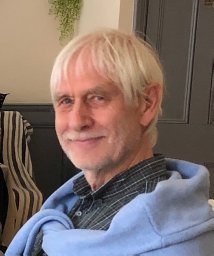 Karl Harlos合作论文数Division of Structural Biology, Henry Wellcome Building of Genomic Medicine2
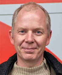 Christian David合作论文数Laboratory for X-ray Nano-Optics, Paul Scherrer Institute2
 Polly Roy合作论文数Department of Infection Biology, Faculty of Infectious and Tropical Diseases, London School of Hygiene & Tropical Medicine2Sarah Sainsbury合作论文数2
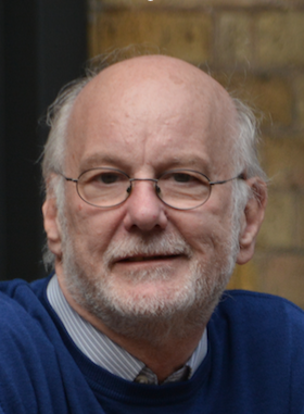 Stuart Siddell合作论文数School of Cellular and Molecular Medicine, University of Bristol2Fauziah Mohd Jaafar合作论文数Institute for Animal Health, Institute for Animal Health2Janne J. Ravantti合作论文数2
 Martin de Porres Walsh合作论文数European Molecular Biology Laboratory, c/o DESY Notkestrasse 85, D-22603 Hamburg, Germany2




