Recent studies highlight the crucial role of the paraspinal musculature (PM), particularly the multifidus (MF), in spinal health and patient outcomes. However, factors associated with PM atrophy and their relative importance remain unclear. To address this gap, we analyzed factors linked to PM atrophy in patients undergoing lumbar fusion using machine learning, aiming to clarify the multifactorial mechanisms underlying this condition. Fatty infiltration (FI) of the lumbar MF was measured as a proxy for muscular atrophy in patients undergoing lumbar spinal fusion. Two machine learning models, logistic regression and extreme gradient boosting (XGBoost), were trained to predict severe FI (> 50
Regional differences affect the reliability of the AO Spine-DGOU Osteoporotic Fracture Classification. European participants showed the highest agreement. This study underscores the need for addressing regional factors to improve the system’s global clinical utility. To evaluate the influence of geographic region on the reliability and reproducibility of the AO Spine-DGOU Osteoporotic Fracture Classification System. This study included 320 participants from various global regions who classified 27 cases of osteoporotic vertebral fractures using the AO Spine-DGOU system which categorizes the fractures to 5 subtypes (OF 1–OF 5). Participants underwent training via an online webinar. Interobserver reliability and intraobserver reproducibility were assessed using Fleiss’ kappa coefficient, and agreement with a gold standard committee was evaluated. The classification system showed moderate to substantial agreement with the gold standard globally (initial kappa 0.58, improving to 0.61). European participants had the highest agreement (kappa 0.64 and 0.66). OF4 fractures were most accurately classified, while OF3 fractures showed the least agreement. Intraobserver reliability was highest among European participants. Post hoc analysis indicated significantly better reliability among German-speaking participants compared to other Europeans (kappa 0.79 vs. 0.70, p = 0.0026). The AO Spine-DGOU Osteoporotic Fracture Classification System demonstrates moderate to substantial reliability and reproducibility, with regional differences influenced by factors such as training and clinical experience. This underlines the necessity of proper education adapted to the regional particularities.
BACKGROUND:Epidural haematoma (EH) is a rare but serious complication of neuraxial anaesthesia. The incidence varies in the published literature, and contemporary procedure- and population-specific estimates are lacking. We estimated the incidence of EH after neuraxial anaesthesia for total joint arthroplasty. We further present an evidence-based algorithm for interdisciplinary management. METHODS:This is a single-centre, retrospective analysis of consecutive patients who underwent primary unilateral or bilateral total hip or knee arthroplasty under spinal, epidural, or combined spinal-epidural anaesthesia from January 2013 to December 2023. EH was identified through a search of two institutional databases (radiology and quality assurance). The medical records of cases with radiologically confirmed EH were reviewed to describe the clinical course and outcomes. The primary outcome was an estimate of the frequency of EH by primary anaesthetic type. RESULTS:Of the total of 96 103 patients, 75 205 patients received spinal anaesthesia and 20 898 patients received either epidural (n=1588) or combined spinal-epidural anaesthesia (n=19 310). We identified one case of symptomatic EH after total hip arthroplasty under combined spinal-epidural anaesthesia requiring urgent lumbar decompression surgery with EH evacuation. We identified one case of asymptomatic EH after epidural anaesthesia, managed conservatively. The estimated population frequencies were 1:10 000 (95% CI: 0.1-3.5:10 000) epidural or combined spinal-epidural anaesthetics and 0:75 000 (95% CI: 0-0.05:10 000) spinal anaesthetics. CONCLUSIONS:We found a low incidence of EH following total joint arthroplasty performed under neuraxial anaesthesia. Although spinal anaesthesia was performed almost four times more frequently, EH was exclusively associated with epidural techniques. Prompt recognition and interdisciplinary collaboration are essential for optimal outcomes.
Study design Literature review with clinical recommendations. Objective To highlight impactful studies on the management of spinal tuberculosis (STB), identified by the AO Spine Knowledge Forum Trauma and Infection group, with recommendations for their integration into clinical practice. Methods Four studies addressing key phases of STB management — imaging diagnosis, laboratory diagnosis, surgical indication and approach, and imaging-based healed status — were reviewed. Consensus recommendations of the KF opinion leaders were graded as strong or conditional following the GRADE methodology. Results Altini et al evaluated the diagnostic value of MRI and FDG-PET/CT in spondylodiscitis including STB; we conditionally recommend FDG-PET/CT as an adjunctive imaging modality when MRI is inconclusive, contraindicated, or limited by artifact. Hu et al developed a multivariable laboratory prediction model based on seven routine indices for early STB diagnosis, achieving an AUC of 0.947; we conditionally recommend this model as an adjunctive screening tool when definitive microbiological testing is delayed or unavailable. Tang et al compared anterior-only, posterior-only, and combined surgical approaches in thoracolumbar STB; we conditionally recommend a posterior-only approach as the preferred surgical strategy in most cases requiring operative treatment. Mittal et al evaluated FDG-PET/CT and contrast MRI for defining healed status and guiding treatment completion; we conditionally recommend FDG-PET/CT as an adjunctive tool when the treatment endpoint remains uncertain or MRI is limited by metallic implants. Conclusion This review provides spine surgeons and clinicians with evidence-based, expert-graded recommendations to enhance standardization and effectiveness across the key clinical decision points in spinal tuberculosis management.
Traumatic fractures of the subaxial cervical spine are relatively common and may lead to devastating consequences. Despite that, there are still a lot of knowledge gaps regarding the classifications of these fractures and their decision-making. This critical review discusses the shortcomings in current classification approaches and the potential knowledge gap in the clinical decision-making of subaxial cervical fractures. From a diagnostic perspective, the most challenging aspect is the lack of consensus on the criteria for discoligamentous injury, which distinguishes between B-type and A-type injury. Careful analysis of Computed Tomography (CT), Plane Radiography, and Magnetic Resonance Imaging (MRI) may provide the most accurate analysis in subtle cases. Deciding which A3/A4 fractures should be treated surgically vs. non-surgically is still debatable, and it's challenged by the scarcity of available literature. The management of unilateral non-displaced facet injuries, whether they should be treated surgically vs. non-surgically, remains a major challenge. While the F modifier of the AO spine classification is helpful, better distinguishing features of F1 vs. F2 are warranted. The current classification schemes do not help to select the surgical approach (whether anterior, posterior, or combined). The choice of surgical approach is more controversial for B-type injuries due to their heterogeneity and variable involvement of anterior and posterior elements. Characterization of the extent of spinal cord injury may add an important dimension to the current classification, aiding to guide the timing of surgical decompression in patients with spinal cord injury.
STUDY DESING:Retrospective cohort study. OBJECTIVE:To characterize the association between paraspinal musculature at C3 and cervical volumetric bone mineral density (vBMD) derived from quantitative computed tomography (QCT) and vertebral bone quality (VBQ) scores from magnetic resonance imaging (MRI). SUMMARY OF BACKGROUND DATA:Osteoporosis and sarcopenia are prevalent among elderly patients and often coexist. Prior studies have shown a positive association between lumbar paraspinal functional cross-sectional area (fCSA) and bone mineral density, but the relationship between cervical paraspinal muscles and cervical bone quality remains unclear. Although dual-energy X-ray absorptiometry (DEXA) is the clinical gold standard for assessing bone density, newer modalities such as QCT and VBQ can evaluate site-specific bone health. Understanding this muscle-bone relationship in the cervical spine may improve preoperative risk stratification and surgical planning. METHODS:Patients with preoperative cervical MRI and CT who underwent anterior cervical discectomy and fusion between 2015 and 2018 were reviewed. Muscles at C3 were categorized into four functional groups: sternocleidomastoid, anterior, posteromedial, and posterolateral. For all groups, cross-sectional area (CSA), fCSA, and fat infiltration (FI) were measured. QCT and VBQ analyses were performed using established methodologies. Multivariable linear regression adjusted for age, sex, and body mass index (BMI) and Benjamini-Hochberg correction were performed. RESULTS:A total of 100 patients (median age, 56.5 years; 38 females) were included. After adjusting, regression analyses demonstrated a significant negative association between fCSA of the anterior group and VBQ scores from C2 to T1, as well as a significant positive association between FI of the same group and VBQ scores. Additionally, a significant positive association was observed between CSA of the posteromedial group and vBMD at C1 and C3. CONCLUSION:Significant associations were observed between cervical paraspinal muscle morphology and vertebral bone quality and density measured using VBQ and QCT, respectively.
Study design Literature Review with clinical recommendations.Objective To highlight impactful studies on subaxial cervical fractures, identified by the AO Spine Knowledge Forum Trauma and Infection group, with recommendations for their integration into clinical practice.Methods Four studies on subaxial cervical fractures that have the potential to shape current practice in subaxial cervical fractures were selected and reviewed. Each study was chosen for its contribution to a critical phase in subaxial fractures management: diagnosis and imaging, surgical vs conservative treatment, and selection of approach.Results Four studies were highlighted. Article 1: Rutsch et al evaluated the sensitivity and specificity of CT, MRI, plain radiography, and LODOX-Statscan in identifying cervical spine injuries. We strongly recommend the use of CT/MRI as gold standard for radiological workup in cervical spine injuries. Article 2: Cirillo et al performed a systematic review of predictor of failure to conservative treatment for isolated unilateral facet fractures. We conditionally recommend surgical treatment in floating lateral mass and greater fragment size. Article 3: Singh et al evaluated the predictors of failure after stand-alone ACDF in subaxial fractures. We conditionally recommend adding posterior fusion in PLL injury, bilateral facet joint dislocation and age above 60 years. Article 4: Kwon et al prospectively randomized and compared anterior vs posterior approach in unilateral facet joint injury. We conditionally recommend anterior surgical fixation in unilateral facet injuries without spinal cord injury.Conclusion This article provides spine surgeons with evidence-based recommendations to enhance standardization and effectiveness of the management of subaxial spine fractures.
Cement discoplasty (CD) is a spine surgery technique involving percutaneous or open polymethylmethacrylate injection into an empty degenerative vacuum disc in the lumbar spine. Although cement acts optimally under compression, the concept of stabilizing without fusing remains uncertain regarding its clinical effectiveness. Despite multiple outcome studies and several meta-analyses, CD continues to be controversial, poorly understood, and not integrated into mainstream surgical practice. Establishing a unified platform is necessary to clarify indications and technique. International peer-reviewed experts in CD were invited to participate in a three-round Delphi process using independent self-administered questionnaires. Fifty statements were developed across five categories: Rationale, Imaging Studies, Technical Aspects, Clinical Indications, and Postoperative Management. Responses were graded on a five-point Likert scale. The survey underwent pilot testing by two experts to ensure face validity and clarity. Consensus was defined as ≥ 80
Frailty might increase the risk for postoperative complications and death. It is not well understood how frailty influences patient-reported outcomes (PROs) in spine surgery. The objective is to investigate on the impact of frailty on PROs 1-year after lumbar fusion. This was a prospective observational study of patients undergoing open posterior lumbar fusion for degenerative conditions with 12-month follow-up (12 M-FU). The modified 5-item frailty index (mFI-5) score was determined preoperatively. PROs included the Oswestry-Disability-Index (ODI), 12-item Short-Form-Healthy-Survey with Physical (PCS-12) and Mental-Component-Score (MCS-12) and Numerical-Rating-Scale (back/leg pain) before surgery and 6 weeks, 3, 6 and 12 months (6 W, 3 M, 6 M, 12 M) postoperatively. PROs were compared between groups stratified as frail (mFI-5 ≥ 2), pre-frail (mFI-5 = 1) and non-frail (mFI-5 = 0). PRO changes at 12 M-FU were evaluated with substantial clinical benefit (SCB) metrics. Regression models were used to determine associations between mFI-5 and PROs. 135 patients (52.6
Study Design Multicenter reliability and validation study.Objectives To adapt and validate the Spinal Instability Spondylodiscitis Score (SISS) to create a practical and reliable classification system for assessing spinal instability in pyogenic spondylodiscitis.Methods The original SISS was modified through structured consensus meetings within the AO Spine Trauma and Infection Knowledge Forum, resulting in the modified SISS (mSISS). The mSISS incorporates four parameters-location, extent of bone lesion, spinal alignment, and mechanical pain-to classify lesions as stable, potentially unstable, or unstable using computed tomography (CT) and clinical data. Fifteen experienced spine surgeons independently evaluated ten representative cases in two rating sessions. Intra- and interrater reliabilities were calculated using intraclass correlation coefficients (ICC) and Fleiss' Kappa. The gold standard was established by consensus of five expert spine surgeons.Results The mSISS demonstrated excellent intrarater reliability for the total score (ICC 0.90, 95% CI 0.84-0.96). Interrater reliability for the total score was 0.87 (95% CI 0.77-0.97) in the first assessment and 0.89 (95% CI 0.80-0.98) in the second. Reliability of individual parameters ranged from moderate to excellent, with spinal alignment showing the highest variability but remaining within acceptable agreement levels. Agreement with the gold standard ratings was high across all parameters.Conclusion The mSISS is a simplified and clinically applicable scoring system for the assessment of spinal instability in pyogenic spondylodiscitis, demonstrating strong reliability among expert spine surgeons. Broader international validation is ongoing to support its integration into clinical decision-making.
Osteoporotic vertebral fractures (OVF) represent a growing clinical challenge due to population aging and the increasing prevalence of osteoporosis. Surgical management of OVF differs substantially from that of traumatic vertebral fractures in younger patients and requires careful consideration of fracture morphology, stability, and patient-related factors. A wide range of surgical techniques is currently available, yet clear treatment algorithms remain limited. A non-systematic narrative review of the literature was performed using PubMed, Scopus, and Google Scholar, supplemented by manual reference screening. The review focuses on contemporary surgical strategies for thoracic and lumbar OVF, with particular emphasis on fracture morphology as defined by the AO Spine-DGOU Osteoporotic Fracture (OF) classification and its implications for treatment selection. Minimally invasive cement augmentation techniques, including vertebroplasty and kyphoplasty, are primarily indicated for stable fracture patterns and provide effective pain relief and early mobilization. Posterior fixation strategies range from short-to long-segment constructs, with stability increasing as construct length increases, particularly in osteoporotic bone. Cement augmentation of pedicle screws significantly improves implant anchorage and reduces mechanical complications. Hybrid stabilization, combining posterior instrumentation with vertebral body augmentation, offers circumferential stabilization with favourable mid-term outcomes and reduced invasiveness compared with combined anterior-posterior surgery. Combined anterior-posterior reconstruction remains the most biomechanically robust option for selected patients with severe instability, neurological deficits, or structural collapse, although its use is limited by patient frailty and surgical morbidity. Surgical treatment of osteoporotic vertebral fractures encompasses a spectrum of techniques tailored to fracture morphology, biomechanical demands, and patient condition. Cement augmentation is suitable for less unstable fractures, whereas posterior fixation-often augmented or combined with hybrid strategies-is preferred in more complex patterns. Individualized treatment selection based on fracture classification and patient factors remains essential to optimize outcomes while minimizing complications.
Study designAn umbrella systematic review.ObjectiveTo identify historically recognized injury characteristics that may affect treatment decisions of traumatic C2 injuries and help improve the description of the "modifiers" presented in the AO Upper Cervical Injury Classification (UCIC).MethodsWe performed an umbrella review of systematic reviews evaluating studies about the management of axis fractures that identify potential modifiers in the treatment of these injuries. These modifiers were grouped according to the new AO UCIC.ResultsEight systematic reviews were included. They were divided into three groups: (1) Axis body fractures - one study, (2) Hangman´s fractures - one study, and (3) Odontoid fractures, six studies. For axis body fractures, most injuries were treated non-operatively, except some Benzel type 3 fractures (AO Type A) with displacement or severe comminution (M1). Hangman´s fractures classified as Effendi I and Levine-Edwards I and II were treated non-operatively with success, with no modifiers identified for non-union or instability. For Levine-Edwards type IIA and III surgery was generally recommended, but these should be classified as AO type B and C respectively without a need for modifiers. For odontoid fractures, fractures in the dens base, with displacement, or in elderly patients were associated with non-union (M1) and patients' specific factors (surgical condition) seem to affect the decision of treatment (M3) for considering surgery.ConclusionsWe identified from the literature some axis injury characteristics that seem to affect the treatment decision in historical series. Knowledge of these modifiers may further enhance the system's clinical utility.
OBJECTIVE:Evidence that individuals with osteoporosis have an increased susceptibility to and frequency of sarcopenia has led to the creation of the term "osteosarcopenia." Given the limitations of dual-energy X-ray absorptiometry in spine bone mineral density assessment, novel techniques such as MRI-based vertebral bone quality (VBQ) are increasingly being utilized to quantify site-specific bone quality in the spine. The relationship between cervical paraspinal muscle morphology and cervical bone quality has not been investigated. This study aimed to explore the relationship between cervical VBQ scores and paraspinal muscles parameters. METHODS:Patients with preoperative cervical MRI who underwent anterior cervical discectomy and fusion between 2015 and 2018 were reviewed. Muscles from C3 to C7 were categorized into 4 functional groups: sternocleidomastoid, anterior, posteromedial, and posterolateral groups. For all groups, the cross-sectional area (CSA), functional CSA (fCSA), and percent fat infiltration (FI) were measured. VBQ scores of the cervical vertebral bodies were performed using prior established methodologies. Multivariable linear regression analyses adjusted for age, sex, and body mass index were performed. The Benjamini-Hochberg procedure was applied to adjust p values. RESULTS:A total of 75 patients (median age 55.4 years; 26 females) were included. A greater VBQ score indicates high fat content of the bone and was significantly associated with the presence of osteopenia/osteoporosis, meaning that high VBQ scores indicate lower bone quality and low VBQ scores indicate better bone quality. After adjustment for age, sex, and body mass index and adjustment of the p values with the Benjamini-Hochberg procedure, regression analysis revealed significant negative associations between fCSA of the anterior muscle group and VBQ scores and a significant positive association between FI of the anterior muscle group with VBQ scores from C2 to T1. CONCLUSIONS:This study presents novel insights into the relationship between cervical vertebral bone mineral quality and paraspinal muscle parameters, particularly at the C3 level, which showed the highest number of significant associations. The findings suggest that measurements of paraspinal muscles at C3 could serve as a proxy for assessing bone quality across the subaxial cervical spine, offering a new perspective in preoperative evaluations for cervical spine surgery. This study is the first to report significant associations between VBQ and paraspinal muscle parameters throughout the subaxial cervical spine.
Study designProspective multicentric study.ObjectiveThoracolumbar fractures without neurologic deficit are challenging situations in terms of treatment decision making. We aimed to analyze the occurrence of adverse events (AEs) after surgical and nonsurgical treatment and its impact on functional outcomes.Methods198 patients from a prospective multicentric database were included. The occurrence of adverse events and treatment failure within 2 years of follow up were recorded. ODI was compared between patients with and without adverse events at six months, 1 year and 2 years follow up. Multivariable regression analysis was conducted to assess the association between post-treatment adverse events and ODI at 1-year follow-up.Results46 adverse events were recorded (23.2%). Higher categories of the Charlson Comorbidity Index (CCI) (P = 0.006), unemployment or retirement (P = 0.027), and current smoking (P = 0.008) were significantly associated with the occurrence of adverse events whereas no significant differences were observed in terms of treatment decision (conservative vs surgical). ODI values were significantly higher in patients with adverse events at the 6-month and 1-year follow-up visits without significant differences at 2 years follow up. Treatment failure was observed in only 5 patients.ConclusionWe found association between the occurrence of AE and higher ODI at 6-months and one-year follow up. Additionally, a higher CCI and smoking status were associated with higher likelihood to develop adverse events in our cohort.
Osteoporosis is a widespread disease with an increasing incidence. In 2018, the “Osteoporotic Fracture working group” affiliated with the German Society for Orthopaedic and Trauma Surgery (DGOU), introduced a novel classification system specifically for osteoporotic thoracolumbar vertebral body fractures. To evaluate the influence of geographic region on the reliability and reproducibility of the AO Spine-DGOU Osteoporotic Fracture Classification System. This study included 320 participants from various global regions who classified 27 cases of osteoporotic vertebral fractures using the AO Spine-DGOU system which categorizes the fractures to 5 subtypes (OF 1 – OF 5). Participants underwent training via an online webinar. Interobserver reliability and intraobserver reproducibility were assessed using Fleiss' kappa coefficient, and agreement with a gold-standard committee was evaluated. The classification system showed moderate to substantial agreement with the gold standard globally (initial kappa 0.58, improving to 0.61). European participants had the highest agreement (kappa 0.64 and 0.66). OF4 fractures were most accurately classified, while OF3 fractures showed the least agreement. Intraobserver reliability was highest among European participants. Post-hoc analysis indicated significantly better reliability among German-speaking participants compared to other Europeans (kappa 0.79 vs. 0.70, p=0.0026). The AO Spine-DGOU Osteoporotic Fracture Classification System demonstrates moderate to substantial reliability and reproducibility, with regional differences influenced by factors such as training and clinical experience. This underlines the necessity of proper education adapted to the regional particularities.
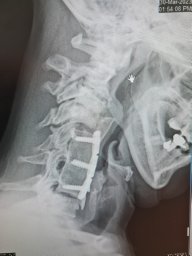
Objective. To report an unusual titanium mesh cage failure after ten years of follow-up. Clinical case. A 40-year-old woman diagnosed with low-grade spinal chondrosarcoma underwent a total en-bloc spondylenctomy (TES) surgery. This technique required a wide resection, anterior reconstruction with a titanium mesh cage (TMC), and posterior stabilization. For several years a favorable clinical outcome was achieved. After 10 years of follow-up, the patient developed acute non-traumatic low back pain without neurological repercussions. The images confirmed a rupture of the titanium cage. After ruling out tumor recurrence, the patient went through spinal instrumentation revision. Conclusion. Isolated TMC rupture is a rare event scarcely reported in the literature. There is a strong recommendation to instrument two levels proximal and distal to the bone defect in 360° reconstructions after a TES. Stability had likely been lost over the years due to the slow resorption of the bone graft. We consider necessary studies with a higher casuistry and level of evidence to analyze the contributing factors for the event of TMC rupture.
Study Design Literature Review with clinical recommendations. Objective To highlight impactful studies on pyogenic spondylodiscitis (PS), identified by the AO Spine Knowledge Forum Trauma and Infection, with recommendations for their integration into clinical practice. Methods Five influential studies on PS that have the potential to shape current practice in spinal infections were selected and reviewed. Each study was chosen for its contribution to a critical phase in PS management: diagnosis, imaging, surgical vs conservative treatment, and antibiotic duration. Recommendations were graded as strong or conditional following the GRADE methodology. Results Five studies were highlighted. Article 1: Pluemer et al introduced the Spinal Infection Treatment Evaluation (SITE) Score, a novel scoring tool for standardizing treatment decision-making. Conditional recommendation to incorporate the SITE Score or SISS Score for improved treatment outcomes. Article 2: Maamari et al conducted a meta-analysis comparing imaging modalities, with conditional recommendation to consider 18F-FDG PET/CT to diagnosis PS as an adjunct to MRI which remains the gold standard. Article 3: Thavarajasingam et al demonstrated the potential survival benefit of early surgery in specific PS cases, leading to a strong recommendation for early intervention in appropriate patients. Article 4: Neuhoff et al compared conservative and surgical treatments in well-resourced settings, concluding a strong recommendation for early surgery in appropriate patients. Article 5: Bernard et al evaluated antibiotic treatment duration, with a conditional recommendation for a 6-week course in confirmed cases, based on comparable efficacy to a 12-week regimen. Conclusions Management of PS remains complex and varied. This perspective provides spine surgeons with evidence-based recommendations to enhance standardization and effectiveness in clinical practice.
ABSTRACT Assessing the bone condition in patients with spinal disease is clinically valuable. However, evaluating bone strength in the presence of spine degenerative changes is challenging. Quantitative computed tomography (QCT) and finite element analysis (FEA) have been proposed as methods for more accurate bone quality assessment. This study investigates the relationship between bone strength predicted by FEA and other relevant biological parameters. This retrospective cross‐sectional study included 127 patients with spinal disease who underwent preoperative CT scans between 2014 and 2020. Baseline patient characteristics, volumetric bone mineral density (vBMD) measured by QCT, and vertebral bone strength predicted by FEA were collected. The degree of degeneration was evaluated by classifying osteophyte formation, disc height narrowing, vertebral sclerosis, and spondylolisthesis into a grading scale ranging from 0 to 2. Multiple linear regression analysis was conducted to assess the effect of each factor on bone strength predicted by FEA. Of 127 patients, 120 patients (median age was 62 years) were included. The median vBMD and vertebral strength were 114.3 mg/cm 3 and 7892.9 N, respectively. After adjusting for age, sex, body mass index, smoking status, diabetes mellitus, vBMD, and degenerative changes, multiple linear regression analysis revealed that sex, vBMD, and degree of degeneration independently increased the vertebral strength measured by FEA. This study suggests that in patients with spinal disease, vertebral bone strength is affected not only by sex and bone mineral density but also by degenerative changes. Thus, bone strength could be predicted more accurately in patients with spinal disease using FEA.
Study Design Cross-sectional survey. Objectives A cornerstone of classification systems is good reliability amongst different groups of classification users. Thus, the aim of this international validation study was to assess the reliability of the new AO Spine DGOU Osteoporotic Fracture Classification (OF classification) stratified by surgical specialty, work-setting, work-experience, and trauma center level. Methods 320 spine surgeons were asked to rate 27 cases according to the OF classification at 2 time points, 4 weeks apart (assessment 1 and 2) in this online-webinar based validation process. The kappa statistic (κ) was calculated to assess the inter-observer reliability and the intra-rater reproducibility. Results A total of 7798 (90.3%) ratings were recorded in assessment 1 and 6621 (76.6%) ratings in assessment 2. Global inter-rater reliability was moderate in both assessments (κ = 0.57; κ = 0.58). Participants with a work-experience of >20 years showed the highest inter-rater agreement in both assessments globally (κ = 0.65; κ = 0.67). Participants from a level-1 trauma center showed the highest agreement (κ = 0.58), whereas participants working at a tertiary trauma center showed higher grade of agreement in the second assessment (κ = 0.66). Participants working in academia showed the highest agreement in assessment 2 (κ = 0.6). Surgeons with academic background and surgeons employed by a hospital showed substantial intra-rater agreement in the second assessment. Conclusions The AO Spine-DGOU Osteoporotic Fracture Classification showed moderate to substantial inter-rater agreement as well as intra-rater reproducibility regardless of work-setting, surgical experience, level of trauma center and surgical specialty.
 Hughes Alexander P合作论文数37Federico P. Girardi合作论文数36
 Andrew A. Sama合作论文数Weill Medical College, Cornell University36Frank P. Cammisa合作论文数36
 Jennifer Shue合作论文数32Jiaqi Zhu合作论文数Hospital for Special Surgery, Spine Care Institute29Lukas Schonnagel合作论文数28
 Erika Chiapparelli合作论文数28Soji Tani合作论文数27
 Matias Petracchi合作论文数26Carlos Solá合作论文数26Marcelo F. Gruenberg合作论文数26
 Thomas Caffard合作论文数25Artine Arzani合作论文数21
 Alfredo Guiroy合作论文数21Ali E. Guven合作论文数19
 Gonzalo Rodrigo Kido合作论文数19
 Matias Pereira Duarte合作论文数Hospital Italiano de Buenos Aires19Krizia Amoroso合作论文数18Sebastian F Bigdon合作论文数Univ Bern, Univ Hosp Bern18
 Andrei Fernandes Joaquim合作论文数Médico Residente de Neurocirurgia;UNICAMP, Universidade de Campinas;UNICAMP, Universidade de Campinas18
 Harvinder Singh Chhabra合作论文数Department of Spine Services, Indian Spinal Injurues16
 Gregory D Schroeder合作论文数The Rothman Institute, Thomas Jefferson University15
 Klaus Schnake合作论文数Zentrums für Wirbelsäulen- und Skoliosetherapie, Malteser Waldkrankenhaus St. Marien15
 Marcelo Valacco合作论文数14Julio Bassani合作论文数13Richard Bransford合作论文数13
 Mariana Bendersky合作论文数12Maximilian Muellner合作论文数12Charlotte Dandurand合作论文数12
 Martin M Estefan合作论文数12
 Lorin M. Benneker合作论文数Klinik Sonnenhof12
 Mohamed M. Aly合作论文数Faculty of Science, El-Menoufia University12Gbolabo Sokunbi合作论文数11Emiliano Vialle合作论文数11
 Henryk Haffer合作论文数11Martin Gagliardi合作论文数11
 William Zelenty合作论文数10Bhatia Nitin N合作论文数Orthopaedic Biomechanics Laboratory, VA Healthcare System10
 John A. Carrino合作论文数School of Medicine, Johns Hopkins University;Department of Radiology and Imaging, Hospital for Special Surgery10
 Mohammad Mostafa El-Sharkawi合作论文数Faculty of Medicine, Assiut University10
 F. C. Oner合作论文数Department of Orthopaedics, University Medical Center Utrecht10
 Ek Tsoon Tan合作论文数Department of Electrical and Computer Engineering, National University of Singapore10
 Alexander R. Vaccaro合作论文数Department of Orthopaedic Surgery, Thomas Jefferson University;Sidney Kimmel Medical College, Thomas Jefferson University;Thomas Jefferson University Hospital10Rishi Kanna合作论文数9
 Jose A Canseco合作论文数Rothman Orthopaed Inst, Thomas Jefferson Univ Hosp9
 Julian Scherer合作论文数Orthopaedic Research Unit, University of Cape Town9Yu-Po Lee合作论文数8Cezar Eugen Popescu合作论文数8




