Estrogen receptor-positive (ER+) breast cancer exhibits a marked propensity for skeletal metastasis; however, the molecular drivers of bone colonization remain incompletely defined. We investigated the tumor suppressor neurofibromin (NF1), a dual repressor for RAS and ER signaling, whose inactivation promotes endocrine therapy (ET) resistance and is associated with inferior relapse-free survival. NF1 copy number loss was detected in 62% of ER+ patients who subsequently developed metastases and was associated with an increased likelihood of bone metastases at initial diagnosis. In mouse xenograft models, NF1-depleted ER+ breast cancer cells demonstrated enhanced dissemination to skeletal sites following surgical resection of primary tumors. Furthermore, after intra-iliac injection, NF1-depleted cells generated significantly greater tumor burden in bone. Transcriptomic profiling revealed enrichment of bone-related gene signatures in NF1-depleted ER+ breast cancer cells, which more potently induced osteoclast differentiation and bone loss in co-culture systems. In parallel, low NF1 expression correlated with repressed T cell functional states in primary breast tumors and bone metastases. Consistent with these clinical observations, NF1-depleted ER+ breast cancer cells more effectively inhibited proliferation, interferon-γ secretion, and cytotoxicity of human primary CD8+ T cells. Collectively, these findings identify NF1 inactivation as a key driver of bone metastasis in a substantial subset of ER+ breast cancers. By amplifying the osteolytic "vicious cycle" and promoting immune evasion, NF1 loss remodels the microenvironment to favor tumor expansion. These results further suggest that NF1 loss functionally links therapy resistance with increased skeletal metastatic potential.
Invariant natural killer T cells (NKTs) have intrinsic antitumor properties that make them promising candidates for chimeric antigen receptor (CAR) immunotherapies. Transgenic cytokine expression can enhance cellular therapy potency, and we hypothesized that co-expressing interleukin-18 (IL-18) alone or with IL-15 would boost CAR-NKT therapeutic potential. To test this, we generated retroviral constructs expressing IL-15 and/or IL-18 with an inducible caspase-9 safety switch and co-transduced them with a GD2-specific CAR into human NKTs. Co-expression of IL-18 or IL-15/IL-18 increased CAR-NKT cytotoxicity, proliferation, and cytokine secretion in vitro compared to IL-15 alone. IL-18 also enhanced GPC3.CAR and CD19.CAR NKT activity against hepatocellular carcinoma and B cell leukemia cells, respectively. In a metastatic neuroblastoma model, IL-18-expressing GD2.CAR-NKTs controlled tumors more effectively than IL-15-only cells, but mice in the IL-15/IL-18 group developed severe toxicities not observed in the IL-18-only group. Mechanistically, IL-18 induced a transcriptional program distinct from IL-15, marked by lower exhaustion signatures and enrichment of metabolic pathways. Finally, targeted metabolomics showed that IL-18 drives broad metabolic reprogramming in CAR-NKTs including increased oxidative phosphorylation, glycolysis, glutaminolysis, and purine metabolism. These findings support the use of IL-18 in developing the next generation of cytokine-armed CAR-NKT cancer immunotherapies.
Abstract Introduction: CDK4/6 inhibitors (CDK4/6i) are the standard of care for hormone receptor-positive/HER2-negative breast cancer (HR+/HER2- BC). Yet resistance is common and often characterized by loss or dysfunction of the Retinoblastoma (Rb) protein or inadequate suppression of its phosphorylation. Circulating tumor cells (CTCs) offer minimally invasive means to monitor tumor biology. Here, we describe the analytical performance of a multiplex immunofluorescence (mIF) assay integrated with an image-analysis pipeline to quantify Rb and phospho-Rb (pRb) expression on CTCs and evaluation of cell recovery efficiency by CTCeptor, a microfluidic CTC isolation platform. Methods: The mIF assay utilized antibodies against Rb (AF595), pRb (AF488), cytokeratin (CK, AF647), and CD45 (AF555), with DAPI for nuclear staining. Antibody specificity and minimal non-specific binding were verified using single-marker staining and isotype controls. Assay performance (sensitivity, specificity, signal-to-noise ratio (SNR), and dynamic range) was evaluated using positive (MCF7, T47D) and negative (T47D-PalboR, MDA-MB-468, or leukocytes) control cell lines. Healthy donor leukocytes were spiked with control cell lines to simulate patient-derived matrices. Various blocking buffers, wash buffers, and cell attachment methods were compared. Cell recovery was evaluated after processing blood spiked with MCF7 cells using CTCeptor followed by staining with anti-CK-AF647, anti-CD45-AF555, and DAPI in HyPICC chamber. Imaging was performed on Leica STED SP8 or Nikon N-STORM microscopes. Quantitative analysis of mean fluorescence intensity (MFI) was conducted using FIJI, and statistical analysis was performed using GraphPad Prism v10. Results: The optimized mIF with integrated image analysis pipeline demonstrated high analytical sensitivity (Rb: 84%, pRb: 83%) and specificity (Rb: 96%, pRb: 100%). SNRs were 5 for Rb and 50 for pRb, with dynamic range exceeding two log units for both markers. Single-marker staining and isotype controls confirmed antibody specificity and minimal non-specific binding. Based on comparative analyses, 1% BSA + 0.5% goat serum was selected as the blocking buffer, and Triton-based buffer as the wash buffer. RareCyte® cell attachment method yielded superior cell retention (79%). Recovery rate after processing the spiked blood with MCF7 cells using CTCeptor followed by staining in HyPICC chamber ranged from 40-60%. Conclusion: We have developed and optimized a quantitative mIF assay and image analysis workflow to detect Rb and pRb expression on CTCs. Future studies will assess the predictive biomarker utility of the assay by measuring Rb and pRb dynamics on CTCs in metastatic HR+/HER2- BC patients before and after CDK4/6i therapy. Acknowledgement: This research was funded by CPRIT (RP210148) and NCATS CTSA pilot grant (1UM1TR004539-01A1). Citation Format: Mantasha Tabassum, Shivaani Suresh Kanna, Wangjia Cao, Suneel Kumar, Mothaffar F. Rimawi, George Miles, Meghana Trivedi. Method development and workflow optimization of a CTC-based biomarker assay to predict response to CDK4/6 inhibitors in HR+/HER2- breast cancer [abstract]. In: Proceedings of the American Association for Cancer Research Annual Meeting 2026; Part 1 (Regular Abstracts); 2026 Apr 17-22; San Diego, CA. Philadelphia (PA): AACR; Cancer Res 2026;86(7 Suppl):Abstract nr 5250.
Proteogenomic analyses have identified an association between LIG1 (DNA Ligase I) loss and chemotherapy resistance in a subset of triple negative breast cancer (TNBC) enriched for TP53 mutations. Here, we demonstrate that co-occurrence of TP53 mutations and LIG1 loss is associated with upregulated DDR activity, including homologous recombination, likely contributing to reduced platinum sensitivity. Unbiased genetic and monotherapy drug screens identified PARP inhibitors (PARPi) as a potential treatment for LIG1-depleted tumors; however, the increase in sensitivity was modest and lower than that observed in TNBC models with homologous recombination deficiency. Subsequently, a screen of PARP inhibition in combination with each of 120 clinically relevant DDR inhibitors revealed that PARPi sensitivity in LIG1-loss cells was significantly enhanced by the addition of an ATR inhibitor (ATRi). Olaparib and ceralasertib demonstrated synergistic cytotoxicity in LIG1-loss cell line models; the combination significantly reduced tumor volume in a LIG1-low PDX model compared to either monotherapy, and showed greater ex vivo cytotoxicity in a LIG1-low PDXO model versus a LIG1-high control. Hence, this study highlights LIG1 status as a stratification factor for ongoing and future clinical trials of DDR-targeted combinations in TNBCs.
Figure S1: Integration of proteogenomics and functional genetics to reveal novel vulnerabilities in TNBC (related to Figure 1).
PURPOSE:Precision oncology trials have generally focused on tumor testing to identify actionable alterations. The National Cancer Institute-Children's Oncology Group Pediatric MATCH trial incorporated return of germline results to assess feasibility of reporting in a cooperative group setting and characterize germline cancer predisposition in patients with refractory cancers. PATIENTS AND METHODS:Tumor and blood DNA from patients 1-21 years of age with treatment-refractory solid tumors, non-Hodgkin lymphomas, or histiocytic disorders underwent cancer gene panel sequencing. Clinical germline reports returned to 151 study sites included pathogenic/likely pathogenic (P/LP) germline variants found in 38 cancer predisposition genes (CPGs). European Society of Medical Oncology (ESMO) recommendations for germline follow-up of tumor variants in CPGs were assessed. RESULTS:Both tumor and germline reports were completed for 1,167 patients (87.5% of enrolled). A total of 295 tumor reports (25%) included 361 CPG variants of which 70 variants (19.4%) were found in the germline sample. Three additional germline-only CPG variants resulted in 73 (6.3%) of 1,167 germline reports containing variants across 21 CPGs previously associated with pediatric and/or adult cancers. Among frequently mutated CPGs in tumors, concurrent germline findings ranged from 8/32 NF1 (25.0%) and 25/163 TP53 (15.3%) to zero of 27 ALK and 18 PTEN tumor variants. ESMO guidelines recommended clinical follow-up for 110 (30.5%) of 361 tumor CPG variants which included 40 (57.1%) of 70 germline variants. CONCLUSION:Coordinated germline and tumor panel testing was feasible and revealed P/LP CPG variants in 6.3% of the Pediatric MATCH cohort. Tumor variant fraction, germline association of CPG with tumor type, and adult-oriented guidelines were not predictive of germline status, emphasizing the need for systematic germline follow-up after tumor genomic testing for pediatric patients.
Proteogenomic analysis is applied to samples from the CALGB 40601 (Alliance) randomized neoadjuvant trial of trastuzumab, lapatinib, or the combination to identify biomarkers associated with pathological response status. Absence of ERBB2 gene amplification and human epidermal growth factor receptor 2 (HER2) protein overexpression by proteogenomics is associated with non-pathological compete response (pCR) (p < 0.05), highlighting potential false positives from standard diagnostics. Pathway analysis in proteogenomics-confirmed HER2+ samples identifies elevated epithelial-mesenchymal transition (EMT) and WNT-β-catenin signaling in non-pCR cases before treatment. Twenty-four pCR-associated proteins reproduce in a second proteomic dataset, and four (GPRC5A, TPBG, SP140L, and NEU1) are significant in a third. A meta-analysis of ten diverse neoadjuvant anti-HER2 treatment regimens from four independent studies confirms that non-pCR cases express higher levels of mRNA for G protein-coupled receptor class C group 5 member A (GPRC5A, p = 0.0002) and trophoblast glycoprotein (TPBG, p = 0.00008). Thus, proteogenomic analysis identifies negative biomarkers for pCR and alternative plasma membrane targets for treatment-resistant HER2+ breast cancer. This trial is registered at clinicaltrials.gov (NCT00770809).
Figure S5: PTPN12-deficient cells exhibit mitotic defects dependent on APCFZR1 hyperactivation (related to Figure 5).
Background: Current medications to prevent breast cancer (BC) are the antiestrogens tamoxifen, raloxifene, anastrozole and exemestane which can prevent 50% of ER+ BC. However, because they can have side effects and require 5 years of continuous use, women who are yet cancer-free are often reluctant to take them. Furthermore, antiestrogens do not prevent ER- BC. Therefore, it is urgent that we develop alternative prevention drugs that require only a short-term treatment and are suitable for preventing both ER+ and ER- BC. No prevention strategies have been developed for ER- or HER2+ subtypes, the most virulent forms of the disease. Ruxolitinib is a small molecule which selectively inhibits the activities of Janus Associated Kinases (JAK); activated JAK phosphorylate STATs leading to modulation of gene expression. In animal models, ruxolitinib prevents progression of premalignant breast lesions to invasive BC. Studies in several human tissue types suggest that there are anticancer “barriers,” namely apoptosis and senescence, which are erected in early lesions to prevent the progression to cancer. We hypothesize that the JAK2-STAT5 pathway in human premalignant lesions promotes the progression of these lesions to malignancy by lowering the apoptosis anticancer barrier; therefore, inhibition of this pro-survival pathway could reduce the load of premalignant lesions in the breast and thus lower BC risk. We are conducting a window-of-opportunity trial through the Translational Breast Cancer Research Consortium (TBCRC) to test whether short-term ruxolitinib treatment of subjects with premalignant lesions will result in increased apoptosis. Trial Design: This is a phase II, double-blind, placebo-controlled clinical trial. Our target population consists of patients with premalignant breast lesions requiring surgical excision. Subjects will be randomized 1:1 to receive either ruxolitinib 20 mg twice daily or placebo for ∼15 days after which they will proceed with standard of care surgical excision within 12 hours of the last dose. Key Eligibility Criteria: 1) Breast biopsy showing atypical ductal hyperplasia, atypical lobular hyperplasia, lobular carcinoma in situ, or ductal carcinoma in situ requiring surgical excision 2) Adequate hematologic and organ function 3) Willing to not use concomitant strong CYP3A4 inhibitors Statistical Methods: The primary endpoint is change in apoptosis, as measured by cleaved caspase-3 staining, in surgical samples relative to baseline biopsy. Lesions of different types will be characterized separately, and the participant’s most advanced lesion present at baseline will be used for the primary analysis. The difference in changes between groups will be evaluated using the Wilcoxon rank sum test. The secondary objectives primarily focused on the effects of ruxolitinib treatment on biomarkers. The secondary endpoints include pre- and post-treatment pSTAT5, TUNEL, Ki67, BCLxL, MCL1, BCL2, PRLR, pSTAT3, and serum prolactin levels. Changes in these markers will be compared between groups using the Wilcoxon rank sum test. Accrual: We expect to enroll 100 subjects from 7 sites in the US in order to obtain 70 evaluable subjects. Currently there are 80 subjects enrolled. Contact Information: For more information on this trial, please visit clinicaltrials.gov NCT02928978 Citation Format: Julie Nangia, Shaun Bulsara, Tao Wang, Amy Ku, Derek Thomas, Ingrid Meszoely, Parijatham S. Thomas, Catherine Parker, Sheldon Feldman, Carla Fisher, Kristalyn Gallagher, Gaorav Gupta, Alastair Thompson, Kristen Otte, Carolina Gutierrez, Chandandeep Nagi, George Miles, Susan Hilsenbeck, Yi Li, C. Kent Osborne, Mothaffar Rimawi. A Randomized Phase II Window-of-opportunity Trial of Ruxolitinib in Patients with High Risk and Premalignant Breast Conditions [abstract]. In: Proceedings of the San Antonio Breast Cancer Symposium 2024; 2024 Dec 10-13; San Antonio, TX. Philadelphia (PA): AACR; Clin Cancer Res 2025;31(12 Suppl):Abstract nr P2-12-19.
Figure S4: PTPN12 deficiency causes aberrant degradation of APCFZR1 substrates (related to Figure 4).
Table S2: Proteogenomic fold-change data (PTPN12 normal vs deficient) (related to Figure 2).
Meningiomas are the most common primary brain tumors in adults but much less frequent in children. Many subtypes exist, including anaplastic (malignant) meningioma, which accounts for less than 20% of pediatric tumors. Meningiomas can arise in association with cancer predisposition syndromes due to germline variants in genes such as NF2, MEN1 and SMARCE1. This report describes a 6-year-old boy diagnosed with anaplastic meningioma who was treated with surgery and focal radiation therapy. The family consented to participate in the Texas KidsCanSeq clinical genomics study. Analysis of germline and tumor samples detected a single germline finding of a CDKN1B pathogenic frameshift variant associated with Multiple Endocrine Neoplasia Type 4 (MEN4) without somatic loss of the other allele. Tumor analysis revealed a YAP1::MAML2 fusion, which has been previously reported in pediatric meningiomas not associated with NF2. YAP1::MAML2 fusion is a known driver for development of meningioma, but the role of the germline CDKN1B variant in the absence of a tumor second hit is unclear. This case highlights the importance of performing combined tumor and germline molecular genetic analysis of rare tumors to help clarify the risk of development of cancer in patients with rare cancer predisposition syndromes.
Triple-negative breast cancer (TNBC) is an aggressive subtype of breast cancer with few effective targeted therapies. Taxanes and other microtubule-targeting agents (MTA) are first-line chemotherapies for TNBC; however, the molecular mechanisms that underlie TNBC taxane sensitivity are largely unknown, preventing selection of taxane-responsive patients and development of more selective therapeutic strategies. In this study, we identified tumor-selective vulnerabilities in TNBC harboring inactivation of the tumor suppressor PTPN12 by integrating proteogenomic characterization and synthetic lethality screening. We discovered that PTPN12 inactivation drives mitotic defects through aberrant hyperactivation of the ubiquitin ligase complex APCFZR1, a critical regulator of the cell cycle. Consistent with the mitotic stress caused by PTPN12 inactivation in TNBC cell lines, tumors harboring loss of PTPN12 exhibit heightened sensitivity to taxane chemotherapy. Collectively, these data suggests that PTPN12 inactivation may drive chromosomal instability and favorable MTA response in TNBC-two prominent features of the disease with unclear mechanistic etiology. SIGNIFICANCE:Many TNBCs respond to MTAs, but the underlying cause(s) of this sensitivity remain poorly understood. Herein, we discover that the tumor suppressor PTPN12 regulates mitotic fidelity and MTA sensitivity in a large subset of patients with TNBC, which has significant implications for the use of MTAs in breast cancer.
Supplementary Figure S1 show shows biochemical staining on MCF10A-PIK3CAH1047R mammary tumors. Supplementary Figure S2 shows expression of ERα in MCF10A-PIK3CAH1047R mammary tumors. Supplementary Figure S3 shows response of MCF10A-PIK3CAH1047R cells to fulvestrant in vitro and in vivo. Supplementary Figure S4 shows droplet digital PCR (ddPCR) analysis. Supplementary Figure S5 shows tolerance of alpelisib administration in mice. Supplementary Figure S6 shows short term alpelisib does not affect lung or bone metastasis from MCF10A-PIK3CAH1047R mammary tumors.
Bone is a common site for metastasis of solid cancers. The diversity of histological and molecular characteristics of bone metastases (BMs) remains poorly studied. Here, we performed single-cell RNA sequencing on 42 BMs from eight cancer types, identifying three distinct ecosystem archetypes, each characterized by an enrichment of specific immune cells: macrophages/osteoclasts, regulatory/exhausted T cells, or monocytes. We validated these archetypes by immunostaining on tissue sections and bioinformatic analysis of bulk RNA sequencing/microarray data from 158 BMs across more than 10 cancer types. Interestingly, we found only a modest correlation between the BM archetypes and the tissues of origin; BMs from the same cancer type often fell into different archetypes, while BMs from different cancer types sometimes converged on the same archetype. Additional analyses revealed parallel immunosuppression and bone remodeling mechanisms, some of which were experimentally validated. Overall, we discovered unappreciated heterogeneity of BMs across different cancers.
PURPOSE To evaluate the relative diagnostic yield of clinical germline genomic tests in a diverse pediatric cancer population. PATIENTS AND METHODS The KidsCanSeq study enrolled pediatric cancer patients across six sites in Texas. Germline analysis included both exome sequencing and a therapy-focused pediatric cancer gene panel. The results were categorized by participants demographics, the presence of pathogenic or likely pathogenic (P/LP) variants, and variants of uncertain significance (VUS) in cancer predisposition genes (CPGs). Pediatric actionable CPGs were defined as those with cancer surveillance recommendations during childhood. RESULTS Cancer P/LP variants were reported by at least one platform in 103 of 578 (17.8%) participants of which 76 were dominant cancer genes (13.1%) with no significant differences by self-described race or Hispanic ethnicity. However, the proportion of participants with VUS was greater in Asian and African American participants ( P = .0029). Diagnostic yield was 16.6% for exome versus 8.5% for panel ( P < .0001) with 42 participants with concordant germline results. Exome-only results included P/LP variants in 30 different CPGs in 54 participants, whereas panel-only results included seven participants with a copy number or structural P/LP variants in CPGs. There was no significant difference in diagnostic yield limited to pediatric actionable CPGs ( P = .6171). CONCLUSION Approximately 18% of a diverse pediatric cancer population had germline diagnostic findings with 50% of P/LP variants reported by only one platform because of CPGs not on the targeted panel and copy number variants (CNVs)/rearrangements not reported by exome. Although diagnostic yields were similar in this diverse population, increases in VUS results were observed in Asian and African American populations. Given the clinical significance of CNVs/rearrangements in this cohort, detection is critical to optimize germline analysis of pediatric cancer populations.
Abstract Introduction: Cyclin-dependent kinases 4/6 inhibitors (CDK4/6i) are used as the first-line treatment of advanced/metastatic or high-risk HR+/HER2- breast cancer (BC) that make up about 60-70% of all cases. However, nearly 50% of patients exhibit drug resistance. Therefore, biomarkers that predict CDK4/6i response are needed to spare patients from toxicities and high costs. Resistance to CDK4/6i [palbociclib, ribociclib, and abemaciclib] is associated with the aberrant or loss of Retinoblastoma (Rb). A decline in phospho-Rb (pRb) levels on therapy is also associated with CDK4/6i response. We aimed to develop a multiplex immunofluorescence (IF) assay to assess Rb and pRb levels that can be used on circulating tumor cells (CTCs) to predict tumor response to CDK4/6i. Here, we report the selection of internal controls for Rb and pRb, assay repeatability and reproducibility, as well as compatibility of the CTC isolation protocol with the assay. Methods: Rb and pRb expression was evaluated by western blotting (WB) and IF in parental (P), estrogen deprivation-resistant (EDR), palbociclib-resistant (PalboR), and EDR/PalboR derivatives of two HR+/HER2- cell lines (MCF7 and T47D), Rb1 KO cells of MCF7 and T47D, as well as triple-negative cell lines, BT549 and MDA-MB-468. IF-stained cells were imaged using Leica STED SP8 confocal microscope and Zeiss Axioscan 7. The concordance between IF and WB results was assessed using a Bland-Altman plot. Selectivity and specificity of the assay as well as repeatability and reproducibility between investigators were also assessed. Blood from healthy subjects was used to test the compatibility of the ScreenCell® filtration system to isolate CTC with the IF assay. Results: Our initial approach utilized indirect IF staining for Rb and pRb using the same primary antibodies as WB. On the Bland-Altman plot, most data points were relatively close to the bias line, suggesting that the difference between the two methods was acceptable. The analytical range for mean Rb and pRb staining intensity was 0.744 - 46.779 and 1.001 - 52.919, respectively. Based on WB and IF data, we nominated T47D PalboR and MDA-MB-468 cells as negative controls for Rb and pRb expression. The selectivity and specificity were 100% and 70%, respectively, for the Rb expression and 99% and 61% for pRb, respectively. The IF assay was repeatable and reproducible between two investigators. The ScreenCell® CTC isolation method was not compatible with the assay, as Rb and pRb expression was either not detected or limited when processed using FC2 or PBTFC buffer, irrespective of the antigen retrieval. Conclusion: We have selected internal cell line controls for Rb and pRb and are in the process of optimizing signal-to-noise ratio and specificity using conjugated antibodies. We are also testing the compatibility of other CTC isolation methods with the IF assay. Citation Format: Mantasha Tabassum, Shivaani Suresh Kanna, Wangjia Cao, Mothaffar Rimawi, George E Miles, Meghana V Trivedi. A multiplexed immunofluorescence assay to assess Rb and phospho-Rb as predictive biomarkers for CDK4/6 inhibitors in HR+/HER2- breast cancer [abstract]. In: Proceedings of the American Association for Cancer Research Annual Meeting 2024; Part 1 (Regular Abstracts); 2024 Apr 5-10; San Diego, CA. Philadelphia (PA): AACR; Cancer Res 2024;84(6_Suppl):Abstract nr 5166.
 Matthew Ellis合作论文数Department of Psychiatry, Washington University School of Medicine In St. Louis29
 Meenakshi Anurag合作论文数Baylor College of Medicine28
 Yongchao Dou合作论文数School of Mathematical Science, Dalian University of Technology26
 Beom-Jun Kim合作论文数Baylor Coll Med, Houston, TX 77030 USA25
 Jonathan Thomas Lei合作论文数Lester and Sue Smith Breast Center, Baylor College of Medicine24
 Bora Lim合作论文数Department of Breast Medical Oncology, The University of Texas MD Anderson Cancer Center22
 Susan Galloway Hilsenbeck合作论文数Smith Breast Center, Baylor College of Medicine;Dan L Duncan Comprehensive Cancer Center, Baylor College of Medicine22
 Bing Zhang (张兵)合作论文数Institute of Remote Sensing and Digital Earth, Chinese Academy of Sciences;Aerospace Information Research Institute, Chinese Academy of Sciences;University of Chinese Academy of Sciences21
 Matthew Holt合作论文数20P. Singh合作论文数19Zheng Ze-Yi合作论文数18Nada Mohamed合作论文数17Emmanuel Bikorimana合作论文数17Hilda Kennedy合作论文数17Diana Fandino合作论文数17
 Jaya Ruth Asirvatham合作论文数17Yunguan Wang合作论文数Quantitat Biomed Res Ctr, Univ Texas Southwestern Med Ctr Dallas17
 Maria Rosario Chica-Parrado合作论文数Lab Biol Mol Canc, Univ Malaga17Anran Chen合作论文数17Dan Ye合作论文数17Peng Jianheng合作论文数17
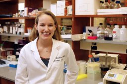 Ariella B Hanker合作论文数Curriculum in Genetics and Molecular Biology, University of North Carolina at Chapel Hill17Arun Sreekumar合作论文数University of Michigan;Medical School;Medical School, University of Michigan17
 Eric C. Chang合作论文数HKU Business School16Michael A Gillette合作论文数10
 Steven A. Carr合作论文数Broad Institute of MIT and Harvard10Eric Jaehnig合作论文数Ludwig Institute for Cancer Research, University of California School of Medicine10
 Mothaffar F. Rimawi合作论文数Dan L Duncan Comprehensive Cancer Center, Baylor College of Medicine10D. R. Mani合作论文数9
 Shankha Satpathy合作论文数Broad Inst MIT & Harvard, Cambridge, MA 02142 USA9
 Karsten Krug合作论文数Broad Inst MIT & Harvard, Cambridge, MA 02142 USA8
 Henry Rodriguez Granobles合作论文数NCI, NIH, Bethesda, MD 20892 USA7
 Chen Huang合作论文数Baylor Coll Med, Lester & Sue Smith Breast Ctr, Houston, TX 77030 USA7
 Donna Marie Muzny合作论文数Human Genome Sequencing Center, Baylor College of Medicine7Tara Hiltke合作论文数7
 Ana I. Robles合作论文数Diagnostics Evaluation Branch, Division of Cancer Treatment and Diagnosis, National Cancer Institute, National Institutes of Health;Office of Cancer Clinical Proteomics Research, National Cancer Institute, National Institutes of Health7Scollon Sarah合作论文数6
 Sharon E Plon合作论文数School of Medicine, Baylor College of Medicine;Dan L Duncan Comprehensive Cancer Center, Baylor College of Medicine;Texas Children's Hospital6
 Bo Wen (文波)合作论文数School of Basic Medical, Fudan University6
 Michael T. Lewis合作论文数The Lester and Sue Smith Breast Center and Department of Molecular and Cellular Biology, Baylor College of Medicine;Room6Lacey E Dobrolecki合作论文数6Ck Osborne合作论文数5
 Li Yi合作论文数5Viktoriya Korchina合作论文数5Tanmayi Vashist合作论文数5Doddapaneni Harshavardhan合作论文数5Ademuyiwa Foluso O合作论文数5
 Donald Williams Parsons合作论文数Department of Molecular and Human Genetics, Baylor College of Medicine5
 Richard A Gibbs合作论文数Human Genome Sequencing Center, Baylor College of Medicine;Dan L Duncan Comprehensive Cancer Center, Baylor College of Medicine5




