George Stewart died unexpectedly in a Florida hospital on 12 October 2017, of an undiagnosed cause after being admitted 10 days earlier. A communication from his wife, Breck, noted that 12 October also marked George’s and her 50th wedding anniversary. Breck was George’s constant companion and ardent supporter of his career endeavors. In memory of George, Gil Castro and John Ubelaker pay tribute to a friend and fellow parasitologist, providing a glimpse of his academic achievements and of him as an individual. This homage also conveys the personal responsibility he took and the effort he put forth to achieve his goals. George had an inclusive academic career and enjoyed a reputation as a noble and industrious individual and scientist. He was known for carving out and providing leadership in his parasitological niche. George obtained rigorous training in preparation for a career in parasitology. He was awarded a Bachelor of Science degree in 1969 from Tulane University and, in 1973, a doctorate degree from Rice University, two bastions of parasitology at the time George was preparing for his vocation. After receiving his doctorate degree, George remained at Rice in the capacity of postdoctoral fellow, research associate, and lecturer until 1976. Gil met George in 1972 soon after taking a position at the University of Texas Medical School at Houston. Gil went to the Biology Department at Rice to visit Clark Read and acquire some of his infected Tribolium flour beetles to be used to start experiments on adult Hymenolepis diminuta. As one would anticipate, meeting with Clark Read to discuss experiments was a pleasure. But adding to this was Gil meeting George Stewart in Read’s laboratory, where they chatted for some time. That fortuitous meeting began a friendship that involved interactions DOI: 10.1645/18-28 J. Parasitol., 104(3), 2018, pp. 342–344 American Society of Parasitologists 2018
On 5 April 2001, Dr. Donald Fairbairn passed away at Kingston General Hospital in Kingston, Ontario. He was 86. He was a highly accomplished individual; a loving husband, father, and grandfather; a scientist; and an outstanding mentor. He was a graduate of Queen’s University and the University of Rochester. After service in World War II, he became a faculty member at McGill University. In 1962 he was appointed head of the Zoology Department at the University of Massachusetts at Amherst, where he established an international center for research on the biochemistry of parasites. Dedicated to his family and his profession, Don Fairbairn was active in university affairs; served as a consultant, advisor, and officer of national and international scientific societies; and was an editor of scientific journals. Don Fairbairn was probably best known in parasitology circles for the impressive training program that he established at the University of Massachusetts that attracted students from around the world. The Fairbairn research enterprise was a drawing card, not only for students, but also for some of the world’s most renowned parasitologists. Within the Amherst setting, his students were treated with visits from the likes of Theodore Von Brand, Ernest Bueding, Elvio Sadun, and Paul Weinstein. These giants of parasitology were both friends and admirers of Don Fairbairn. He had many admirable qualities, as many graduate students, postdoctoral fellows, and faculty members who trained with him can attest. First and foremost, Don Fairbairn was an unparalleled expert on the biochemistry of parasites, making ground-breaking discoveries on the chemical structure and function of helminths. His contributions to our understanding of the energy metabolism, and lipid and nucleic acid metabolism in helminths, helped explain, both physiologically and genetically, adaptations in parasites, both within and outside their host. Von Brand, in his classic book, Biochemistry of Parasites, cited Fairbairn 92 times. These citations reflect the meritorious nature of Fairbairn’s work. That work was always based on hypotheses derived from visions that extended well beyond those of his contemporaries. Experiments from his laboratories were characterized by novelty, depth, technological expertise, and scientific rigor. Experimental designs were intricate and involved various approaches, as well as replications, that led to the same conclusion. His interpretations of experimental results were expressed with uncommon insight and integration of knowledge and were written with the skill and eloquence that characterized the works of Alfred Russel Wallace or Arthur C. Guyton. Don Fairbairn was a scientist and scholar of the highest caliber. Under his tutelage, students were not to be lectured or tested on the ethical conduct of research. This was learned from the questions that he asked and the standards that were reflected in his personal integrity. It was not necessary to schedule formal laboratory meetings to update him on activities. He was that great coach, always sitting on the same bench as his players and was always there when needed. He came to each student’s laboratory every day to discuss experimental designs, procedures, and new technologies and to determine individual needs. Working under the tutelage of Don Fairbairn involved learning on a daily basis. Laboratory activities were planned and carried out knowing that he would come sometime during the day to discuss one’s progress and to suggest ways to continuously improve performance and productivity. His daily visits to the laboratories were anticipated with a sense of excitement by trainees and with considerable preparation. His insight and acumen were great resources for students. He was a taskmaster in the sense that students knew his expectations were high—not because he stated so, but because of the example he set forth. In short, tasks were self-imposed. He worked continuously throughout the day. Although he had a laserlike focus on achievement of experimental results, he was also driven to help students develop professionally. To him, these two ends went hand-in-hand. In these efforts, he had tremendous stamina and patience. He was a gentleman who never raised his voice to make a point. He was kind, generous, and courteous to everyone, from the laboratory dishwasher to the professor. He could usually point out, in very subtle ways, a shortcoming in one’s way of thinking about their work, but always tempered this by mentally searching and supporting one’s primary interests and recognizing individual contributions. Because the balance was always tipped toward the latter, and because of the kind smile with which he ended these one-on-one teaching sessions, students knew their work was appreciated. Because of this, students were receptive to his suggestions and motivated to work beyond their limits. His method of teaching was contagious. Students learned from Don Fairbairn that the key to long-term succession in a laboratory is teaching—teaching one another through precise documentation and through word of mouth. Don Fairbairn’s students benefitted from his teaching, the quality of his work, and his reputation in that they were actively recruited upon completion of their training to fill academic positions throughout the United States and abroad. Don Fairbairn loved his work. He also loved his family, dividing his time and devotion to both until retirement from his scientific career. His postretirement years, spent initially at his cottage on a small farm in Vermont, were fulfilling. He and his wife Mary were inseparable companions. As an avid and highly skilled angler, he very much enjoyed trout fishing, whether alone in the stream that meandered through his farm or with friends in the Arctic Circle. Woodworking was his avocation. His skills were self-taught, and products of his efforts in his wood shop were comparable in quality to products from his laboratory. In all his endeavors, he was a perfectionist. This was evident in the dedication and precision that was brought to bear on everything he did. In an essay, ‘‘The Decline of Heroes,’’ Arthur M. Schlessinger, Jr., notes that ‘‘individuals have lived who did what no substitute could have done to set history on one path rather than another.’’ When future students ponder the history of parasitology, Don Fairbairn will emerge as such an individual. Those who knew him, particularly his students, will always cherish their role in history, and even more, the opportunity to train with an individual whose greatest attribute was his character. He will remain a giant in the field of parasitology and a giant in our memories.
The hypothesis that lactoferrin protects mice against lethal effects of bacterial lipopolysaccharide (LPS) is the subject of experimental investigations described in this article. Lipopolysaccharide is a powerful toxin produced by Gram negative bacteria that when injected into humans or experimental animals reproduce many of the pathophysiologic and immune responses caused by live bacteria. Lactoferrin administered intraperitoneally 1 hr prior to injection of LPS significantly enhanced the survival of mice, reducing LPS-induced mortality from 83.3% to 16.7%. Changes in locomotor and other behavioral activities resulting from LPS injection were not present in mice treated with lactoferrin. Also, histological examination of intestine revealed remarkable resistance to injury produced by LPS if mice were pretreated with lactoferrin. Severe villus atrophy, edema and epithelial vacuolation were observed in LPS-treated animals but not in lactoferrin-treated counterparts. Electrophysiological parameters were used to assess secretory and absorptive functions in the small intestine. In mice treated with LPS, transmural electrical resistance was reduced and absorption of glucose was increased. Lactoferrin treatment had no significant influence on basal electrophysiological correlates of net ion secretion or glucose absorption nor on changes induced by LPS. Collectively, these results suggest that lactoferrin attenuates the lethal effect of LPS and modulates behavioral and histopathological sequela of endotoxemia.
The gastrointestinal tract may be viewed as an ecologic system in which a balance between the host and bacterial flora exists. Two major host components appear to be involved in maintaining this balance. The first is a non-specific structural barrier provided by the epithelial layer of the gastrointestinal mucosae. The second component involves functional immunological elements found in the mucosal and submucosal compartments, e.g., gut associated lymphoid tissue. When gut integrity is disrupted by invasive pathogens or by trauma, a myriad of pro-inflammatory mediators are released from cells in the gut wall that exert actions in the tissue or gut lumen1. One of these mediators is lactoferrin, an iron binding protein found in high concentration in most human exocrine secretions. Despite controversies on its physiological role, evidence is emerging that lactoferrin plays an important role in host defense against toxic metabolites and antigenic components of potential pathogens2–4. This manuscript is intended to provide an overview of work related to lactoferrin’s modulatory roles in inflammation, and to present observations from experimental studies on the preservation of intestinal structure and function by lactoferrin during intestinal inflammation. The possibility that lactoferrin limits the autodestructive inflammatory responses presents a new alternative for the future management of systemic inflammation.
Abstract Adaptation is a word with many connotations (Prosser and Brown 1961; Fairbairn 1970), but, in general, refers to the fitness of an organism for its environment (Gould 1982; Keymer and Read, Chapter 3; Dobson and Merenlender, Chapter 5; May, Chapter 6; Harvey et al., Chapter 17, this volume). Physiologists view ‘adaptation’ as the process of adjustment by an organism to a changing environment. This view includes adjustments that occur within the life of an individual, and therefore represents acclimation or acclimatization, i.e. adjustments to single or multiple environmental factors, respectively, that contribute to homeostasis (Prosser and Brown 1961). However, because coping with environmental changes enhances survival and reproductive capacity in the long term, physiological adaptations relate to ‘adaptations’ in the evolutionary sense (Dobzhansky 1951). This holds true for both parasites and their hosts.
Parasite infections and gastrointestinal motility Supplement 16. Handbook of Physiology, The Gastrointestinal System, Motility and Circulation Gilbert A. Castro, Gilbert A. Castro Department of Physiology and Cell Biology, Medical School, University of Texas Health Science Center, Houston, TexasSearch for more papers by this author Gilbert A. Castro, Gilbert A. Castro Department of Physiology and Cell Biology, Medical School, University of Texas Health Science Center, Houston, TexasSearch for more papers by this author Published online: 1 January 2011 https://doi.org/10.1002/cphy.cp060130 Read the full textAboutPDF ToolsRequest permissionExport citationAdd to favoritesTrack citation ShareShare Give accessShare full text accessShare full-text accessPlease review our Terms and Conditions of Use and check box below to share full-text version of article.I have read and accept the Wiley Online Library Terms and Conditions of UseShareable LinkUse the link below to share a full-text version of this article with your friends and colleagues. Learn more.Copy URL Share a linkShare onFacebookTwitterLinked InRedditWechat Abstract The sections in this article are: 1 Parasite-Induced Alterations 1.1 Esophagus 1.2 Stomach 1.3 Small Intestine 1.3.1 Manometric Measurements 1.3.2 Transit Measurements 1.3.3 Myoelectric Measurements 1.3.4 In Vitro Measurements Of Altered Responsiveness 1.4 Cecum, Colon, and Rectum 2 Basis For Motility Changes 2.1 Parasite-Derived Substances 2.2 Gastrointestinal Hormone Imbalances 2.3 Inflammation 3 Summary Comprehensive PhysiologyBrowse other articles of this reference work:BROWSE TABLE OF CONTENTSBROWSE BY TOPICBROWSE A-Z RelatedInformation
Antigens derived from Trichinella spiralis were used to challenge, in vitro, sensitized jejunum from infected guinea-pigs while monitoring ion transport properties of the tissue. Antigen challenge resulted in dose-dependent increases in trans-epithelial electrical potential difference and short circuit current. Both antigen-stimulated electrical alterations and Schultz-Dale contractions were demonstrated in small intestinal tissue after the passive transfer of immune serum containing anti-trichinella homocytotropic antibodies.
Epithelium of isolated small intestinal segments were studied in Ussing-type chambers to detect physiological changes associated with rapid, immune rejection of Trichinella spiralis infective larvae. Electrophysiological parameters associated with Na+-coupled hexose transport were measured. Changes in transepithelial electrical potential difference (PD), resistance, and short circuit current (Isc) due to the addition of actively absorbed beta-methyl-D-glucoside (BMG) to the mucosal solution were determined. Measurements were made prior to and 30 min after primary and secondary infections. Animals were infected by intraduodenal inoculation. As the infective larval dose in primarily infected (nonimmunized) rats increased from 50 to 2000 larvae the magnitude of the rise in Isc elicited by BMG decreased in a dose-dependent fashion, with 50 larvae per rat having no effect. In previously infected (immunized) rats challenged with a secondary inoculum, all doses, ranging from 50 to 2000 larvae per rat, decreased the BMG-stimulated change in Isc by approximately 50%. The effect of 50 worms per rat in immunized hosts was equivalent to that produced by approximately 1600 worms in nonimmunized animals. Measurements of 14C-BMG mucosa-to-serosa flux confirmed that Na+-BMG cotransport was responsible for observed changes in Isc. Results support the conclusion that changes in intestinal epithelial function are associated with larval challenge of immune rats.
This investigation was designed to test the hypothesis that alterations in small-bowel motility are associated with total parenteral nutrition (TPN). Motility, as reflected by intestinal transit, was studied in rats maintained by TPN for 7–10 days and compared to that of rats fed an oral diet isocaloric with the intravenously administered solution. Transit was measured by injecting radioactively labeled chromium (Na251CrO4) into the duodenum via permanently implanted catheters. Fifteen minutes after injection of the label, animals were killed and the linear distribution of the isotope in the gut was determined. The leading edge of radioactivity traversed 75–87% (95% confidence limit) of the gut length in enterally fed rats and 83–97% in rats on TPN. The difference between the average position of these fronts for the two groups was not statistically significant (P>0.05). In addition, the regression of percent radioactivity traversing or present in a given segment on gut length yielded a slope with 95% confidence limits of −10.48 to −15.02 for orally fed control rats and −9.83 to −12.87 for rats on TPN. Differences between these slopes were not statistically significant (P>0.05). Results support the conclusion that factors which regulate small-bowel motility are not altered significantly by TPN during the time that other functional and structural changes reportedly occur.
Small intestinal morphologic and biochemical changes were studied following jejuno-ileal bypass for obesity after body weight stabilization had occurred. Four patients underwent biopsy of in-continuity and bypassed jejunal and ileal segments of the small intestine 11 to 22 months after the bypass operation. Microscopically, marked mucosal villus hypertrophy of the in-continuity bowel was observed, especially in the ileum. Bypassed jejunal mucosa underwent atrophy compared with pre-bypass jejunum, whereas bypassed ileum appeared similar microscopically to pre-bypass ileum. The specific activities of mucosal disaccharidase enzymes (maltase, sucrase, lactase and trehalase) in units per mg protein remained similar to pre-bypass levels in segments of the in-continuity jejunum and the bypassed jejunum and ileum. On the other hand, elevated mucosal disaccharidase levels were measured in biopsy specimens of the in-continuity ileum. Total enzyme activity per unit length of intestine, however, was estimated to be elevated in both in-continuity jejunum and ileum secondary to mucosal villus hypertrophy. These data indicate that following small bowel bypass: (1) the in-continuity ileum undergoes greater biochemical and morphologic adaptation than the jejunum; and (2) intraluminal nutrients and chyme appear to be essential to maximal intestinal adaptation.
 Marian L. Kruzel合作论文数6Yael Harari合作论文数4Chung-Ying Chen合作论文数3
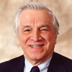 Stanley Dudrick合作论文数Department of Surgery, Yale School of Medicine;Saint Mary’s Hospital, Yale University2Graham R. Bullick合作论文数2Rl Bowers合作论文数1N W Weisbrodt合作论文数1Weisbrodt Norman W合作论文数1
 Raymond A. Frizzell合作论文数Cystic Fibrosis Research Center, Departments of Pediatrics and Cell Biology, University of Pittsburgh1
 Edward M. Copeland合作论文数Department of Surgery, College of Medicine, University of Florida1
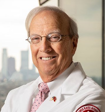 John M. Daly合作论文数Lewis Katz School of Medicine, Temple University1John E. Ubelaker合作论文数1




