BACKGROUND: Breast cancer patients have a high risk of brain and central nervous system metastases (BrM) and therefore have poor outcomes. This study aimed to assess the impact of clinico-genomic factors on BrM risk in breast cancer and develop validated nomograms for risk prediction. METHODS: Clinico-genomic data on breast cancer patients were obtained from the AACR GENIE Biopharma Collaborative. These data were split into training and testing sets by academic institution. Using the training set, risk of BrM was evaluated through multivariable Fine-Gray subdistribution hazard modeling, implementing LASSO penalization for feature selection. Prediction metrics were assessed internally on the training set through bootstrap-cross validation, and externally on the testing set. Calibration curves, AUC, and Brier scores were calculated to assess prediction and discrimination. A nomogram for 2-, 3.5-, and 5- year risk of BrM was constructed. RESULTS: Synchronous bone metastases, HER+/HR- breast cancer subtype, and TP53 alterations were selected as top predictors of BrM risk. The final multivariable model showed good discriminate capability at 2- (AUC: 68.7; Brier: 5.3), 3.5- (AUC: 65.9, Brier: 12.9), and 5-years (AUC 63.0, Brier: 17.4). This model was externally validated on the testing set and performed well at 2- (AUC 72.5; Brier: 10.5), 3.5- (AUC: 73.3; Brier 13.6), and 5-years (AUC: 74.9; Brier: 16.5). CONCLUSIONS: Nomograms that calculate individualized BrM risk probabilities for patients with breast cancer can provide clinical utility informing patients and their health care team members on risk of BrM development. An interactive webtool for individualized BrM risk probabilities can be found here: https://gcioffi.shinyapps.io/BrCA_nomo_brm_risk/ .
BACKGROUND: Individuals with breast cancer are at high risk for developing brain and central nervous system metastases (BrM), which are aggressive and clinically challenging. This study aimed to assess the impact of clinico-genomic factors on BrM survival in breast cancer and to develop a validated nomogram for survival probability. METHODS: Clinico-genomic data on breast cancer patients were obtained from the AACR GENIE Biopharma Collaborative. These data were split into training and testing sets by academic institution. Using data from the training set, BrM survival was evaluated through multivariable Cox proportional hazards modeling, with LASSO penalization for feature identification. Performance metrics were assessed internally on the training set through bootstrapcross-validation, and externally on the testing set. Calibration curves, AUC, and Brier scores were generated to evaluate model performance. A nomogram for 12-, 18-, and 24- month survival probability was constructed. RESULTS: HER2+/HR+ subtype, ARID1A alterations, and a lower number of prior cancer-directed regimens were top predictor associated with improved survival after BrM development. This model showed strong discriminate capability at 12- (AUC: 79.7; Brier: 18.2), 18- (AUC: 78.5, Brier: 19.9) and 24-months (AUC 82.0, Brier: 17.9). After external validation, the model performed similarly at 12- (AUC: 73.5; Brier: 21.1), 18- (AUC: 76.2; Brier 20.1), and 24-months (AUC: 79.5; Brier: 17.9). CONCLUSIONS: Nomograms that calculate individualized BrM survival probabilities for patients with breast cancer offer clinical value by informing scientists and healthcare professionals about risk factors for survival after BrM diagnosis. A webtool for individualized BrM survival probabilities can be found here: https://gcioffi.shinyapps.io/BrCA_nomo_brm_survival/
Figure S4 shows sex-specific hazard of death based on atopic trait
Background: In cancer, age and sex are often studied individually, but the impact of the intersection of these factors on cancer incidence and survival remains unclear. Using population-level data, we provide an up-to-date analysis of the impact of sex and age on cancer incidence and survival. Methods: Using data from the United States Cancer Statistics public use research database and the Centers for Disease Control and Prevention’s National Program of Cancer Registries Survival database, we assessed sex and age differences in the incidence and survival of malignant cancers diagnosed from 2001 to 2020. Results: Males experienced higher cancer incidence than females in all sites and age groups, excluding 20–29- and 30–39-year-olds. The highest Male-to-female (M:F) age-adjusted incidence rates (IRR) were observed in mesothelioma within ages 80+ (IRR: 5.48; 95% CI: 5.25–5.71; p < 0.001), and lowest in endocrine cancer within ages 20–29 years (M:F IRR: 0.20; 95% CI: 0.20–0.21; p < 0.001). Among all sites and age groups, excluding 0–9 years, males experienced worse survival than females, particularly within ages 20–29 years (Hazard Ratio (HR): 2.19; 95% CI: 2.15–2.23; p < 0.001). Highest M:F HRs were observed in endocrine system cancers within ages 20–29 (HR: 3.52; 95% CI: 3.15–3.94; p < 0.001), and lowest among lymphomas within ages 0–9 (HR: 0.74; 95% CI: 0.63–0.87; p < 0.001). Conclusions: Significant age and sex differences in cancer incidence and survival were observed across the US from 2001 to 2020. Males had a higher cancer incidence compared to females, with notable exceptions for younger age groups among certain types, suggesting age may be a critical component in further understanding the biology of sex differences in cancer.
BACKGROUND:US incidence rates of nonmalignant brain tumors are 3-fold higher in highest versus lowest incidence states. A county-level analysis was conducted to assess whether geographic variation in nonmalignant meningioma (NMM) incidence is related to demographics, cancer registry, health care, and other factors. METHODS:Age-adjusted incidence rates of NMM in US counties during 2010-2019 were modeled with data from the Central Brain Tumor Registry of the United States. Demographic, geographic, cancer registry, environmental, health care, health, lifestyle, and socioeconomic factors at the county level were drawn from numerous data sources. Bayesian index regression models were fit containing spatial random effects. RESULTS:Three domains were significantly associated with rates of NMM at the county level: cancer registry practices (funding source and % radiographically confirmed), socioeconomic status index (higher levels with percent working in white-collar occupations as an important contributor), and demographics (% Black and % female). No associations were observed for general health or environmental factors. In the fully adjusted model, the number of counties with significantly elevated and lowered spatial random effects decreased by 33% and 28%, respectively, compared to a no-covariate model. CONCLUSIONS:Although general health and environmental factors cannot be ruled out in explaining the geographic variation in NMM incidence rates, results suggest that socioeconomic factors, certain demographic characteristics, and cancer diagnosis and registry practices may all play a significant role in driving such variation. These results may have implications for other tumor types diagnosed primarily radiographically or outside hospital settings, where variation in detection and reporting may affect incidence rates.
BACKGROUND:With the significant shift in the classification, risk stratification, and standards of care for gliomas, we sought to understand how the overall survival of patients with these tumors is impacted by molecular features, clinical metrics, and treatment received. METHODS:We assembled a cohort of patients with histopathologically diagnosed glioma from The Cancer Genome Atlas (TCGA), Project Genomics Evidence Neoplasia Information Exchange, and Dana-Farber Cancer Institute/Brigham and Women's Hospital. This incorporated retrospective clinical, histological, and molecular data alongside a prospective assessment of patient survival. RESULTS:Of 4400 gliomas were identified: 2195 glioblastomas, 1198 IDH1/2-mutant astrocytomas, 531 oligodendrogliomas, 271 other IDH1/2-wild-type gliomas, and 205 pediatric-type glioma. Molecular classification updated 27.2% of gliomas from their original histopathologic diagnosis. Examining the distribution of molecular alterations across glioma subtypes revealed mutually exclusive alterations within tumorigenic pathways. Non-TCGA patients had significantly improved overall survival compared to TCGA patients, with 26.7%, 55.6%, and 127.8% longer survival for glioblastoma, IDH1/2-mutant astrocytoma, and oligodendroglioma, respectively (all P < .01). Several prognostic features were characterized, including NF1 alteration and 21q loss in glioblastoma, and EGFR amplification and 22q loss in IDH1/2-mutant astrocytoma. Leveraging the size of this cohort, nomograms were generated to assess the probability of overall survival based on patient age, the molecular features of a tumor, and the treatment received. CONCLUSIONS:By applying modern molecular criteria, we characterize the genomic diversity across glioma subtypes, identify clinically applicable prognostic features, and provide a contemporary update on patient survival to serve as a reference for ongoing investigations.
Background: Previous studies have described sex-specific patient subtyping in glioblastoma. The cluster labels associated with these “legacy data” were used to train a predictive model capable of recapitulating this clustering in contemporary contexts. Methods: We used robust ensemble machine learning to train a model using gene microarray data to perform multi-platform predictions including RNA-seq and potentially scRNA-seq. Results: The engineered feature set was composed of many previously reported genes that are associated with patient prognosis. Interestingly, these well-known genes formed a predictive signature only for female patients, and the application of the predictive signature to male patients produced unexpected results. Conclusions: This work demonstrates how annotated “legacy data” can be used to build robust predictive models capable of multi-target predictions across multiple platforms.
Table S4 provides results from univariate survival and cumulative incidence analyses
Table S6 provides results by timing of brain metastasis (at diagnosis or after diagnosis)
Table S7 provides results for those without metastatic disease at diagnosis
Table S5 provides adjusted results from survival and cumulative incidence analyses
Figure S1 illustrates the data filtering process used to build the analytic dataset
Table S3 provides an overview of characteristics of included individuals by atopic disease status
BACKGROUND:Development of new melanoma therapies has increased survival, and more patients are living to develop brain metastasis (BrM). Identifying those at increased risk of BrM is of significant public health importance, and our objective was to assess the relationship between atopy and survival or reduced BrM cumulative incidence (CI) in melanoma. METHODS:This retrospective study was conducted in individuals (≥66 years) in linked Surveillance, Epidemiology, and End Results and Medicare data. History of atopy diagnosed prior to melanoma was ascertained using International Classification of Diseases-9/10 codes. Associations between atopy, CI of BrM, and overall survival were assessed using cox proportional hazards models to estimate hazard ratios and P values. RESULTS:A total of 23,508 cutaneous melanoma cases were identified. Overall, 6.1% developed BrM, and 38% had history of atopy. Atopy was associated with an 18% decrease in death (P < 0.001). Among those without metastasis at diagnosis, atopy decreased BrM CI by 16% (P = 0.006). Among those with metastasis at diagnosis (any site), only those who received checkpoint inhibitors had a suggestive but nonsignificant survival with atopy. CONCLUSIONS:Atopy confers improved survival and decreased BrM CI. History of atopy in the elderly may identify those with more robust immune function that may be more responsive to treatment. IMPACT:Elderly individuals with prior diagnosis of atopy had significantly improved survival and decreased incidence of BrM as compared with individuals without atopy. This suggests that history of atopy may identify a subgroup within melanoma with improved response to treatment and a more robust immune system.
Abstract BACKGROUND The Stupp protocol was accepted as standard of care for glioblastoma in 2005, and has led to significant increases in overall survival. Prior analyses have identified additional survival gains post-Stupp, though the source of these is unknown. In this analysis, we leveraged two large datasets to identify trends in survival and potential treatment factors associated with these improvements. MATERIAL AND METHODS Provider-side commercial claims data were obtained from IQVIA for adults (18+ years) diagnosed with malignant brain tumor (ICD-9/ICD-10:191.0-191.9/C71.0-C71.9) from 2015-2021 who received biopsy/resection and temozolomide within 60 days of diagnosis. Treatment patterns were determined using ICD-9/ICD-10 procedure and HCPCS/CPT codes. Death was determined by CPT codes or last claim followed by ≥30 days of no claims. Elixhauser comorbidity score was generated prior to diagnosis. Population-based survival data were obtained from the National Program of Cancer Registries (NPCR) from 2004-2018 for microscopically-confirmed adult (18+ years) glioblastoma cases receiving surgery and radiation. Median survival was estimated using Kaplan-Meier regression, and cox proportional hazards models were used to assess potential prognostic factors. Survival analyses were censored at 36 months. RESULTS The IQVIA dataset had 18,883 individuals and NPCR had 92,540 individuals identified as being diagnosed with glioblastoma and meeting treatment criteria. Median survival among IQVIA cases was 13.9 months (95%CI=13.7-14.1), and 14 months (95%CI=14-14) in NPCR. Median survival in NPCR increased from 11 months in 2004-2006, to 14 months in 2016-2018. Within IQVIA, 19% of individuals had ≥1 claim for Bevacizumab and 15.5% had ≥1 claim for tumor treating fields (TTFields). Use of TTFields and/or Bevacizumab in treatment were associated with significantly improved survival after adjustment for age, sex, and comorbidities. CONCLUSION s: Survival in glioblastoma continues to improve over time, which may be due to developments in therapeutic approaches. Assessment of population-level survival patterns is essential for understanding the impact of treatment advancements.
Table S1 shows codes used to classify atopic disease types
Sex differences in cancer are well-established. However, less is known about sex differences in diagnosis of brain metastasis and outcomes among patients with advanced melanoma. Using a United States nationwide electronic health record-derived de-identified database, we evaluated patients diagnosed with advanced melanoma from 1 January 2011–30 July 2022 who received an oncologist-defined rule-based first line of therapy (n = 7969, 33% female according to EHR, 35% w/documentation of brain metastases). The odds of documented brain metastasis diagnosis were calculated using multivariable logistic regression adjusted for age, practice type, diagnosis period (pre/post-2017), ECOG performance status, anatomic site of melanoma, group stage, documentation of non-brain metastases prior to first-line of treatment, and BRAF positive status. Real-world overall survival (rwOS) and progression-free survival (rwPFS) starting from first-line initiation were assessed by sex, accounting for brain metastasis diagnosis as a time-varying covariate using the Cox proportional hazards model, with the same adjustments as the logistic model, excluding group stage, while also adjusting for race, socioeconomic status, and insurance status. Adjusted analysis revealed males with advanced melanoma were 22% more likely to receive a brain metastasis diagnosis compared to females (adjusted odds ratio [aOR]: 1.22, 95% confidence interval [CI]: 1.09, 1.36). Males with brain metastases had worse rwOS (aHR: 1.15, 95% CI: 1.04, 1.28) but not worse rwPFS (adjusted hazard ratio [aHR]: 1.04, 95% CI: 0.95, 1.14) following first-line treatment initiation. Among patients with advanced melanoma who were not diagnosed with brain metastases, survival was not different by sex (rwOS aHR: 1.06 [95% CI: 0.97, 1.16], rwPFS aHR: 1.02 [95% CI: 0.94, 1.1]). This study showed that males had greater odds of brain metastasis and, among those with brain metastasis, poorer rwOS compared to females, while there were no sex differences in clinical outcomes for those with advanced melanoma without brain metastasis.
The Central Brain Tumor Registry of the United States (CBTRUS), in collaboration with the Centers for Disease Control and Prevention (CDC) and National Cancer Institute (NCI), is the largest aggregation of histopathology-specific population-based data for primary brain and other central nervous system (CNS) in the US. CBTRUS publishes an annual statistical report which provides critical reference data for the broad neuro-oncology community. Here, we summarize the key findings from the 2022 CBTRUS annual statistical report for healthcare providers.Incidence data were obtained from the CDC's National Program of Cancer Registries (NPCR) and NCI's Surveillance, Epidemiology, and End Results Program for 52 central cancer registries (CCRs). Survival data were obtained from 42 NPCR CCRs. All rates are per 100 000 and age-adjusted using the 2000 US standard population. Overall median survival was estimated using Kaplan-Meier models. Survival data for selected molecularly defined histopathologies are from the National Cancer Database. Mortality data are from the National Vital Statistics System.The average annual age-adjusted incidence rate of all primary brain and other CNS tumors was 24.25/100 000. Incidence was higher in females and non-Hispanics. The most commonly occurring malignant and predominately non-malignant tumors was glioblastoma (14% of all primary brain tumors) and meningioma (39% of all primary brain tumors), respectively. Mortality rates and overall median survival varied by age, sex, and histopathology.This summary describes the most up-to-date population-based incidence, mortality, and survival, of primary brain and other CNS tumors in the US and aims to serve as a concise resource for neuro-oncology providers.
 Jill S. Barnholtz-Sloan合作论文数Winship Cancer Institute, Emory University;Department of Biostatistics and Bioinformatics, Rollins School of Public Health, Emory University102
 Kristin A Waite合作论文数54
 Quinn Ostrom合作论文数Basic Science Departments, School of Medicine, Duke University;Duke Cancer Institute, School of Medicine, Duke University53Carol Kruchko合作论文数52Corey Neff合作论文数33Mackenzie Price合作论文数28
 Nirav Patil合作论文数Dept Biomed Engn, Case Western Reserve Univ23
 Andrew E. Sloan合作论文数Department of Neurosurgery, School of Medicine, Case Western Reserve University;Piedmont Healthcare;Department of Neurological Surgery University Hospitals Cleveland Medical Center12Mantas Dmukauskas合作论文数9Salama April K合作论文数9Joel Saltzman合作论文数8
 Iorgulescu J Bryan合作论文数Brigham & Womens Hosp7
 Richard Lee合作论文数Department of Stem Cell & Regenerative Biology, Harvard University;Harvard Medical School;Brigham and Women's Hospital7
 Asrar Alahmadi合作论文数6
 Justin D. Lathia合作论文数Department of Cardiovascular and Metabolic Sciences, Lerner Research Institute, Cleveland Clinic;Department of Molecular Medicine, School of Medicine, Case Western Reserve University6
 Patrick Y. Wen合作论文数Department of Neurology, Harvard Medical School;Center For Neuro-Oncology, Dana-Farber Cancer Institute6Aman Opneja合作论文数Med Ctr, Univ Hosp Cleveland5
 Haley Gittleman合作论文数Division of Biometrics V, Office of Translational Sciences, Center for Drug Evaluation and Research, FDA5Hia Ghosh合作论文数4J Eva Selfridge合作论文数4David L Bajor合作论文数4
 David Bajor合作论文数4
 Jacob Edelson合作论文数Department of Biomedical Data Sciences, Stanford University School of Medicine4
 Ruchit Patel合作论文数Brigham and Women's Hospital, Harvard Medical School4
 Varun Bhave合作论文数Harvard Med Sch, Boston, MA 02115 USA4
 David Meredith合作论文数Dept Pathol, Brigham & Womens Hosp4
 Liam F Spurr合作论文数The Pritzker School of Medicine, The University of Chicago4Hersh Gupta合作论文数Dana-Farber Brigham and Women's Cancer Center, Dana-Farber Cancer Institute and Harvard Medical School4Frank Dubois合作论文数Broad Inst MIT & Harvard, Cambridge, MA 02142 USA4
 Samantha E Hoffman合作论文数Dept Neurosurg, Brigham & Womens Hosp4
 Noah F Greenwald合作论文数Center for Skull Base and Pituitary Surgery, Department of Neurosurgery, Brigham and Women’s Hospital, Harvard Medical School, Boston, USA4
 Tihan Tarik合作论文数Department of Pathology, School of Medicine, The University of California, San Francisco4
 Wenya Linda Bi合作论文数Department of Neurosurgery, Brigham and Women's Hospital;Harvard Medical School4
 David Reardon合作论文数Center for Neuro-Oncology, Dana-Farber Cancer Institute;Harvard Medical School4
 Sandro Santagata合作论文数Laboratory of Systems Pharmacology, Harvard Medical School;Department of Pathology, Brigham and Women's Hospital;Dana-Farber Cancer Institute4
 Mehdi Touat合作论文数Drug Development Department (DITEP), University Paris Sud4
 Ankit Mangla合作论文数Department of Medicine, John H. Stroger Jr. Hospital of Cook County4
 Maury Eduardo A合作论文数4
 Kerstetter-Fogle Amber合作论文数Case Western Reserve University4
 Eleanor L. Woodward合作论文数4
 E. Antonio Chiocca合作论文数Department of Neurosurgery, Brigham and Women's Hospital;Institute for the Neurosciences, Brigham and Women's Hospital;Harvard Medical School;Center for Neuro-oncology, Dana-Farber Cancer Institute4Prashanthi N. Thota合作论文数4
 Keith L. Ligon合作论文数Center for Patient Derived Models, Dana-Farber Cancer Institute;Division of Neuropathology, Brigham and Women's Hospital;Medical School, Harvard University;Travera;Broad Institute, Massachusetts Institute of Technology;Boston Children's Hospital4
 Joshua B. Rubin合作论文数School of Medicine, Washington University in St. Louis;Division of Hematology & Oncology, Washington University in St. Louis4
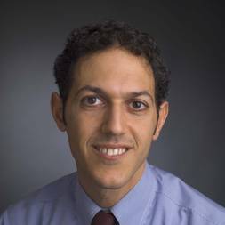 Rameen Beroukhim合作论文数Division of Medical Sciences, Harvard Medical School;Dana-Farber Cancer Institute4
 Yvonne Li合作论文数Meyerson Laboratory, Dana-Farber Cancer Institute4Terri S. Armstrong合作论文数4
 Sanford Markowitz合作论文数Department of Medicine, School of Medicine, Case Western Reserve University;Department of Genetics and Genome Sciences, School of Medicine, Case Western Reserve University;Department of Molecular Biology and Microbiology, School of Medicine, Case Western Reserve University;Case Comprehensive Cancer Center, Case Western Reserve University3Brittany Cordeiro合作论文数3




