NKRs assist CTLs to recognize and conjugate cancer cells expressing low-level MHCI/peptide complexes.
Abstract Background: Psychological distress, including perceived stress, anxiety, and depression, can activate the sympathetic nervous system and increase norepinephrine (NE) and epinephrine (E). These catecholamines may impair anti-tumor immunity through β2-adrenergic effects on CD8+ T-cells and myeloid-derived suppressor cells (MDSCs). Antidepressants that alter NE reuptake may further modify sympathetic signaling and immune regulation in cancer survivors. Methods: This analysis included 152 cancer survivors who completed the Perceived Stress Scale (PSS), PROMIS Anxiety (PROMIS), and Center for Epidemiological Studies Depression (CES-D) surveys and reported clinical diagnosis of anxiety or depression and current antidepressant use. Plasma NE, E, 3,4-dihydroxyphenylglycol (3,4-DHPG), and dopamine (DA) were quantified by LC-MS/MS, and CD8+ T-cell and MDSC subsets were assessed by flow cytometry as percent cell-subset events over total cell type events. Antidepressant use was classified as none (N=120), selective serotonin reuptake inhibitor (SSRI, N=11), or serotonin-NE or NE-DA reuptake inhibitors (SNRI/NDRI, N=21); five clinically diagnosed individuals not taking antidepressants were excluded. Associations of distress measures, clinical diagnoses, and medication class with catecholamines and immune profiles were evaluated using ANCOVA adjusted for age, education, and smoking history, with reciprocal adjustment for PROMIS or CES-D in clinical diagnosis models. Results: Catecholamine levels were not associated with PSS, PROMIS, or CES-D. Higher perceived stress was associated with altered CD8+ T-cell distributions: naïve CD8+ T-cells were 30.7% higher in the highest vs lowest quartile (62.2 vs 47.6%, p=0.03), while memory CD8+ T-cells were 28.5% lower (36.6 vs 51.2%, p=0.01). Clinical anxiety (N=23) was not related to catecholamine levels, but clinical depression (N=24) was associated with higher NE (22.2%; 663.0 vs 542.2 pg/mL, p=0.01) and DA (41.8%, 20.5 vs 14.5 pg/mL, p=0.002). Neither diagnosis was associated with CD8+ T-cells or MDSCs. Antidepressant class showed expected physiologic patterns: SNRI/NDRI users had higher NE (37.7%; 485.3 vs 352.4, p=0.04), lower 3,4-DHPG (19.9%; 912.7 vs 1139.4 pg/mL, p=0.0004), and higher DA (64.5%, 16.1 vs 9.8 pg/mL, p=0.001) than SSRI users. PMN-MDSCs trended 32.2% higher (1.15 vs 0.87%, p=0.11) and M-MDSCs 54.8% higher (4.32 vs 2.79%, p=0.09) among SNRI/NDRI users vs SSRI users, consistent with increased adrenergic-linked immunosuppressive myeloid activity. Conclusion: Clinical depression was associated with higher sympathetic catecholamines, and NE-reuptake-inhibiting antidepressants produced expected catecholamine shifts. Corresponding increases in PMN-MDSC and M-MDSC among SNRI/NDRI users suggest adrenergic signaling may contribute to immunosuppressive myeloid abundance in cancer survivors. Citation Format: Ji E. Park, Andrew Ray, Rikki Cannioto, Kathryn Glaser, Han Yu, Karen Hulme, Susan LaValley, Orla Maguire, Hans Minderman, Krystin Mantione, Nicolas Schlecht, Shipra Gandhi, Elizabeth Bouchard, Elizabeth Repasky, Christine Ambrosone, Chi-Chen Hong. Psychological distress, antidepressants, and sympathetic-immune profiles in cancer survivors [abstract]. In: Proceedings of the American Association for Cancer Research Annual Meeting 2026; Part 1 (Regular Abstracts); 2026 Apr 17-22; San Diego, CA. Philadelphia (PA): AACR; Cancer Res 2026;86(7 Suppl):Abstract nr 7565.
Abstract Background: Chronic psychosocial distress may accelerate breast cancer progression by altering immune, and inflammatory pathways, yet its genome-wide transcriptional effects in breast tumors remain unclear. This study uses RNA sequencing (RNA-seq) to characterize how distress influences transcriptional programs within the breast tumor microenvironment (TME). Methods: Tumor samples from the Women’s Health after Breast Cancer Study were analyzed for genome-wide transcriptional effects of distress. Participants completed the Perceived Stress Scale (PSS) and the Center for Epidemiologic Studies Depression Scale (CES-D) at diagnosis, including total scores and subscales (somatic symptoms, depressive affect, interpersonal problems, and positive affect). RNA-seq was performed on FFPE tumors from 195 women (152 ER+, 43 ER−). Principal component analysis (PCA) identified distress domains contributing to transcriptional variability. Participants were classified using PSS (>14 vs 0-14) and CES-D somatic symptoms (>3 vs ≤3). Differential gene-expression and gene-set enrichment analyses evaluated high-stress/low-somatic symptoms and low-stress/high-somatic symptoms groups vs a common low-stress/low-somatic symptoms reference, adjusting for age and education. Statistical significance was defined as FDR<0.05. Results: PCA indicated subtype-specific distress signatures, with CES-D somatic symptoms explaining the most transcriptional variance in ER+ tumors and PSS explaining more variance in ER− tumors. In ER+ tumors, high stress with low somatic symptoms upregulated immune-activation pathways, including B-cell signaling, interferon responses, complement, and antigen presentation (top NES ∼2.1-2.7, FDR<0.05). Key genes (FLG, IGLV3-16, IGKV3D-15, RPS7P3) mapped to immune-activation and interferon pathways. In ER− tumors, high stress showed enrichment of PD-1 co-inhibition and MHC-I antigen-presentation (NES=2.36 and 2.15, respectively, FDR<0.03), with suppression of neuronal, metabolic, mitochondrial, and protein-synthesis signaling (NES −1.4 to −2.1, FDR<0.05). High somatic symptoms in ER+ tumors enriched translational and ribosomal pathways (NES=3.0, FDR<0.01). In ER− tumors, high somatic symptoms were associated with increased keratinization, leptin, WNT, and IGF signaling (NES∼1.8-2.7, FDR<0.02), and reduced chromatin-regulation, DNA replication, RNA-processing, translation pathways, and MHC-I antigen presentation (NES ∼1.8 to -1.9, FDR<0.006). Conclusions: Psychological distress shapes breast-tumor transcriptional programs in a subtype-specific manner. Stress and depressive domains map to distinct immune, metabolic, and biosynthetic pathways in ER+ vs ER− disease, suggesting that different forms of distress engage different processes in the TME. This information could be leveraged to design new treatments for patients. Citation Format: Shipra Gandhi, Sayeeda Yasmeen, Spencer Rosario, Wiam Bshara, Thaer Khoury, Hans Minderman, Orla Maguire, Zhihong Gong, Ayana T. Ruffin, Megan Meek Wyatt, Mahmoud Abdelbary, Chrystal Mary Paulos, Elizabeth Repasky, Pawel Kalinski, Christine Ambrosone, Song Yao, Chi-Chen Hong. Distinct breast cancer gene signatures by estrogen receptor status associated with psychological distress [abstract]. In: Proceedings of the American Association for Cancer Research Annual Meeting 2026; Part 1 (Regular Abstracts); 2026 Apr 17-22; San Diego, CA. Philadelphia (PA): AACR; Cancer Res 2026;86(7 Suppl):Abstract nr 3583.
DC-primed MART-1-specific CTLs express high levels of NKRs, but recognize and kill cancer cells in a strictly TCR-dependent manner.
DNAM-1 and NKG2D expression is correlated with long term survival and cytotoxic genes in tumor samples from melanoma patients (TCGA).
Human peripheral blood CD8+ T cells require NKR-mediated co-stimulation for optimal effector response to weak TCR stimulation.
Expression of DNAM-1 and DNAM-1-competing (inhibitory) receptors reflects CTL cytotoxic function.
Immunotherapy with checkpoint inhibitors targeting the PD1/PD-L1 and CTLA4 pathways has limited activity in patients with microsatellite stable (MSS) colorectal adenocarcinoma (CRC). In a prior study, the combination of cetuximab and pembrolizumab failed to improve outcomes for patients with advanced RAS wild-type (RAS(wt)) CRC. In this post hoc secondary analysis, we show that the cetuximab and pembrolizumab-treated patients with TP53 mutant (p53(mt)) tumors had significantly higher progression-free survival (PFS) and a decrease in tumor burden compared to patients with TP53 wild-type (p53(wt)) tumors but no difference in overall survival compared to patients with p53(wt) tumors. The gene set enrichment analysis showed a uniform upregulation of multiple metabolic and immune gene sets, including NK-mediated immunity and IL-12 pathway, while the IL6 pathway was downregulated. There were no overlapping transcriptional alterations between the p53(mt) and p53(wt) groups with treatment that remain constant despite the therapeutic intervention. Functional overlap with treatment in both groups in the proliferative, immune, and metabolic pathways were identified. In the baseline tumor samples, the number of PD-L1(+) tumor cells was significantly higher in p53(mt) tumors while the number of OX40(-)/AE1_AE3(-)/PD-L1(-) non-tumor cells, positive for either LAG3, CTLA4 or TIM3, was significantly higher in p53(wt) tumors. In conclusion, TP53 status was prognostic of improved PFS with cetuximab plus pembrolizumab in RAS(wt) CRC. Future studies evaluating immune-oncology agents in patients with MSS, RAS(wt) CRC should include TP53 as an integrated biomarker and evaluate its performance as a positive predictive biomarker (ClinicalTrials.gov NCT02713373).
565 Background: Psychological distress is common among women with BC, yet tumor-level biological correlates remain poorly defined. We used tumor transcriptomics to identify pathways associated with patient-reported stress and depression symptoms. Methods: RNA sequencing was conducted on FFPE tumor samples in 195 women with stage I-III BC (152 ER+, 43 ER–) from the Women’s Health after Breast Cancer cohort. At diagnosis, participants completed Perceived Stress Scale (PSS-10) and Center for Epidemiologic Studies Depression (CES-D) Scale. High-low stress (PSS-10 >14 vs 0-14) and high-low somatic symptoms (CES-D somatic > 3 vs ≤ 3) were defined. Differential expression and gene-set enrichment analyses compared high-stress/low-somatic-symptom (HS/LSS) and low-stress/high-somatic-symptom (LS/HSS) groups with low-stress/low-somatic-symptom controls, stratified by estrogen receptor (ER) status, adjusted for age and education. Caris Life Sciences CODEai evaluated overall survival (OS) from tissue collection to last contact. Results: In ER+BC, HS/LSS showed increased humoral-immune activity and checkpoint signatures, with reduced neuroendocrine activation. LS/HSS enriched for a neuroendocrine state, ribosome biogenesis, immune and cell-cycle pathways, with loss of epithelial regulatory programs. In ER– BC, HS/LSS showed growth-factor-driven proliferation with reduced epithelial differentiation and greater immune-checkpoint engagement. LS/HSS exhibited metabolic-stress features with loss of epithelial/immune programs and increased keratinization, consistent with impaired antigen presentation and altered differentiation. Table outlines key up- and down-regulated signatures. Using CODEai, in ER+ (n=11309), higher PHOX2A (41 m vs 38.4 m) and TDGF1 ( 42 m vs 37.4 m), and in ER– (n=6402), higher SCL5A11 (24 m vs 20.7 m) and KRT40 (23.5 m vs 21.1 m) were associated with better OS, all p < 0.01. Conclusions: Patient-reported stress and depressive somatic symptoms map to distinct ER-specific tumor transcriptional signatures involving immune regulation, proliferation, metabolism and differentiation. Concordant survival associations support potential clinical relevance and warrant validation to identify actionable targets. ER+ ER– HS/LSS Genes (log 2 FC) Up IGLV3-16 (3.3) , IGHV3-30 (2.5) IGF2 (4.2) Down CHGA (–21.5) KRT40 (–18.4) , PADI3 (–24.1) Pathways (NES) Fcγ-receptor (2.7), B-cell receptor (BCR) (2.7), PD-1 co-inhibition (2.1) PD-1 co-inhibition (2.4), MHC-I (2.1), ribosome (–2.1) LS/HSS Genes (log 2 FC) Up PCSK1 (4.0) IGFBP1 (7.6), HSD11B1 (4.6), OSGIN2 (3.0) Down PHOX2A (–6.0) , TDGF1 (–5.4) IGHV7-4-1 (–18.9) , MUC2 (–21.5) , SLC5A11 (–21.1) , PADI3 (–22.3) Pathways (NES) Ribosome biogenesis (3.1), IFNα (2.7), G1/S (2.7), G2/M (2.6), BCR (2.6) Keratinization (2.7), cornified envelope (2.3), antigen presentation (–1.8), G1/S (–1.7)
CD28-driven costimulatory signal 2 allows naïve CD8+ T cells to selectively respond to tumor-associated antigens (TAA) presented by dendritic cells (DC), but not by non-professional antigen-presenting cells. In a sharp contrast, DC-primed cytotoxic T lymphocytes (CTL) bearing the same T cell receptor (TCR) as their naïve precursors target cancer cells in a CD28-independent fashion, due to the lack of CD28 ligands in non-hematological tumors. It remains elusive whether effector CTLs utilize alternative costimulatory pathways to enhance the antigen sensitivity and selectivity of their TCR. Here, we report that NK receptors (NKR) DNAM-1 and NKG2D replace CD28 in delivery of signal 2 to costimulate TCR-driven anti-cancer response of human effector CTLs. We found that DNAM-1, and to a lesser extend NKG2D, facilitated CTL activation by suboptimal TCR stimuli, resulting in enhanced CTL effector response and polyfunctionality. Using MART-1 and NY-ESO-1 as model TAAs, we showed that blockade of NKRs significantly impaired the CTL recognition and killing of antigen-low cancer cells. Analysis of melanoma samples further revealed that CTL-expression of NKRs was associated with effector status of CD8+ tumor-infiltrating lymphocytes and long-term survival. Enhancing NKR engagement by T cell genetic modifications strongly enhanced the killing of cancer cells, providing new means to develop more effective and selective adoptive T cell therapies. This work was supported by the NIH/NCI grants 1P01CA234212, 2P30A016056, 2P50CA159981, R50CA211108 and 1P50CA254865, DOD Grant BL4 W81XWH-19-1-0674, Rustum Family Foundation, Jacobs Family Foundation, Roswell Alliance Foundation and Roswell Park Institutional Funds. Tumor Immunology: Cellular Responses and Tumor Microevironment (TIME)
Background: Melanoma metastasis, driven by tumor microenvironment (TME)-mediated crosstalk facilitated by extracellular vesicles (EVs), remains a major therapeutic challenge. A critical barrier to clinical translation is the overlap in protein cargo between tumor-derived and healthy cell EVs. Objective: To address this, we developed Scaffold-free Functional Deconvolution (SFD), a novel computational approach that leverages a comprehensive healthy cell EV protein database to deconvolute non-oncogenic background signals. Methods: Beginning with 1915 proteins (identified by MS/MS analysis on an Orbitrap Fusion Lumos Mass Spectrometer using the IonStar workflow) from melanoma EVs isolated using REIUS, SFD applies four sequential filters: exclusion of normal melanocyte EV proteins, prioritization of metastasis-linked entries (HCMDB), refinement via melanocyte-specific databases, and validation against TCGA survival data. Results: This workflow identified 21 high-confidence targets implicated in metabolic-associated acidification, immune modulation, and oncogenesis, and were analyzed for reduced disease-free and overall survival. SFD’s versatility was further demonstrated by surfaceome profiling, confirming enrichment of H7-B3 (CD276), ICAM1, and MIC-1 (GDF-15) in metastatic melanoma EV via Western blot and flow cytometry. Meta-analysis using Vesiclepedia and STRING categorized these targets into metabolic, immune, and oncogenic drivers, revealing a dense interaction network. Conclusions: Our results highlight SFD as a powerful tool for identifying clinically relevant biomarkers and therapeutic targets within melanoma EVs, with potential applications in drug development and personalized medicine.
Renal cell carcinoma with sarcomatoid features (sRCC) is a highly aggressive tumor type yet preferentially responds to immune checkpoint blockade (ICB). To better understand microenvironmental mediators of this paradoxical immune sensitivity, we performed single-cell analyses of human sRCC tumors compared against clear cell RCC (ccRCC), with validation spatially and in bulk transcriptomic datasets totaling over 3,000 RCC tumors. We describe a robust immune network in sRCC using these orthogonal approaches: tumor-infiltrating T cells in sRCC are more activated, and subsequently exhausted, while being enriched for CXCL13 expression. Congruently, tertiary lymphoid structures are pervasive in sRCC, paralleling functional enrichment of humoral immune activity. Tumor clone analysis revealed increased iron-associated programs in sRCC, presenting a potential vulnerability. We furthermore leveraged the paradoxical biology of sRCC to derive a genomic dedifferentiation signature (GDS) that, while negatively prognostic, identifies patients most likely to benefit from ICB across cohorts and tumor types.
599 Background: Well-differentiated NETs are highly vascular and hence, inhibition of angiogenesis is of interest. VEGF, a key molecule in promoting angiogenesis, has also been shown to promote an immunosuppressive tumor microenvironment. Nintedanib, an oral inhibitor of FGFR1–3, VEGFR1–3 and PDGFRA, has previously been evaluated in a phase II clinical trial in well-differentiated, non-pancreatic NETs, where 83% of the evaluable patients were progression-free at 16 weeks and median PFS was 11 months. In preclinical studies, nintedanib has been shown to increase PD-L1 expression in tumor cells, which then results in improved outcomes with immune checkpoint inhibition. There are multiple indicators of T-cell exhaustion, including PD-L1 and LAG-3. LAG-3+ T-cells have been found to correlate with poor response to treatment in neuroendocrine neoplasms. Given these findings, this study evaluates serum VEGF and LAG-3 expression to determine potential biomarkers of response in patients treated with nintedanib. Methods: 27 sets of paired samples collected at baseline and 8-weeks post treatment with nintedanib, from clinical trial NCT02399215 were used for this study. With IRB approval, we tested for serum VEGFa and serum LAG-3 using Luminex test kits: HCKP1-11K-01 (LAG-3) and HCYTA-60K-01 (VEGFa). We compared the baseline, 8-week, and percentage change in serum VEGF and serum LAG-3 levels with Rd Cd8+ pAkt+ T-cells (activated cytotoxic T-cells) as a marker of immune activation, Rd CD25+ pAkt+ T-cells (regulatory T-cells) as a marker of immune suppression, and PFS. Results: The median VEGF levels at baseline and 8-weeks were 10.50 pg/mL and 11.90 pg/mL respectively. The median LAG-3 level at baseline and 8-weeks were 59000 pg/mL and 62900 pg/mL respectively. Higher VEGFa levels in the post-treatment samples were associated with poorer progression free survival (HR= 1.009, 95% HR CI: [0.999, 1.019], with P= 0.0760). Higher levels of VEGF at baseline were also associated with lower numbers of Rd CD8+ pAkt + cells (Pearson coefficient = -0.42, p-value 0.04) at baseline. Increased baseline CD25+ pAkt+ levels were positively correlated with percent change in serum LAG-3 levels between baseline and post-treatment samples. (Pearson Co-efficient: 0.45; p=value: 0.02). Conclusions: Elevated serum VEGFa levels at 8-weeks post treatment correlated with lower PFS. Serum VEGFa level is a potential indicator of effector T-cell suppression, as evidenced by low number of activated cytotoxic T-cells in patients with higher VEGFa levels at baseline. Higher baseline regulatory T-cells were associated with a greater increase in serum LAG-3 levels, suggesting that serum LAG-3 may be an indicator of an immunosuppressive tumor microenvironment.
Identifying plasma biomarkers early after allo-HCT may become crucial to prevent and treat severe aGvHD. We utilized samples from 203 allo-HCT patients selected from the Blood & Marrow Transplant Clinical Trials Network (BMT CTN) to identify new biomarker models to predict aGvHD and overall mortality. Two new biomarkers (Gal-3 and LAG-3), and previously identified biomarkers (ST2/IL33R, IL6, Reg3A, PD-1, TIM-3, TNFR1) were screened. Increased Gal-3 levels measured at Day +7 post-transplant predicted the development of aGvHD (grade 2–4) in the total population [AUC: 0.602; P = 0.045] while higher Day +14 levels predicted overall mortality due to toxicity among patients receiving reduced intensity conditioning [P = 0.028] but not myeloablative conditioning. Elevated LAG-3 levels (Day +21) were associated with less severe aGvHD [159.1 ng/mL vs 222.0 ng/mL; P = 0.046]. We developed a model utilizing Gal-3, LAG-3, and PD-1 levels at Days +14 and +21 with an improved performance to predict aGvHD and overall non-relapse mortality. We confirmed four informative biomarkers (Reg3A, ST2, TIM-3, and TNFR1) predict severe aGvHD at day +14 and day +21 (grade 3–4). In conclusion, the combination of Gal-3 alone or in combination with LAG-3, and PD-1 is a new informative model to predict aGvHD development and overall non-relapse mortality after allo-HCT.
Abstract Background: Responsiveness or ignorance of antigens presented to naïve T cells by dendritic cells (DC) involves the CD28-driven “signal 2”, but it is unclear how activated CTLs respond to the same non-mutated tumor-associated antigens (TAAs) presented by cancer versus healthy cells to avoid autoimmunity. Since CD8+ T cells express activating NK receptors (NKRs) and NKR ligands are commonly expressed by cancer cells but not healthy tissues, we evaluated the crosstalk between NKRs and TCR during recognition of cancer cells by DC-primed TAA-specific CTLs. Methods: Associations between DNAM-1, NKG2D, CTL markers, and patients’ survival was analyzed using TCGA. In vitro sensitization was used to induce human TAA-specific CTLs by TAA-loaded DCs. The roles of DNAM-1 and NKG2D in CTL recognition and killing were tested using IFN-γ ELISpot, conjugate formation, degranulation, and cytotoxicity assays. The mechanisms and molecular effects of NKR-mediated costimulation were analyzed using IsoPlexis, calcium flux, western blot, and RNA sequencing. Double-transduction of T cells with TCR and NKR constructs or pre-treatment of cancer cells by chemotherapy was used to manipulate the levels of NKRs on T cells or their ligands on cancer cells. Results: TCGA analysis revealed that DNAM-1 and NKG2D are strongly associated with intratumoral CD8+ T cells rather than NK cells, being critical for the long-term survival of melanoma patients. Human DC-primed CTLs significantly upregulated DNAM-1 and NKG2D compared to naïve CD8+ T cells, but retained strict dependence on TCR in cancer cell recognition. Unexpectedly, blockade of DNAM-1 and NKG2D prevented the TCR-mediated CTL recognition and killing of cancer cells expressing low-TAA levels, but were redundant in the recognition of high-TAA-expressing cancer cells. DNAM-1, and to a lesser extent NKG2D, lowered TCR activation threshold and enhanced proximal TCR signaling and CTL polyfunctionality. NKR overexpression in TCR-transgenic T cells or chemotherapy-driven elevation of NKR ligands on cancer cells allowed effective recognition and killing of weakly immunogenic cancer cells. Conclusions: Reduced TCR activation threshold in the presence of NKR costimulation enables CTL activation by low levels of cognate MHC I-peptide complexes on cancer cells. Our data helps explain the ability of non-mutated “self” antigens to mediate tumor rejection in the absence of autoimmunity, and provides new tools to enhance the effectiveness of cancer therapies. Citation Format: Bowen Dong, Nataša Obermajer, Takemasa Tsuji, Junko Matsuzaki, Cindy Bonura, Henry Withers, Mark Long, Colin Chavel, Scott H. Olejniczak, Hans Minderman, Robert P. Edwards, Walter J. Storkus, Pedro Romero, Pawel Kalinski. NK receptors enable TCR-mediated CTL recognition and killing of cancer cells expressing non-mutated tumor-associated antigens [abstract]. In: Proceedings of the American Association for Cancer Research Annual Meeting 2024; Part 1 (Regular Abstracts); 2024 Apr 5-10; San Diego, CA. Philadelphia (PA): AACR; Cancer Res 2024;84(6_Suppl):Abstract nr 3966.
CTL recognition of non-mutated tumor-associated antigens (TAA), present on cancer cells but also in healthy tissues, is an important element of cancer immunity, but the mechanism of its selectivity for cancer cells and opportunities for its enhancement remain elusive. In this study, we found that CTL expression of the NK receptors (NKR) DNAM-1 and NKG2D was associated with the effector status of CD8+ tumor-infiltrating lymphocytes (TIL) and long-term survival of melanoma patients. Using MART-1 and NY-ESO-1 as model TAAs, we demonstrated that DNAM-1 and NKG2D regulate T-cell receptor (TCR) functional avidity and set the threshold for TCR activation of human TAA-specific CTLs. Superior costimulatory effects of DNAM-1 over CD28 involved enhanced TCR signaling, CTL killer function and polyfunctionality. Double transduction of human CTLs with TAA-specific TCR and NKRs resulted in strongly enhanced antigen sensitivity, without a reduction in the antigen specificity and selectivity of killer function. In addition, the elevation of NKR-Ligand expression on cancer cells by chemotherapy also increased CTL recognition of cancer cells expressing low levels of TAA. Our data help to explain the ability of self-antigens to mediate tumor rejection in the absence of autoimmunity and support the development of dual-targeting adoptive T cell therapies that use NKRs to enhance the potency and selectivity of recognition of TAA-expressing cancer cells.
Abstract Background: SOR is an active agent and has favorable immunomodulatory effects in HCC. Combinations of immune checkpoint inhibitors with SOR may enhance effector T-cell function and lead to improved clinical outcomes for patients with HCC. We tested this hypothesis in an open-label, multicenter study of SOR and PEM in patients with advanced HCC. Methods: Pts with advanced or metastatic HCC, CP Class A, ECOG PS of 0/1 were included. One prior therapy was allowed. Pts were treated with SOR alone for 4 weeks (lead-in) at a stable dose (minimum 200 BID), followed by SOR plus PEM 200mg IV q3 weeks. The phase Ib part included the first 6 pts who completed the SOR lead-in and began SOR+PEM. Treatment continued until disease progression or unacceptable toxicity. Disease assessment was performed q6 weeks using the RECIST 1.1 criteria. The primary endpoint was overall response rate (ORR). The study used an exact one-stage design. With 27 evaluable pts, the study had 80% power to detect an ORR ≥20% with SOR/PEM vs. 5% with SOR alone (a=0.05). Peripheral blood mononuclear cells were collected at baseline and on-treatment for correlative analyses (flow cytometry). Results: Of the 37 total pts enrolled, 27 were evaluable (9 female). Median age was 68 years. Forty-four percent of the pts had viral hepatitis. Four pts were pre-treated with atezolizumab/bevacizumab (n=3) or tivozanib/durvalumab (n=1). The ORR was 33% (95% CI: 18 - 52%) p= 0.08. Best response was PR in 9 patients (33%), 12 patients SD (44%), and 6 (22%) had PD. One patient had a near CR. Median progression-free (PFS) and overall survival (OS) were 4.8 (95% CI: 3.4 - 16.3) and 28.5 (95% CI: 15.2 - 57.7) months respectively. Fifty-two percent of patients had a decrease in AFP >50%. One atezolizumab/bevacizumab pretreated pt with progressive disease as best response had a near-complete response to SOR+PEM. The most common grade ≥3 treatment-related adverse events (AEs) in the safety population (n=37) were hypertension (16%), immune-related AEs (11%), fatigue (8%) and diarrhea (8%). CD8+ T-cells increased by 5.5% (-35.6 - 36.9%; p=0.035) and Tregs decreased by 14.73% (-378.6 - 61%, p=0.049) from baseline to C1D1. The increase in activated Tregs prior to PEM initiation was associated with worse OS (HR: 1.8, p=0.03) while a higher Teff/Treg at C1D1 (prior to PEM initiation) was associated with improved PFS (HR: 0.4, p=0.036). An increase in the percentage of Tregs between baseline and C4D1 was associated with worse PFS (HR: 2; p=0.03). Conclusions: SOR+PEM is a safe and active treatment for pts with advanced HCC. The results of our study confirm the favorable immunomodulatory effects of SOR and support further exploration of this regimen for patients with advanced HCC. Further tissue and blood correlative analyses are ongoing. NCT03211416. Citation Format: Renuka Iyer, Sahithi Sonti, Devalingam Mahalingam, Sarbajit Mukherjee, Sayan Chakraborty, Kristopher Attwood, Anthony George, Orla Maguire, Hans Minderman, Christos Fountzilas. Phase Ib/II study of sorafenib (SOR) and pembrolizumab (PEM) in patients (pts) with advanced hepatocellular carcinoma (HCC) [abstract]. In: Proceedings of the American Association for Cancer Research Annual Meeting 2024; Part 2 (Late-Breaking, Clinical Trial, and Invited Abstracts); 2024 Apr 5-10; San Diego, CA. Philadelphia (PA): AACR; Cancer Res 2024;84(7_Suppl):Abstract nr CT210.
 Orla Maguire合作论文数40
 O'Loughlin Kieran L合作论文数31
 Maria R. Baer合作论文数School of Medicine, University of Maryland18
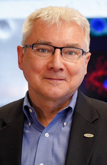 Pawel Kalinski合作论文数Department of Immunology, Roswell Park Comprehensive Cancer Center18
 Paul Wallace合作论文数Department of Primary Care and Population Health, University College London18Mr Baer合作论文数17
 Renuka Iyer合作论文数Roswell Park Cancer Institute17Kl O'Loughlin合作论文数16
 Kristopher Attwood合作论文数Department of Biostatistics, School of Pharmacy and Pharmaceutical Sciences, University at Buffalo;Roswell Park Cancer Institute, University at Buffalo15
 Mark Long合作论文数13William R. Greco合作论文数Department of Biostatistics, Roswell Park Cancer Institute13Junko Matsuzaki合作论文数Institute for Genetic Medicine, Hokkaido University12Bowen Dong合作论文数Dept Med, Roswell Park Canc Inst10
 Scott H Olejniczak合作论文数Department of Immunology, Roswell Park Cancer Institute10
 Walter J. Storkus合作论文数Department of Dermatology, School of Medicine, University of Pittsburgh;Department of Immunology, School of Medicine, University of Pittsburgh10
 Pedro Romero合作论文数Département d'Oncologie, Centre Hospitalier Universitaire Vaudois, Université de Lausanne10Henry G. Withers合作论文数10
 Takemasa Tsuji合作论文数Center for Immunotherapy, Roswell Park Comprehensive Cancer Center10
 Natasa Obermajer合作论文数Faculty of Pharmacy, University of Ljubljana10Colin Chavel合作论文数9Y M Rustum合作论文数9Abrams Scott I合作论文数9
 Alan Hutson合作论文数Department of Biostatistics & Bioinformatics, Roswell Park Comprehensive Cancer Center9Attaya Suvannasankha合作论文数Department of Medicine, Roswell Park Cancer Institute9Rocco C. Venuto合作论文数9
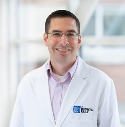 Jason B. Muhitch合作论文数Department of Immunology, Roswell Park Cancer Institute8
 John M. Kirkwood合作论文数Division of Malignant Hematology and Medical Oncology, Department of Medicine, School of Medicine, University of Pittsburgh;Department of Pathology, School of Medicine, University of Pittsburgh8K. O'Loughlin合作论文数7Rp Edwards合作论文数7
 Sarbajit Mukherjee合作论文数Roswell Park Comprehensive Cancer Center7Kathleen M. Tornatore合作论文数7
 Paul Klenerman合作论文数Nuffield Department of Medicine, Green Templeton College, University of Oxford7Joel Saltzman合作论文数7
 David Bajor合作论文数6T. George合作论文数6Fountzilas Christos合作论文数6
 Babiker Hani M合作论文数Division of Hematology and Oncology, The University of Arizona Cancer Center;Division of Hospital Medicine, Mayo Clinic;Division of Hospital Medicine, The University of Arizona Cancer Center6
 Cara Haymaker合作论文数Translational Molecular Pathology, University of Texas MD Anderson Cancer Center, Houston, Texas, USA6
 Erkut Borazanci合作论文数Oncology Research Division, HonorHealth Research Institute;Translational Genomics Research Institute, City of Hope;College of Medicine, University of Arizona6
 Kunle Odunsi合作论文数University of Chicago Comprehensive Cancer Center;Department of Obstetrics and Gynecology, The University of Chicago6
 Eunice S. Wang合作论文数Department of Medicine, Jacobs School of Medicine & Biomedical Sciences, University at Buffalo;Roswell Park Comprehensive Cancer Center, Jacobs School of Medicine & Biomedical Sciences, University at Buffalo;Department of Tumor Immunology, Roswell Park Cancer Institute6
 Patrick Boland合作论文数Rutgers Cancer Institute of New Jersey6
 Igor Puzanov合作论文数Roswell Park Comprehensive Cancer Center6
 Chantale Bernatchez合作论文数Cell Therapy Manufacturing Center6Udo Vanhoefer合作论文数6P LINSSEN合作论文数6
 Vivek Subbiah合作论文数Sarah Cannon Research Institute6Karen A. Hicks合作论文数5Cheryl L. Allen合作论文数5




