Background Data: Abdominal (AAA) or thoracic (TAA) aortic aneurysms are a significant health concern traditionally treated by open surgical repair. ESG therapy may offer protection from aneurysm rupture with a reduction in procedure morbidity and mortality.Methods: Over a 10-year period, 817 patients were treated with ESGs for AAA (723) or TAA (94). Patients received 1 of 12 different stent graft devices. Technical and clinical success of ESGs was reviewed, and the incidence of procedure-related complications was analyzed.Results: The mean age was 74.3 years (range, 25-95 years); 678 patients (83%) were men; 86% had 2 or more comorbid medical illnesses, 67% of which included coronary artery disease. Technical success, on an intent-to-treat basis was achieved in 93.8% of patients. Primary clinical success, which included freedom from aneurysm-related death, type I or III endoleak, graft infection or thrombosis, rupture, or conversion to open repair was 65 +/- 6% at 8 years. Of great importance, freedom from aneurysm rupture after ESG insertion was 98 +/- 1% at 9 years. There was a 2.3% incidence of perioperative mortality. One hundred seventy five patients died of causes not related to their aneurysm during a mean follow-up of 15.4 months.Conclusions: Stent graft therapy for aortic aneurysms is a valuable alternative to open aortic repair, especially in older sicker patients with large aneurysms. Continued device improvements coupled with an enhanced understanding of the important role of aortic pathology in determining therapeutic success will eventually permit ESGs to be a more durable treatment of aortic aneurysins.
INTRODUCTION:Preoperative parathyroid adenoma localization allows for a minimally invasive surgical approach. Current methods used for adenoma localization include ultrasound, sestamibi scan, and occasionally magnetic resonance imaging (MRI). In cases in which an adenoma is not localized after a radiologic work-up, the surgeon must perform a four-gland exploration. Preoperative selective venous sampling (SVS) has been described as a safe and clinically effective method to localize a parathyroid adenoma after failed exploration. The purpose of this study was to evaluate the efficacy of SVS as a primary localization technique in cases where conventional localization techniques have failed.MATERIALS AND METHODS:Fourteen patients with nonlocalizing parathyroid adenomas after ultrasonography, sestamibi scanning, and MRI were retrospectively reviewed. Eight patients underwent SVS prior to surgery and six patients underwent a four-gland neck exploration without preoperative SVS. The two groups were assessed for accuracy of the SVS in localizing the adenoma, operative time, length of hospitalization, and complications.RESULTS:All of the patients in the study underwent successful uncomplicated surgical parathyroidectomy. The SVS proved accurate in preoperatively localizing the adenoma in 7 of 8 patients and allowed for a minimally invasive approach in 87% of patients. The mean operative time was 33 minutes for the SVS group and 67 minutes in the non-SVS group. None of the patients in either group experienced procedural complications.CONCLUSION:This study suggests that preoperative SVS may represent a safe and effective method of preoperatively localizing the nonlocalized parathyroid adenoma. In cases where conventional radiologic techniques fail to localize an adenoma, SVS may obviate the need for a four-gland exploration.
BACKGROUND: Advanced abdominal pregnancy is a rare, life-threatening condition that presents a number of challenges. CASE: A 29-year-old primigravida with 10 years of secondary infertility and a previous tuboplasty had a 21-week abdominal pregnancy treated with preoperative arterial embolization before laparoscopically assisted fetal delivery. Postoperatively, 4 cycles of methotrexate were administered at 50 mg/m2 intramuscularly every 3 weeks for the retained abdominal placenta. Subsequent spontaneous conception occurred, and a live, full-term infant was delivered by cesarean delivery 17 months later. No adverse sequelae were found during long-term follow-up. CONCLUSION: This report demonstrates successful minimally invasive management of an advanced abdominal pregnancy with a multimodal approach that included preoperative arterial embolization, laparoscopically assisted delivery, and judicious use of postoperative methotrexate.
Objective: On November 23, 1992, the first endovascular stent graft (ESG) repair of an aortic aneurysm was performed in North America. Following the treatment of this patient, we have continued to evaluate ESG over the past 10 years in the treatment of 817 patients. Summary and Background Data: Abdominal (AAA) or thoracic (TAA) aortic aneurysms are a significant health concern traditionally treated by open surgical repair. ESG therapy may offer protection from aneurysm rupture with a reduction in procedure morbidity and mortality. Methods: Over a 10-year period, 817 patients were treated with ESGs for AAA (723) or TAA (94). Patients received 1 of 12 different stent graft devices. Technical and clinical success of ESGs was reviewed, and the incidence of procedure-related complications was analyzed. Results: The mean age was 74.3 years (range, 25–95 years); 678 patients (83%) were men; 86% had 2 or more comorbid medical illnesses, 67% of which included coronary artery disease. Technical success, on an intent-to-treat basis was achieved in 93.8% of patients. Primary clinical success, which included freedom from aneurysm-related death, type I or III endoleak, graft infection or thrombosis, rupture, or conversion to open repair was 65 ± 6% at 8 years. Of great importance, freedom from aneurysm rupture after ESG insertion was 98 ± 1% at 9 years. There was a 2.3% incidence of perioperative mortality. One hundred seventy five patients died of causes not related to their aneurysm during a mean follow-up of 15.4 months. Conclusions: Stent graft therapy for aortic aneurysms is a valuable alternative to open aortic repair, especially in older sicker patients with large aneurysms. Continued device improvements coupled with an enhanced understanding of the important role of aortic pathology in determining therapeutic success will eventually permit ESGs to be a more durable treatment of aortic aneurysms.
PURPOSE: To prospectively evaluate the efficacy and safety of reteplase with percutaneous transluminal angioplasty (PTA) in the treatment of thrombosed polytetrafluoroethylene hemodialysis arteriovenous grafts (AVGs).MATERIALS AND METHODS: Forty-two patients were entered into the study. Sixty-two procedures in 43 grafts were performed. One unit of reteplase and 4,000 units of heparin were administered into the AVGs. Routine venography and percutaneous transluminal angioplasty (PTA) was then performed. Patients were transferred for hemodialysis immediately after the procedure.RESULTS: Technical success was achieved in 92% of the cases. Four cases involved intentional repeat thrombosis because of poor outflow and/or need for a new graft site. Minor complications occurred in 6.5% of the cases. No major complications occurred. The mean procedure time for experienced versus less-experienced interventionalists was significantly shorter (p < .001). Primary patency rates were 50%, 34%, and 34% at 30, 90, and 180 days, respectively.CONCLUSION: Reteplase in conjunction with heparin and PTA is a safe and effective means of thrombolysis of AVGs. Its efficacy is comparable to that of other available thrombolytic drugs.
A patient with acute type B dissection and a tube configuration of the intimal flap presented with signs of advanced mesenteric and renal ischemia as well as decreased pulses in the lower extremities. The patient was referred for emergency percutaneous fenestration of the abdominal aorta as a salvage procedure and a possible bridge to later surgery. After fenestration, femoral pulses became transiently stronger and then disappeared. The patient died after exploratory laparotomy. Postmortem examination demonstrated dehiscence of the infrarenal abdominal aortic intima with occlusion of the aortic bifurcation.
PURPOSE:To evaluate prospectively the efficacy of treating thrombosed hemodialysis arteriovenous polytetrafluoroethylene (PTFE) grafts using tissue-type plasminogen activator (tPA) and percutaneous transluminal angioplasty (PTA).MATERIALS AND METHODS:Forty-two sequential thrombosed PTFE dialysis grafts in 33 patients presented for declotting. All 42 grafts were treated with a modified lysis and PTA technique with use of 2 mg tPA and 3,000-5,000 U heparin in a total volume of 5 mL, administered into the graft via an angiocatheter. The elapsed time from tPA injection until completion was recorded. Prospective data collection included demographic information, technical details of the procedure, immediate outcomes, complications, and patency rates.RESULTS:Technical success, defined as complete graft recanalization with a palpable thrill after treatment plus successful hemodialysis, was achieved in all cases, except five. These five cases were deliberate graft closures due to inadequacy of the outflow veins to support an arteriovenous graft after successful lysis. Mean lysis time was 40.8 minutes and mean room procedure time after the lysis period was 65.4 minutes. Eight procedure-related complications occurred (two major and six minor). The follow-up period was 4-241 days, with an estimated mean of 157 days. The 30-day and 90-day primary patency rates were 57% and 50%, respectively.CONCLUSIONS:Treatment of thrombosed PTFE dialysis grafts with use of 2 mg tPA and 3,000 U of heparin is safe and effective. Use of this modified lysis and PTA technique allows an expeditious procedure in the angiography suite. However, this technique precludes imaging of the outflow veins before treatment, so that grafts entering diffusely diseased veins may need to be closed after successful lysis.
The purpose of this study was to describe our experience with balloon and self-expanding endovascular grafts for the management of thoracic aortic lesions. Between February 1997 and June 1998, 20 endovascular grafts were implanted in 14 patients for the treatment of thoracic aortic aneurysms and pseudoaneurysms. Endovascular procedures were performed using one of four different devices: (1) Dacron-covered balloon-expandable Palmaztrade mark stent, (2) balloon-expandable Palmaz stent-PTFE graft prosthesis (BE-PS), (3) self-expanding internally supported Nitinol Dacron prosthesis (Vanguardtrade mark SE-V), and (4) self-expanding externally supported Nitinol PTFE prosthesis (Excludertrade mark SE-E). The results show that endovascular grafting represents a potentially important alternative therapy to open repair of the thoracic aorta. Self-expanding devices were, in our experience, easier to use and more accurately deployed.
Major breakthroughs in catheter and guidewire design as well as improvements in angiographic x-ray equipment currently allow interventional radiologists to diagnose massive life-threatening upper and lower GI hemorrhage and to stop the bleeding safely and effectively using superselective catheterization and microcoil embolization. For chronic or recurrent GI bleeding, when endoscopy is unrevealing or equivocal, barium studies, CT scanning, nuclear medicine studies, and angiography can help determine the cause of bleeding. A multidisciplinary approach, including the gastroenterologist, radiologist, and surgeon, is extremely helpful in managing GI bleeding, particularly in high-risk patients or patients presenting as diagnostic dilemmas.
OBJECTIVESTo define the utility of intraoperative transeophageal echocardiography (TEE) during endovascular thoracic aortic repair.DESIGNRetrospective study.MATERIALSFive patients underwent six transluminal endovascular stent-graft procedures for repair of thoracic aortic disease.METHODSAfter induction of anaesthesia, a multiplane or biplane TEE probe was placed to obtain views of the diseased aorta. Both transverse and longitudinal planes of the aortic arch and descending thoracic aortic segments were imaged. The aortic pathology was confirmed by TEE and the proximal and distal extents of the intrathoracic lesion were defined. Doppler and colour-flow imaging was used to identify flow patterns through the aorta before and after stent-graft deployment.RESULTSVisualisation and confirmation of the aortic pathology by ultrasonography was accomplished in all patients. TEE was able to confirm proper placement of the endograft relative to the aortic lesion after deployment and was able to confirm exclusion of blood flow into the aneurysm sacs.CONCLUSIONSTEE may facilitate repair by confirming aortic pathology, identifying endograft placement, assessment of the adequacy of aneurysm sack isolation, as well as dynamic intraoperative cardiac assessment.
To describe abscess formation complications in desmoid tumors of patients with Gardner's syndrome and percutaneous drainage.
 Victoria Teodorescu合作论文数5
 Michael L Marin合作论文数Department of Surgery, Mount Sinai School of Medicine;The Lauder Family Cardiovascular Ambulatory Center, Mount Sinai4
 Abigail Falk合作论文数4
 Robert Lookstein合作论文数Icahn School of Medicine at Mount Sinai3
 Larry H. Hollier合作论文数LSU Health Sciences Center3J Guller合作论文数3
 Antonio Carroccio合作论文数Department of Health Promotion Sciences, University of Palermo2Jaime Uribarri合作论文数Icahn School of Medicine at Mount Sinai2
 Randall B Griepp合作论文数Department of Cardiovascular Surgery, Icahn School of Medicine at Mount Sinai2Oak Juliana合作论文数2
 Joseph A Vassalotti合作论文数Department of Medicine, Mount Sinai School of Medicine2F. Scott Nowakowski合作论文数Department of Vascular and Interventional Radiology Klingenstein Pavillion, Mount Sinai Medical Center2D Spielvogel合作论文数2Ronald A. Kahn合作论文数2Claudie M Sheahan合作论文数1Michael Minor合作论文数1NJ Morrissey合作论文数1
 Perry Gerard合作论文数Division of Cardiology, New York Medical College at Westchester Medical Center1
 Larry Harold Hollier合作论文数Baylor College of Medicine;Department of Plastic Surgery, Texas Children's Hospital1Eloy Jean Anderson合作论文数1Farr R. Nezhat合作论文数Weill Cornell Medical College, Cornell University;NewYork-Presbyterian Hospital1Eric M. Genden合作论文数1
 Richard L. Berkowitz合作论文数Department of Obstetrics and Gynecology, Columbia University1William Forman合作论文数1Tikva Jacobs合作论文数1Michael Kaplan合作论文数1David M Moskowitz合作论文数1Jiyong Ahn合作论文数1Claudia M Sheahan合作论文数1Sreedhar Gaddipati合作论文数1Nicholas Morrissey合作论文数1Jamal Rahaman合作论文数1
 Mitchell S. Cappell合作论文数Gastroenterology & Hepatology, Beaumont Hospital, Royal Oak1Catherine Maldjian合作论文数1Z. Lefkovitz合作论文数1Alan Garten合作论文数1
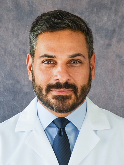 Sharif Ellozy合作论文数Department of Surgery, Weill Cornell Medicine, Cornell University1S N Konstadt合作论文数1Richard E. Parsons合作论文数1Marcus D'Ayala合作论文数1Kristina S. Chae合作论文数1




