The molecular mechanisms by which extracellular guidance cues regulate axonal morphology are not fully understood. Recent findings suggest that increased activity of the protein kinase Akt promotes dendritic branching and elongation in hippocampal neurons. We tested whether expression of constitutively active Akt (CA-Akt) in primary sensory neurons would promote axonal branching and whether targeting CA-Akt to lipid rafts, common sites of Akt function, would differentially regulate axonal morphology. Biolistic transduction of sensory neurons induced a rapid expression of CA-Akt, resulting in increased axonal branching, cell hypertrophy, and growth cone expansion. Additionally, we found that targeting of CA-Akt to lipid rafts significantly potentiated growth cone expansion compared with expression of CA-Akt throughout the neuron. Because lipid rafts are concentrated within the growth cone, this finding suggests that signaling of expansion is likely regulated locally. We found that CA-Akt-mediated growth cone expansion, but not axonal branching, was attenuated by coexpression of dominant-negative Rac1. In contrast, blockade of mammalian target of rapamycin (mTOR) prevented axonal branching and hypertrophy in response to CA-Akt, but not growth cone expansion. These data indicate that Akt activity can regulate growth cone expansion via localized Rac1 signaling and regulate axonal branching and soma size via activation of mTOR. (C) 2009 Wiley-Liss, Inc.
Neurotrophins promote sprouting and elongation of central nervous system (CNS) axons following injury. Consequently, it has been suggested that neurotrophins could be used to repair the CNS by inducing axonal sprouting from nearby intact axons, thereby compensating for the loss of recently injured axons. We tested whether long-term overexpression of neurotrophins in the rat cortex would induce sprouting of cortical serotonergic axons following a neurotoxic injury. After a single subcutaneous injection of para-chloroamphetamine (PCA; 9 mg/ml) that lesions the majority of serotonergic axons in the rat cortex, we injected adenoviral vectors containing cDNAs for brain-derived neurotrophic factor (Adv.BDNF), neurotrophin-3 (Adv.NT-3), or nerve growth factor (Adv.NGF) into the rat frontal cortex. Nine days later, we measured significant increases in the concentration of the respective neurotrophins surrounding the vector injection sites, as measured by ELISA. Immunohistochemical localization of serotonin revealed a fourfold increase in the density of serotonergic fibers surrounding the injection sites of Adv.BDNF and Adv.NT-3, corresponding to a 50% increase in cortical serotonin concentration, compared with a control vector containing the cDNA for enhanced green fluorescent protein (Adv.EGFP). In contrast, there was no difference in serotonergic fiber density or cortical serotonin concentration surrounding the injection of Adv.NGF compared with Adv.EGFR These data demonstrate that localized overexpression of BDNF or NT-3, but not NGF, is sufficient to promote sprouting of serotonergic axons in the cortex following an experimental neurotoxic injury. (c) 2005 Wiley-Liss, Inc.
We reported recently that overexpression of neurotrophin‐3 (NT‐3) by motoneurons in the spinal cord of rats will induce sprouting of corticospinal tract (CST) axons (Zhou et al. [2003] J. Neurosci. 23:1424–1431). We now report that overexpression of brain‐derived neurotrophic factor (BDNF) or glial cell‐derived neurotrophic factor (GDNF) in the rat sensorimotor cortex near the CST neuronal cell bodies together with overexpression of NT‐3 in the lumbar spinal cord significantly increases axonal sprouting compared to that induced by NT‐3 alone. Two weeks after unilaterally lesioning the CST at the level of the pyramids, we injected rats with saline or adenoviral vectors (Adv) carrying genes coding for BDNF (Adv.BDNF), GDNF (Adv.GDNF) or enhanced green fluorescent protein (Adv.EGFP) at six sites in the sensorimotor cortex, while delivering Adv.NT3 to motoneurons in each of these four groups on the lesioned side of the spinal cord by retrograde transport from the sciatic nerve. Four days later, biotinylated dextran amine (BDA) was injected into the sensorimotor cortex on the unlesioned side to mark CST axons in the spinal cord. Morphometric analysis of axonal sprouting 3 weeks after BDA injection showed that the number of CST axons crossing the midline in rats treated with Adv.BDNF or Adv.GDNF were 46% and 52% greater, respectively, than in rats treated with Adv.EGFP or PBS (P < 0.05). These data demonstrate that sustained local expression of neurotrophic factors in the sensorimotor cortex and spinal cord will promote increased axonal sprouting after spinal cord injury, providing a basis for continued development of neurotrophic factor therapy for central nervous system damage. © 2003 Wiley‐Liss, Inc.
 J. Clay Goodman合作论文数11
 Brian Baumgartner合作论文数Department of Biochemistry;Texas A&M University;Department of Biochemistry, Texas A&M University11MH Grider合作论文数9Slc Woo合作论文数8
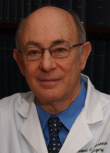 Robert G. Grossman合作论文数Department of Neurosurgery, Methodist Hospital Neurological Institute8
 Leroy Hood (莱诺·伊·胡德)合作论文数Department of Immunology, University of Washington;The Hood-Price Lab for Systems Biomedicine, Institute for Systems Biology;Providence St. Joseph Health;Department of Microbiology & Immunology, University of British Columbia;Keck Graduate Institute of Applied Life Sciences7Q. Chen合作论文数5
 Carol Readhead合作论文数5
 George M. Smith合作论文数Temple University5
 Todd Trask合作论文数4SH Chen合作论文数4
 Brian J Popko合作论文数Feinberg School of Medicine, Northwestern University4Estuardo Aguilar-Cordova合作论文数4Ahmet Çolak合作论文数3RG GROSSMAN合作论文数3SLC WOO合作论文数3Rl Sidman合作论文数3LR McGowen合作论文数3SH CHEN合作论文数3X Zheng合作论文数3
 Lijun Zhou (周利君)合作论文数Zhongshan School of Medicine, Sun Yat-Sen University3Philip R. Wyde合作论文数3E D Rabinovsky合作论文数2Js Garvey合作论文数2DA CARRIER合作论文数2
 Andrea Nedergaard Jensen合作论文数Department of Public Health, Faculty of Health and Medical Sciences, University of Copenhagen2
 Frank Gannon合作论文数Springfield City Group;Health Translation Queensland2
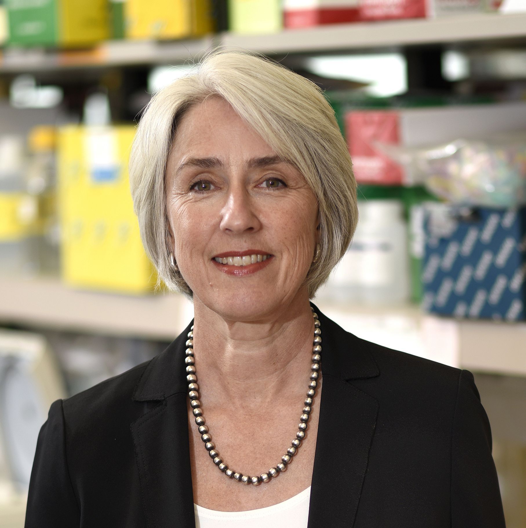 Margaret A. Goodell合作论文数Department of Molecular and Cellular Biology, Baylor College of Medicine;Stem Cells and Regenerative Medicine Center, Baylor College of Medicine2
 Weidong Le合作论文数Sichuan Academy of Medical Sciences2
 Shu-Hsia Chen合作论文数2
 Malcolm K Brenner合作论文数Center for Cell and Gene Therapy, Baylor College of Medicine;Blood, Marrow and Cell Therapy Center, Houston Methodist Hospital2Nagahide Takahashi合作论文数Department of Psychiatry, Nagoya University Graduate School of Medicine2Szu-Ting Chen合作论文数Biochemistry & Molecular Biology, University of Southern California2J. Regino Perez-Polo合作论文数2
 James L Mcmanaman合作论文数2
 Alan R Davis合作论文数2E LAI合作论文数1Z. Gugala合作论文数1C T Freund合作论文数1L Hassinger合作论文数1K D Carey合作论文数1PA PASKEVICH合作论文数1D.G. Kieback合作论文数1X W Tong合作论文数1WJ Hamilton合作论文数1S. L. C. Woo合作论文数1Ra Saavedra合作论文数1Stephen W. Hunt合作论文数1Richard L. Sidman合作论文数1




