BackgroundWe previously identified RasGRP3 as a regulator of erythropoietin-mediated reduced umbilical cord blood (UCB) CD34+ cell transmigration.1 To examine the effects of manipulating RasGRP3 expression on hematopoietic stem and progenitor cell (HSPC) homing, engraftment, long term reconstitution, and self-renewal, we utilized a mouse model for UCB transplantation.MethodsUCB derived hematopoietic cells were lentiviral transduced with shRNA constructs targeting RasGRP3, leading to reduced expression, or with overexpression constructs leading to stabilized expression of RASGRP3. On the day of transplantation and 2 days following lentiviral transduction, UCB units were sorted for CD34+, fluorophore+ cells. These selected cells were then transplanted to immune deficient mice (NSG) which had been sublethally irradiated 1 day prior. Mice were bled every 4 weeks to examine human cell donor chimerism in the PB. For secondary transplantations, we used BM pooled from all primary recipients. This pooled BM was transplanted to immune deficient mice (NSG) or transgenic immune deficient mice that express a trio of human cytokines (NSGS) which had been sublethally irradiated 1 day prior. To examine BM homing, mouse BM was harvested 3 hours after transplantation.ResultsAt weeks 12, 16, and 20 post transplantation, there were consistent but statistically insignificant trends demonstrating reduced human cell chimerism in the PB when RasGRP3 was knocked down. At weeks 16 and 20, RasGRP3 overexpression trended towards leading to increased human cell chimerism in the PB. Interestingly, both knockdown and overexpression of RasGRP3 trended toward decreased human chimerism in recipient mouse BM 20 weeks post transplantation. There were decreased percentages of primitive HSPC in the BM of mice receiving RasGRP3 shRNA compared to control. At week 16 post-secondary transplantation, mice that received RasGRP3 knockdown cells showed statistically significant decreases in human cell chimerism in the PB of NSG mice, and a similar trend in NSGS mice. Similarly, RasGRP3 recipients receiving RasGRP3 overexpressed cells showed trends toward increased PB chimerism at week 16 in secondary mice. Interestingly, there were no significant differences in human cell chimerism in the BM of either NSG or NSGS receiving cells with RasGRP3R expression manipulation. Mice receiving RasGRP3 knockdown UCB trended toward having lower numbers of total human cell in the BM 3-hours post transplantation, suggesting RasGRP3 expression levels affect in vivo homing.ConclusionsRasGRP3 expression levels play a role in UCB homing and engraftment, but the main phenotypic effects appear to be on PB hematopoietic and hematopoietic derived cells.
Lipid droplets (LDs) are cellular organelles critical for lipid homeostasis, with intramyocyte LD accumulation implicated in metabolic disorder-associated heart diseases. Here we identify a human long non-coding RNA, Lipid-Droplet Transporter ( LIPTER ), essential for LD transport in human cardiomyocytes. LIPTER binds phosphatidic acid and phosphatidylinositol 4-phosphate on LD surface membranes and the MYH10 protein, connecting LDs to the MYH10-ACTIN cytoskeleton and facilitating LD transport. LIPTER and MYH10 deficiencies impair LD trafficking, mitochondrial function and survival of human induced pluripotent stem cell-derived cardiomyocytes. Conditional Myh10 deletion in mouse cardiomyocytes leads to LD accumulation, reduced fatty acid oxidation and compromised cardiac function. We identify NKX2.5 as the primary regulator of cardiomyocyte-specific LIPTER transcription. Notably, LIPTER transgenic expression mitigates cardiac lipotoxicity, preserves cardiac function and alleviates cardiomyopathies in high-fat-diet-fed and Lepr db/db mice. Our findings unveil a molecular connector role of LIPTER in intramyocyte LD transport, crucial for lipid metabolism of the human heart, and hold significant clinical implications for treating metabolic syndrome-associated heart diseases.
Multiple myeloma (MM) is an incurable malignancy of plasma cells. To identify targets for MM immuno-therapy, we develop an integrated pipeline based on mass spectrometry analysis of seven MM cell lines and RNA sequencing (RNA-seq) from 900+ patients. Starting from 4,000+ candidates, we identify the most highly expressed cell surface proteins. We annotate candidate protein expression in many healthy tissues and validate the expression of promising targets in 30+ patient samples with relapsed/refractory MM, as well as in primary healthy hematopoietic stem cells and T cells by flow cytometry. Six candidates (ILT3, SEMA4A, CCR1, LRRC8D, FCRL3, IL12RB1) and B cell maturation antigen (BCMA) present the most favor-able profile in malignant and healthy cells. We develop a bispecific T cell engager targeting ILT3 that shows potent killing effects in vitro and decreased tumor burden and prolonged mice survival in vivo, suggesting therapeutic relevance. Our study uncovers MM-associated antigens that hold great promise for immune -based therapies of MM.
Cord blood (CB) has been proven to be an alternative source of haematopoietic stem cells (HSCs) for clinical transplantation and has multiple advantages, including but not limited to greater HLA compatibility, lower incidence of graft-versus-host disease (GvHD), higher survival rates and lower relapse rates among patients with minimal residual disease. However, the limited number of HSCs in a single CB unit limits the wider use of CB in clinical treatment. Many efforts have been made to enhance the efficacy of CB HSC transplantation, particularly by ex vivo expansion or enhancing the homing efficiency of HSCs. In this chapter, we will document the major advances regarding human HSC ex vivo expansion and homing and will also discuss the possibility of clinical translation of such laboratory work.
Abstract Introduction Cord blood banking has consistently outpaced the utilization of cord blood units (CBUs). Thus, the average duration of cryopreservation among banked CBUs will likely continue to increase. It remains unclear how long cryopreserved CBUs remain functional, and it is critical to determine whether duration of cryopreservation should be used as an exclusionary criterion during selection for clinical use or if alternative post-thaw metrics can identify potent cryopreserved CBUs regardless of age. Objectives Our goal was to determine whether long-term (27-year) cryopreserved CBUs retain viable and functional hematopoietic stem (HSCs) and progenitor cells (HPCs). We further sought to leverage differences in HSC/HPC function (measured by in vivo engraftment) to demonstrate the utility of using omics approaches to identify candidate genes for use as molecular potency markers. Methods We performed comprehensive ex vivo, in vivo, and molecular analyses on the numbers, viability, and function of three 27-year cryopreserved CBUs using 3-year cryopreserved and fresh CBUs for comparison. Assays included viability staining, immunophenotyping by flow cytometry, primary and secondary colony forming unit (CFU) assays, ex vivo expansion of immunophenotypic HSCs/HPCs/CFUs, limiting dilution transplantations into immune-deficient mice, secondary transplantations, and RNA-sequencing of sorted HSCs and multipotent progenitor cells. Results Compared to fresh and recently cryopreserved CBU controls, long-term cryopreserved CBUs yield statistically similar numbers of viable immunophenotypic HSCs, multipotent HPCs, and committed myeloid and lymphoid HPCs. They retain highly functional cells, demonstrating similar primary and secondary CFU numbers and expansion capacity compared to controls, as well as robust engraftment, SCID repopulating cell frequency, and secondary engraftment capacity in mouse models of transplantation. Transcriptomic modelling revealed 18 genes, including MALT1 and MAP2K1, and several gene programs, including lineage determination programs and oxidative stress responses, that are strongly enriched in high engrafting HSCs/HPCs. Discussion CBUs cryopreserved for up to 27 years retain highly functional HSCs/HPCs. Thus, duration of cryopreservation alone is not an ideal exclusionary criteria for selection of CBUs. Preserving older CBUs may help to maintain a large and diverse pool of donors for clinical selection. Further, transcriptomics can identify candidate genes associated with engraftment for elucidation of possible CBU potency markers regardless of the duration of cryopreservation.
Experimental hematopoietic stem cell transplantation (HSCT) is an invaluable tool in determining the function and characteristics of hematopoietic stem cells (HSC) from experimental mouse and human donor groups. These groups could include, but are not limited to, genetically altered populations (gene knockout/knockin models), ex vivo manipulated cell populations, or in vivo modulated cell populations. The basic fundamentals of this process involve taking cells from a mouse/human donor source and putting them into another mouse (recipient) after preconditioning of the recipient with either total body irradiation (TBI) for mouse donor cells or into sublethally irradiated immune-deficient mice for human donor cells. Then, at pre-determined time points post-transplant, sampling a small amount of peripheral blood (PB) and at the termination of the evalaution, bone marrow (BM) to determine donor contribution and function by phenotypic analysis. Exploiting the congenic mouse strains of C57BL/6 (CD45.1- CD45.2+), BoyJ (CD45.1+ CD45.2-), and their F1-crossed hybrid C57BL/6 × BoyJ (CD45.1+ CD45.2+), we are able to quantify donor, competitor, and recipient mouse cell contributions to the engraftment state. Human donor cell engraftment (e.g., from the cord blood [CB], mobilized PB, or BM) is assessed by human cell phenotyping in sublethally irradiated immune-deficient mouse recipients (e.g., NOD scid gamma mice that are deficient in B cells, T cells, and natural killer cells and have defective dendritic cells and macrophages). Engraftment of cells from primary mouse recipients into secondary mice allows for an estimation of the self-renewal capacity of the original donor HSC. This chapter outlines concepts, methods, and techniques for mouse and human cell models of HSCT and for assessment of donor cells collected and processed in hypoxia versus ambient air.
Umbilical cord blood transplantation is a life-saving treatment for malignant and non-malignant hematologic disorders. It remains unclear how long cryopreserved units remain functional, and the length of cryopreservation is often used as a criterion to exclude older units. We demonstrate that long-term cryopreserved cord blood retains similar numbers of hematopoietic stem and progenitor cells compared with fresh and recently cryopreserved cord blood units. Long-term cryopreserved units contain highly functional cells, yielding robust engraftment in mouse transplantation models. We also leverage differences between units to examine gene programs associated with better engraftment. Transcriptomic analyses reveal that gene programs associated with lineage determination and oxidative stress are enriched in high engrafting cord blood, revealing potential molecular markers to be used as potency markers for cord blood unit selection regardless of length of cryopreservation. In summary, cord blood units cryopreserved for extended periods retain engrafting potential and can potentially be used for patient treatment.
Little is known about a regulatory role of CaMKK2 for hematopoietic stem (HSC) and progenitor (HPC) cell function. To assess this, we used Camkk2−/− and wild type (WT) control mouse bone marrow (BM) cells. BM cells were collected/processed and compared under hypoxia (3% oxygen; physioxia) vs. ambient air (~21% oxygen). Subjecting cells collected to ambient air, even for a few minutes, causes a stress that we termed Extra Physiological Shock/Stress (EPHOSS) that causes differentiation of HSCs and HPCs. We consider physioxia collection/processing a more relevant way to assess HSC/HPC numbers and function, as the cells remain in an oxygen tension closer physiologic conditions. Camkk2−/− cells collected/processed at 3% oxygen had positive and negative effects respectively on HSCs (by engraftment using competitive transplantation with congenic donor and competitor cells and lethally irradiated congenic recipient mice), and HPCs (by colony forming assays of CFU-GM, BFU-E, and CFU-GEMM) compared to WT cells processed in ambient air. Thus, with cells collected/processed under physioxia, and therefore never exposed and naïve to ambient air conditions, CaMKK2 not only appears to act as an HSC to HPC differentiation fate determinant, but as we found for other intracellular mediators, the Camkk−/− mouse BM cells were relatively resistant to effects of EPHOSS. This information is of potential use for modulation of WT BM HSCs and HPCs for future clinical advantage.
Hematopoietic stem cells (HSCs) manifest impaired recovery and self-renewal with a concomitant increase in differentiation when exposed to ambient air as opposed to physioxia. Mechanism(s) behind this distinction are poorly understood but have the potential to improve stem cell transplantation. Single-cell RNA sequencing of HSCs in physioxia revealed upregulation of HSC self-renewal genes and downregulation of genes involved in inflammatory pathways and HSC differentiation. HSCs under physioxia also exhibited downregulation of the epigenetic modifier Tet2. Tet2 is a-ketoglutarate, iron-and oxygen-dependent dioxygenase that converts 5-methylcytosine to 5-hydroxymethylcytosine, thereby promoting active transcription. We evaluated whether loss of Tet2 affects the number and function of HSCs and hematopoietic progenitor cells (HPCs) under physioxia and ambient air. In contrast to wild-type HSCs (WT HSCs), a complete nonresponsiveness of Tet2(-/-) HSCs and HPCs to changes in oxygen tension was observed. Unlike WT HSCs, Tet2(-/-) HSCs and HPCs exhibited similar numbers and function in either physioxia or ambient air. The lack of response to changes in oxygen tension in Tet2(-/-) HSCs was associated with similar changes in self-renewal and quiescence genes among WT HSC-physioxia, Tet2(-/-) HSC-physioxia and Tet2(-/-) HSC-air. We define a novel molecular program involving Tet2 in regulating HSCs under physioxia.
Regulation of hematopoietic stem and progenitor cell numbers and function as well as that of their more mature offspring within the bone marrow is critical for normal hematopoiesis. In this article, we mainly describe the role of the chemokine stromal cell-derived factor-1 (SDF-1)/CXCL12 and its receptor, CXCR4, in maintenance of these cell populations within the bone marrow. This includes mechanisms in which CXCL12 and CXCR4 expression is regulated under homeostatic or inflammatory conditions, and how this alters the balance between myeloid and lymphoid cell development.
Hematopoietic cells are regulated in part by extracellular cues from cytokines. Leukemia inhibitory factor (LIF) promotes survival, self-renewal, and pluripotency of mouse embryonic stem cells (mESC). While genetic deletion of LIF affects hematopoietic progenitor cells (HPCs), the direct effect of LIF protein exposure on HPC survival is not known. Furthermore, post-translational modifications (PTM) of LIF and their effects on its function have not been evaluated. We demonstrate that treatment with recombinant LIF preserves mouse and human HPC numbers in stressed conditions when growth factor addition is delayed ex vivo. We show that Lif is upregulated in response to irradiation-induced stress. We reveal novel PTM of LIF where it is cleaved twice by dipeptidyl peptidase 4 (DPP4) protease so that it loses its 4 N-terminal amino acids. This truncation of LIF down-modulates LIF's ability to preserve functional HPC numbers ex vivo following delayed growth factor addition. DPP4-truncated LIF blocks the ability of full-length LIF to preserve functional HPC numbers. This LIF role and its novel regulation by DPP4 have important implications for normal and stress hematopoiesis, as well as for other cellular contexts in which LIF and DPP4 are implicated.
Aryl hydrocarbon receptor (AhR) antagonism is known to expand human hematopoietic stem cells (HSCs). However, its regulatory effect on the lineage-skewed differentiation of HSCs has not been sufficiently studied. Here, we investigate the effect of the AhR-selective antagonist CH223191 on the regulation of HSC differentiation. Consistent with the well-known effects of AhR antagonists, CH223191 treatment increase phenotypic HSCs (Lin-CD34 +CD38-CD90 +CD45RA-) and preserves their functionality. On the other hand, CH223191 leads to an overall expansion of megakaryocyte (MK)-lineage populations, such as MK progenitors (MKps, CD34 + CD41 +), immature MKs (CD41 + CD42b-), and mature MKs (CD41 + CD42b +), and it also activates MK/platelet-associated signaling pathways. Furthermore, CH223191 expands MKps, mature MKs, and p-selectin (CD62p)-positive platelet-like particles in immune thrombocytopenia (ITP) patient bone marrow (BM). These results highlight the numerical expansion of human MK-lineage progeny through AhR antagonism with CH223191. This approach using CH2231291 may be applicable in the development of auxiliary treatment regimens for patients with abnormal thrombopoiesis.
GSK983, a novel tetrahydrocarbazole, inhibits the replication of a variety of unrelated viruses in vitro with EC50 values of 5–20nM. Both replication of the adenovirus Ad-5 and the polyoma virus SV-40, and episomal maintenance of human papillomaviruses (HPV) and Epstein-Barr virus (EBV) are susceptible to GSK983. The compound does not inhibit all viruses; herpes simplex virus (HSV-1), human immunodeficiency virus (HIV), and lytic replication of EBV were not susceptible at concentrations below 1μM. GSK983 does inhibit the growth of cell lines immortalized by HTLV-1, EBV, HPV, SV40 and Ad-5, with EC50 values in the range of 10–40nM. Depending on the cell line, the compound induces either apoptosis or cytostasis at concentrations over 20nM. GSK983 also inhibits cell lines immortalized by non-viral mechanisms, but has little effect on primary cells. The CC50 values for keratinocytes, fibroblasts, lymphocytes, endothelial, and bone marrow progenitor cells are all above 10μM. The pattern of inhibition, which includes diverse viruses as well as growth of immortalized cells of varied origins, suggests the target is a host cell protein, rather than a viral protein. Preliminary mechanism studies indicate that GSK983 acts by inducing a subset of interferon-stimulated genes.
Neutrophils, the most abundant white blood cells in circulation, are closely related to cancer development and progression. Healthy primary neutrophils present potent cytotoxicity against various cancer cell lines through direct contact and via generation of reactive oxygen species. However, due to their short half-life and resistance to genetic modification, neutrophils have not yet been engineered with chimeric antigen receptors (CARs) to enhance their antitumor cytotoxicity for targeted immunotherapy. Here, we genetically engineered human pluripotent stem cells with synthetic CARs and differentiated them into functional neutrophils by implementing a chemically defined platform. The resulting CAR neutrophils present superior and specific cytotoxicity against tumor cells both in vitro and in vivo. Collectively, we established a robust platform for massive production of CAR neutrophils, paving the way to myeloid cell-based therapeutic strategies that would boost current cancer-treatment approaches.
Enhancing the efficiency of hematopoietic stem cell (HSC) homing and engraftment is critical for cord blood (CB) hematopoietic cell transplantation (HCT). Recent studies indicate that N6-methyladenosine (m6A) modulates the expression of mRNAs that are critical for stem cell function by influencing their stability. Here, we demonstrate that inhibition of RNA decay by regulation of RNA methylation, enhances the expression of the homing receptor chemokine C-X-C receptor-4 (CXCR4) in HSCs. We show that YTH N6-methyladenosine RNA binding protein 2 (YTHDF2), a m6A reader and FTO α-ketoglutarate dependent dioxygenase (FTO), a m6A eraser play an opposite role in this process. Through screening, we identified several FDA-approved compounds that regulate the expression of YTHDF2 and FTO in CB CD34+ cells. We show that transient downregulation of YTHDF2 or activation of FTO by using these compounds inhibits CXCR4 decay in CB HSCs and promotes their homing and engraftment. Our results demonstrate a novel regulation strategy to enhance the function of CB HSCs and provide a translational approach to enhance the clinical efficacy of HCT.
Bone marrow (BM) hematopoietic stem cells (HSCs) become dysfunctional during aging (i.e., they are increased in number but have an overall reduction in long-term repopulation potential and increased myeloid differentiation) compared with young HSCs, suggesting limited use of old donor BM cells for hematopoietic cell transplantation (HCT). BM cells reside in an in vivo hypoxic environment yet are evaluated after collection and processing in ambient air. We detected an increase in the number of both young and aged mouse BM HSCs collected and processed in 3% O2 compared with the number of young BM HSCs collected and processed in ambient air (~21% O2). Aged BM collected and processed under hypoxic conditions demonstrated enhanced engraftment capability during competitive transplantation analysis and contained more functional HSCs as determined by limiting dilution analysis. Importantly, the myeloid-to-lymphoid differentiation ratio of aged BM collected in 3% O2 was similar to that detected in young BM collected in ambient air or hypoxic conditions, consistent with the increased number of common lymphoid progenitors following collection under hypoxia. Enhanced functional activity and differentiation of old BM collected and processed in hypoxia correlated with reduced "stress" associated with ambient air BM collection and suggests that aged BM may be better and more efficiently used for HCT if collected and processed under hypoxia so that it is never exposed to ambient air O2.
We wish to suggest the possibility there is a link between the brain and hematopoiesis in the bone marrow and that in the future it may be possible to use such information for better understanding of the regulation of hematopoiesis, and for efficacious treatment of hematopoietic disorders.
Hematopoietic and nervous systems are linked via innervation of bone marrow (BM) niche cells. Hematopoietic stem/progenitor cells (HSPCs) express neurotransmitter receptors, such as the γ-aminobutyric acid (GABA) type B receptor subunit 1 (GABBR1), suggesting that HSPCs could be directly regulated by neurotransmitters like GABA that directly bind to GABBR1. We performed imaging mass spectrometry and found that the endogenous GABA molecule is regionally localized and concentrated near the endosteum of the BM niche. To better understand the role of GABBR1 in regulating HSPCs, we generated a constitutive Gabbr1-knockout mouse model. Analysis revealed that HSPC numbers were significantly reduced in the BM compared with wild-type littermates. Moreover, Gabbr1-null hematopoietic stem cells had diminished capacity to reconstitute irradiated recipients in a competitive transplantation model. Gabbr1-null HSPCs were less proliferative under steady-state conditions and upon stress. Colony-forming unit assays demonstrated that almost all Gabbr1-null HSPCs were in a slow or noncycling state. In vitro differentiation of Gabbr1-null HSPCs in cocultures produced fewer overall cell numbers with significant defects in differentiation and expansion of the B-cell lineage. To determine whether a GABBR1 agonist could stimulate human umbilical cord blood (UCB) HSPCs, we performed brief ex vivo treatment prior to transplant into immunodeficient mice, with significant increases in long-term engraftment of HSPCs compared with GABBR1 antagonist or vehicle treatments. Our results indicate a direct role for GABBR1 in HSPC proliferation, and identify a potential target to improve HSPC engraftment in clinical transplantation.
Radiation-induced bystander effects (RIBEs) is a neglected, but crucial, area of radiation response. In this issued of Blood, Hu et al(1) have provided important new information and mechanistic insights into RIBE-impairment of hematopoietic stem (HSC) and progenitor (HPC) cells in hematopoietic cell transplantation (HCT), with implications for the mitigation of RIBEs.
 Scott Cooper合作论文数Departments of Medicine (Hematology/Oncology), Indianapolis, IN 46223, U.S.A.278Giao Hangoc合作论文数161L Lu合作论文数124
 Charlie Mantel合作论文数119Maegan L. Capitano合作论文数58
 Louis M. Pelus合作论文数School of Medicine, Indiana University49De Williams合作论文数40
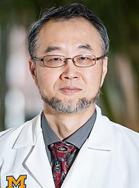 Chang Kim合作论文数Immunology & Hematopoiesis Lab, Department of Pathology, Medical School, University of Michigan;Mary H. Weiser Food Allergy Center, Medical School, University of Michigan;Department of Comparative Pathobiology, Purdue University39Mas Moore合作论文数38
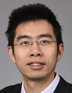 Xinxin Huang (黄鑫欣)合作论文数Institutes of Biomedical Sciences, Fudan University37RN SHEN合作论文数32S Vadhan-Raj合作论文数30Robert Hromas合作论文数Hematology Malignancy Program and Stem Cell Transplantation, Indiana University School of Medicine30
 Popoola Ralph合作论文数29
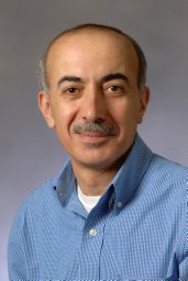 Edward F. Srour合作论文数School of Medicine, Indiana University29Young-June Kim合作论文数28Zh Li合作论文数27Y Ge合作论文数26Akihiko Gotoh合作论文数26Se Braun合作论文数24
 Keisuke Miyazawa合作论文数First Department of Internal Medicine, Tokyo Medical University24H. Scott Boswell合作论文数23Man Ryul Lee合作论文数21Kenneth Cornetta合作论文数21Yj Kim合作论文数20
 Mervin Yoder合作论文数Department of Surgery, School of Medicine, University of Pittsburgh20
 Stephen E. Braun合作论文数New England Primate Research Center, Harvard Medical School20Heather A. O'Leary合作论文数Robert C. Byrd Health Sciences Center, West Virginia University School of Medicine20
 Bin Guo (郭滨)合作论文数College of Basic Medical Sciences, Shanghai Jiao Tong University;Shanghai General Hospital19
 James Ropa合作论文数19Mang Xiao合作论文数19T Tauchi合作论文数18BS KWON合作论文数17
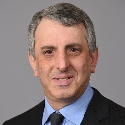 Mark H. Kaplan合作论文数Department of Microbiology and Immunology, School of Medicine, Indiana University17
 D. W. Clapp合作论文数Department of Pediatrics, School of Medicine, Indiana University17Timothy B. Campbell合作论文数16Y Lee合作论文数16Jay Gaddy合作论文数16
 Seiji Fukuda合作论文数16Xuan Ou合作论文数15Reuben Kapur合作论文数Herman B Wells Center for Pediatric Research, Indiana University School of Medicine15Bs Youn合作论文数14A Ritchie合作论文数14
 Gen-Sheng Feng (冯根生)合作论文数Shenzhen Bay Laboratory14N JACOBSEN合作论文数13G Risdon合作论文数13M Horie合作论文数13
 Steven Messina-Graham合作论文数13Steven Gillis合作论文数13




