Introduction Managing open lower extremity fractures is challenging, with potential complications such as amputation and infection. The aim of the study was to determine whether the time delay and initial treatment of the patients treated in a non -specialized hospital before being transferred to a dedicated level I trauma center led to a worse outcome. Methods Retrospective data from 44 patients (37 males and seven females) undergoing free tissue transfer for lower extremity open fractures from January 2017 to December 2022 were analyzed. Group A received primary care externally and was later transferred for definitive treatment (n=17, 38.6%), while group B received initial care at a level I trauma center (n=27, 61.4%). Surgical outcomes, complications, the duration of the hospital stay, and assessment times were compared. Various demographic variables, co -morbidities, prior interventions, and flap types were analyzed. Results Average age (A: 55.1 +/- 16.7; B: 38.7 +/- 19.8 years; p=0.041), overall hospitalization (A: 55.7 +/- 22.8; B: 42.8 +/- 21.3 days; p=0.041), and time to soft tissue reconstruction differed significantly between groups (A: 30.7 +/- 12.2; B: 18.9 +/- 9.3 days; p=0.013). Overall, 31.8% had multiple injuries without statistical differences between groups A and B (29.4% vs. 33.3%; p>0.05). There were no statistical differences between the groups in terms of major and minor complications and bone healing characteristics. Limb salvage was successful overall in 93.2% (A: 94.1%; B: 92.6%; P>0.05). Major complications occurred in 9.1%; three patients underwent major amputation (A: n=2; B: n=1). Minor complications were observed in 43.2% of patients (partial flap necrosis, wound dehiscence and non-union; A: 41.2%; B: 44.4%; p>0.05). Overall, 65.9% of patients (A: 64.7%; B: 66.7%; p>0.05) experienced uneventful bone healing, while 18.2% of patients (A: 23.5%; B: 14.8%; p>0.05) experienced delayed healing. Flaps used were mostly musculocutaneous (71.7%). Various assessed demographic characteristics, including age and presence of polytrauma, showed no significant influence on complications (p>0.05). Conclusion Although there is a significant difference in the time course of externally treated patients with open fractures, prolonged treatment is not associated with a higher complication rate or compromised bone healing outcome. Despite the findings, it is important to avoid delays and strive for interdisciplinary collaboration.
Introduction The incidence of deep surgical site infections following high tibial osteotomy (HTO) ranges between 0.4 to 4.7%. It is a severe complication with a high risk for poor clinical outcome. The aim of this study was to proof that a salvage algorithm for infected HTO with unstable bone situation leads to an infection-free status and bone union of the osteotomy and that correct limb alignment can be restored with good knee function. Materials and Method The study included seven patients with peri-implant infections following HTO. Infections occurred 83 +/- 58.9 days (range: 24-191) after HTO. All patients underwent the "RESTORE" algorithm: patients received (1) REmoval of the HTO hardware and extensive debridement; (2) the osteotomy was STabilized with a hexapod external fixator (Taylor Spatial Frame, TSF); (3) the osteotomy gap was Opened; and (4) the alignment was REconstructed using the TSF, aiming for the intended limb alignment of the initial HTO. Patient-reported outcomes were assessed 22-36 months after removal of the TSF. Results After 24 weeks (range: 11-35), an infection-free status and bone healing were achieved. In all cases, the limb was saved, and the previously targeted mechanical axis of the lower limb was restored. All patients reached full extension of the knee joint and at least 110 degrees of flexion. For KOOS: Symptoms 67.86 +/- 18.1, Pain 73.41 +/- 16.58, ADL 78.99 +/- 21.32, Sports 52.14 +/- 25.96, and QoL 41.96 +/- 24.66. OKS 35.71 +/- 8.8, SF-12 Physical Health 38.89 +/- 10.3, and SF-12 Mental Health 46.86 +/- 13.76. Conclusion The "RESTORE" algorithm is a safe and effective salvage procedure. The concept allows for saving the limb and obtaining the previously planned limb alignment. Patient-reported outcome measures showed slightly lowered values than healthy samples, but substantially better values than patients awaiting HTO. Due to the possibility of initial full weight-bearing, the risk of higher morbidity caused by immobilization is minimized.
Diabetes mellitus (DM) is associated with impaired wound healing, partly driven by excessive neutrophil extracellular trap (NET) formation mediated by peptidyl-arginine deiminase 4 (PADI4). While circulating NET markers predict poor healing outcomes, they likely reflect established tissue damage and offer limited opportunity for early intervention. We therefore investigated the association between PADI4 haplotypes, PADI4 expression, NET formation, and clinical outcomes, namely infections and delayed wound and bone healing, in 687 surgical patients (44.7% with DM). Pre-surgical PADI4 expression was 9.4-fold higher in patients with DM, particularly in those who developed wound healing complications. Neutrophils carrying the PADI4 minor haplotype showed increased PADI4 mRNA and protein expression and produced larger quantities of NETs more rapidly than those with the major haplotype. Clinically, patients with DM carrying the minor haplotype had the highest rates of delayed wound healing and infections. Together, these results demonstrate that PADI4 genetic variation influences neutrophil behavior and clinical outcomes. PADI4 haplotyping may provide a clinically actionable biomarker to identify patients with DM at high risk for wound healing complications and guide early preventive strategies. ### Competing Interest Statement The authors have declared no competing interest. ### Funding Statement This study received funding from the German Research Council (EH471/5-1) and the Bundesministerium für Bildung und Forschung (13GW0661D). C.L. was supported by the Studienstiftung des deutschen Volkes. ### Author Declarations I confirm all relevant ethical guidelines have been followed, and any necessary IRB and/or ethics committee approvals have been obtained. Yes The details of the IRB/oversight body that provided approval or exemption for the research described are given below: The ethics committee of the University Hospital Tübingen ("Ethik-Kommission an der Medizinischen Fakultät der Eberhard-Karls-Universität und am Universitätsklinikum Tübingen") gave ethical approval for this work (#666/2018BO2, approved 20.09.2018). I confirm that all necessary patient/participant consent has been obtained and the appropriate institutional forms have been archived, and that any patient/participant/sample identifiers included were not known to anyone (e.g., hospital staff, patients or participants themselves) outside the research group so cannot be used to identify individuals. Yes I understand that all clinical trials and any other prospective interventional studies must be registered with an ICMJE-approved registry, such as ClinicalTrials.gov. I confirm that any such study reported in the manuscript has been registered and the trial registration ID is provided (note: if posting a prospective study registered retrospectively, please provide a statement in the trial ID field explaining why the study was not registered in advance). Yes I have followed all appropriate research reporting guidelines, such as any relevant EQUATOR Network research reporting checklist(s) and other pertinent material, if applicable. Yes All data produced in the present work are contained in the manuscript.
Early and reliable detection of infection is vital for successful treatment. Serum markers such as C-reactive protein (CRP) and procalcitonin (PCT) are known to increase with a time lag. Azurocidin 1 (AZU1) has emerged as a promising marker for septic patients, but its diagnostic value in orthopedic and trauma patients remains unexplored. Between July 2020 and August 2023, all patients necessitating inpatient treatment for periprosthetic joint infection (PJI), peri-implant infection (II), soft tissue infection, chronic osteomyelitis, septic arthrodesis, bone non-union with and without infection were enrolled. Patients undergoing elective total joint arthroplasty (TJA) served as the control group. Blood samples were collected and analyzed for CRP, white blood cell count (WBC), PCT, and AZU1. Based on the inclusion and exclusion criteria 222 patients were included in the study (trauma = 38, soft tissue infection = 75, TJA = 33, PJI/II = 39, others = 37). While sensitivity and specificity were comparably high for AZU1 (0.734/0.833), CRP and PCT had higher specificity (0.542/1 and 0.431/1, respectively), and WBC a slightly higher sensitivity (0.814/0.455) for septic conditions. Taken together, the area under the curve (AUC) showed the highest accuracy for AZU1 (0.790), followed by CRP (0.776), WBC (0.641), and PCT (0.656). The Youden-Index was 0.57 for AZU1, 0.54 for CRP, 0.27 for WBC, and 0.43 for PCT. Elevated AZU1 levels effectively distinguished patients with a healthy condition from those suffering from infection. However, there is evidence suggesting that trauma may influence the release of AZU1. Additional research is needed to validate the diagnostic value of this new biomarker and further explore its potential clinical applications.
This paper discusses the development and application of an augmented reality (AR) system for assisting in nail implantation procedures for complex tibial fractures. Traditional procedures involve extensive X-ray usage from various angles, leading to increased radiation exposure and prolonged surgical times. The study presents a method using pre- and post-operative computed tomography (CT) data sets and a convolutional neural network (CNN) trained on segmented bone and metal objects. The augmented reality system overlays accurate 3D representations of bony fragments and implants onto the surgeon's view, aiming to reduce radiation exposure and intervention time. The study demonstrates successful segmentation of bone and metal objects in cases of heavy metal artifacts, achieving promising results with a relatively low number of training sets. The integration of this system into the clinical workflow could potentially improve surgical outcomes, significantly reduce radiation time, and therefore improve patient safety.
Background Contact teaching was prohibited by nationwide lockdowns during the COVID-19 pandemic. The scientific literature contains no concrete figures concerning e-learning via webinars in the subject of orthopedics and trauma surgery in the context of the COVID-19 pandemic. Objective This research study was established to collect facts and figures about webinars as a representative part of e-learning in the subject of orthopedics and trauma surgery in the context of the COVID-19 pandemic. Material and methods German-speaking non-commercial and edited webinars, produced by suppliers from Germany were identified using common search engines. Structured interviews with questionnaires about provider, format, and number of participants in the webinars over time, were offered to the operating companies. Results The study included four suppliers of webinars (AO Online Campus, BVOU Study Club, OU TO GO and WebDGU). There was approval of all operating companies to participate in the interviews and to disclose facts and figures about their webinars. All suppliers showed an increased supply and demand for webinars during the COVID-19 pandemic. Discussion OU TO GO and BVOU Study Club already offered webinars prior to the COVID-19 pandemic. AO online Campus was designed in addition to the existing portfolio. The supply of WebDGU was newly established. Limitations of this study are the exclusion of commercial suppliers, non-German operating companies and all other types of e-learning except webinars. The increased supply and demand for webinars during the COVID-19 pandemic gives hope for further improvement of e-learning in the subject of orthopedics and trauma surgery.
The reconstruction of posttraumatic defects of bone and soft tissue, as well as residual deformities of the bone is often a lengthy and complicated procedure. Multiple surgical interventions are necessary to reconstruct the affected extremity and restore its functionality. To achieve an optimal result it often takes months or years and requires great patience and compliance of the patient. This treatment should be carried out in centers with the appropriate instrumental and human resources. Since the pathologies are often complex with bony deformities or loss of bone, as well as infections and soft tissue defects, the treatment should be carried out by very experienced surgeons to successfully manage these complex reconstructions. This often requires interdisciplinary cooperation, especially with experienced plastic surgeons. A soft tissue reconstruction for better blood circulation in the exposed bony structures, as well as the bony defects themselves, can be reconstructed at the same time.
Die Rekonstruktion posttraumatischer Defekte von Knochen und Weichteilen sowie verbliebener Deformitäten ist ein oft langwieriges und kompliziertes Verfahren. Mehrfache operative Eingriffe sind notwendig, um nach Monaten bis Jahre ein optimales Ergebnis zu erreichen und letztendlich die Funktionsfähigkeit der betroffenen Extremität wieder herzustellen. Diese Behandlung sollte durch sehr erfahrene Operateure durchgeführt werden, die auch über die entsprechenden instrumentellen und personellen Ressourcen verfügen, um diese komplexen Rekonstruktionen erfolgreich durchzuführen. Hier bedarf es nicht selten einer interdisziplinären Zusammenarbeit insbesondere mit versierten plastischen Chirurgen. Eine weichteilige Deckung zur besseren Durchblutung der freiliegenden knöchernen Strukturen als auch knöcherne Defekte an sich, können so gemeinsam wieder rekonstruiert werden.
ZusammenfassungPseudarthrosen nach Sternotomien sind rar und meist gut im Rahmen operativer Revisionen zu behandeln. Bei fehlgeschlagenen Revisionen ist ein differenziertes Vorgehen notwendig, das sowohl die biologischen wie auch biomechanischen Rahmenbedingungen berücksichtigt. Es wird über den Fall einer therapierefraktären atrophen Pseudarthrose nach auswärtig erfolgloser Revision berichtet. Diese wurde durch ein Therapievorgehen, orientiert an den Kriterien des Non Union Scoring System (NUSS), erfolgreich behandelt. Die Versorgungsstrategie wie auch der erfolgreiche Behandlungsverlauf werden dargestellt.
Contact teaching was prohibited by nationwide lockdowns during the COVID-19 pandemic. The scientific literature contains no concrete figures concerning e‑learning via webinars in the subject of orthopedics and trauma surgery in the context of the COVID-19 pandemic.This research study was established to collect facts and figures about webinars as a representative part of e‑learning in the subject of orthopedics and trauma surgery in the context of the COVID-19 pandemic.German-speaking non-commercial and edited webinars, produced by suppliers from Germany were identified using common search engines. Structured interviews with questionnaires about provider, format, and number of participants in the webinars over time, were offered to the operating companies.The study included four suppliers of webinars (AO Online Campus, BVOU Study Club, OU TO GO and WebDGU). There was approval of all operating companies to participate in the interviews and to disclose facts and figures about their webinars. All suppliers showed an increased supply and demand for webinars during the COVID-19 pandemic.OU TO GO and BVOU Study Club already offered webinars prior to the COVID-19 pandemic. AO online Campus was designed in addition to the existing portfolio. The supply of WebDGU was newly established. Limitations of this study are the exclusion of commercial suppliers, non-German operating companies and all other types of e‑learning except webinars. The increased supply and demand for webinars during the COVID-19 pandemic gives hope for further improvement of e‑learning in the subject of orthopedics and trauma surgery.HINTERGRUND: Im Verlauf der COVID-19-Pandemie haben offizielle Schutzmaßnahmen traditionelle Präsenzfortbildungen zum Erliegen gebracht. Für das Fach Orthopädie und Unfallchirurgie (O&U) gibt es bisher bezüglich der Angebots- und Nachfragesituation von E‑Learning per Webinar im Kontext der COVID-19-Pandemie keine belastbaren Zahlen.Das Ziel der vorliegenden Arbeit ist die quantitative Beschreibung von Angebot und Nachfrage deutschsprachiger Online-Fortbildungen mit Webinar-Charakter aus der Orthopädie und Unfallchirurgie (O&U) im zeitlichen Zusammenhang mit der COVID-19-Pandemie.Über gängige Suchmaschinen wurden deutschsprachige, nichtkommerzielle, editierte und wissenschaftlich fundierte Fortbildungen in Form von Webinaren im Bereich O&U von Anbietern mit Sitz in Deutschland identifiziert und interviewt.Alle 4 eingeschlossenen Anbieter (AO Online Campus, BVOU Study Club, OU TO GO, WebDGU) nahmen an den strukturierten Interviews teil und stimmten einer Offenlegung der Teilnehmerzahlen zu. Das Angebot von Webinaren stieg im zeitlichen Zusammenhang mit der COVID-19-Pandemie an. Ebenfalls erhöhten sich bei allen 4 Anbietern die Teilnehmerzahlen.Während OU TO GO und der BVOU Study Club bereits vor der Pandemie auf E‑Learning-Formate spezialisiert waren, wurde das Kursangebot der AO durch den neu etablierten AO online Campus erweitert und WebDGU ganz neu konzipiert. Eine Limitation der Studie ist die exklusive Ausrichtung auf Webinare und nichtkommerzielle Anbieter. Die Ergebnisse lassen auf positive Entwicklungen in O&U im Bereich E‑Learning hoffen.
Introduction There is a tremendous scope of hardware and software development going on in augmented reality (AR), also in trauma and orthopaedic surgery. However, there are only a few systems available for intra-operative 3D imaging and guidance, most of them rely on peri- and intra-operative X-ray imaging. Especially in complex situations such as pelvic surgery or multifragmentary multilevel fractures, intra-operative 3D imaging and implant tracking systems have proven to be of great advantage for the outcome of the surgery and can help reduce X-ray exposure, at least for the surgical team (Ochs et al. in Injury 41:1297 1305, 2010). Yet, the current systems do not provide the ability to have a dynamic live view from the perspective of the surgeon. Our study describes a prototype AR-based system for live tracking which does not rely on X-rays. Materials and methods A protype live-view intra-operative guidance system using an AR head-mounted device (HMD) was developed and tested on the implantation of a medullary nail in a tibia fracture model. Software algorithms that allow live view and tracking of the implant, fracture fragments and soft tissue without the intra-operative use of X-rays were derived. Results The implantation of a medullar tibia nail is possible while only relying on AR-guidance and live view without the intra-operative use of X-rays. Conclusions The current paper describes a feasibility study with a prototype of an intra-operative dynamic live tracking and imaging system that does not require intra-operative use of X-rays and dynamically adjust to the perspective of the surgeons due to an AR HMD. To our knowledge, the current literature does not describe any similar systems. This could be the next step in surgical imaging and education and a promising way to improve patient care.
BACKGROUND. Lower extremity external fixators have complex geometries that induce pronounced metal artifact on CT. Iterative metal artifact reduction (iMAR) algorithms help reduce such artifact, although no dedicated iMAR preset exists for external fixators. OBJECTIVE. The purpose of our study was to compare iMAR presets for CT examinations in terms of quantitative metal artifact burden and subjective image quality in patients with external fixators for complex lower extremity fractures. METHODS. This retrospective study included 72 CT examinations in 56 patients (20 women, 36 men; mean age, 56 ± 18 [SD] years) with lower extremity external fixators (regular, hybrid, or monotube). Examinations were reconstructed without iMAR (hereafter referred to as "noMAR") and with three iMAR presets (iMARspine, iMARhip, iMARextremity). A radiology resident quantified metal artifact burden using software. Two radiology residents independently assessed overall image quality and diagnostic confidence using 4-point scales (4 = excellent [highest quality or highest confidence]). Techniques were compared using Bonferroni-corrected post hoc tests. Interreader agreement was assessed by intraclass correlation coefficients (ICCs). A post hoc multinomial regression model was used for predicting overall image quality. RESULTS. Mean quantitative metal artifact burden was 100,816 ± 45,558 for noMAR, 88,889 ± 44,028 for iMARspine, 82,295 ± 41,983 for iMARhip, and 81,956 ± 41,890 for iMARextremity. Overall image quality yielded an ICC of 0.94 or greater. Using pooled reader data, median overall image quality score for the regular fixator was 2 (noMAR), 3 (iMARspine and iMARhip), and 4 (iMARextremity); for the hybrid fixator, 1 (noMAR), 2 (iMARspine), and 3 (iMARhip and iMARextremity); and for the monotube fixator, 2 (noMAR), 3 (iMARspine and iMARhip), and 4 (iMARextremity). Metal artifact burden was lower and overall image quality was higher (p < .05) for iMARhip and iMARextremity than noMAR and iMARspine for all fixators (aside from image quality of iMARhip and iMARextremity vs iMARspine for regular fixators) but were not different (all, p > .05) between iMARhip and iMARextremity. Median diagnostic confidence was 4 for all fixators and reconstructions. Independent predictors of overall quality relative to noMAR were iMARspine (odds ratio [OR] = 1.92-5.51), iMARhip (OR = 5.56-31.10), and iMARextremity (OR = 7.07-38.21). All iMAR presets introduced new reconstruction artifacts for all examinations for both readers. CONCLUSION. For the three fixator types, iMARhip and iMARextremity achieved greatest metal artifact burden reduction and highest subjective image quality, although both introduced new reconstruction artifacts. CLINICAL IMPACT. CT using the two identified iMAR presets may facilitate perioperative management of external fixators.
Zusammenfassung Hintergrund Im Verlauf der COVID-19-Pandemie haben offizielle Schutzmaßnahmen traditionelle Präsenzfortbildungen zum Erliegen gebracht. Für das Fach Orthopädie und Unfallchirurgie (O&U) gibt es bisher bezüglich der Angebots- und Nachfragesituation von E‑Learning per Webinar im Kontext der COVID-19-Pandemie keine belastbaren Zahlen. Fragestellung Das Ziel der vorliegenden Arbeit ist die quantitative Beschreibung von Angebot und Nachfrage deutschsprachiger Online-Fortbildungen mit Webinar-Charakter aus der Orthopädie und Unfallchirurgie (O&U) im zeitlichen Zusammenhang mit der COVID-19-Pandemie. Material und Methoden Über gängige Suchmaschinen wurden deutschsprachige, nichtkommerzielle, editierte und wissenschaftlich fundierte Fortbildungen in Form von Webinaren im Bereich O&U von Anbietern mit Sitz in Deutschland identifiziert und interviewt. Ergebnisse Alle 4 eingeschlossenen Anbieter (AO Online Campus, BVOU Study Club, OU TO GO, WebDGU) nahmen an den strukturierten Interviews teil und stimmten einer Offenlegung der Teilnehmerzahlen zu. Das Angebot von Webinaren stieg im zeitlichen Zusammenhang mit der COVID-19-Pandemie an. Ebenfalls erhöhten sich bei allen 4 Anbietern die Teilnehmerzahlen. Diskussion Während OU TO GO und der BVOU Study Club bereits vor der Pandemie auf E‑Learning-Formate spezialisiert waren, wurde das Kursangebot der AO durch den neu etablierten AO online Campus erweitert und WebDGU ganz neu konzipiert. Eine Limitation der Studie ist die exklusive Ausrichtung auf Webinare und nichtkommerzielle Anbieter. Die Ergebnisse lassen auf positive Entwicklungen in O&U im Bereich E‑Learning hoffen. Graphic abstract
The purpose of this study was to assess changes in health-related quality of life (HRQL) and work intensity following double-level knee osteotomy (DLO). It was hypothesized that postoperative HRQL would be comparable to that of the general population and that work intensity can be restored in the short term. Twenty-four patients (28 varus knees; mechanical tibiofemoral angle: −11.0 ± 3.0° (−6.0 to −17.0), age: 49.1 ± 9.5 (31–65) years) who underwent DLO were included. The duration the patients were unable to work was evaluated. HRQL was measured with the SF-36 questionnaire, which consists of a physical (PCS) and mental component summary score (MCS). The pre- to postoperative changes in the PCS and MCS were analysed. The PCS and MCS were also compared to those of the general population, who has a reference score value of 50 points. The work intensity measured with the REFA classification and the Tegner activity scale were assessed preoperatively and at the final postoperative follow-up examination (18.0 ± 10.0 (5–43) months). The duration that the patients were unable to work was 12.2 ± 4.4 (6–20) weeks. The PCS improved from 32.1 ± 11.3 (14.5–53.3) preoperatively to 54.6 ± 8.5 (25.2–63.7) (p < 0.001) at the final follow-up, and the MCS improved from 53.9 ± 11.1 (17.1–67.7) to 57.2 ± 3.1 (47.3–61.7) (n.s). The preoperative PCS was significantly lower than the reference score of the general population (p < 0.001), whereas the preoperative MCS was similar between the two groups (n.s.). At follow-up, no significant differences were observed between the PCS and the MCS of the patient group and those of the general population. Five patients who were unable to work prior to surgery due to knee symptoms returned to work with moderate (four patients) or even very heavy (one patient) workloads. The Tegner activity scale increased significantly from a median of 2.0 (0.0–5.0) to 4.0 (2.0–7.0) (p < 0.001). Our results demonstrate an improvement in quality of life and return to working activity following DLO in the short term. The HRQL can be improved by DLO in patients with varus knee osteoarthritis to the level of the general population. These results can assist surgeons in discussing realistic expectations when considering patients for DLO. Study type: therapeutic, IV.
Motivation: Aim of this project is the automatic classification of total hip endoprosthesis (THEP) components in 2D X-ray images. Revision surgeries of total hip arthroplasty (THA) are common procedures in orthopedics and trauma surgery. Currently, around 400.000 procedures per year are performed in the United States (US) alone. To achieve the best possible result, preoperative planning is crucial. Especially if parts of the current THEP system are to be retained. Methods: First, a ground truth based on 76 X-ray images was created: We used an image processing pipeline consisting of a segmentation step performed by a convolutional neural network and a classification step performed by a support vector machine (SVM). In total, 11 classes (5 pans and 6 shafts) shall be classified. Results: The ground truth generated was of good quality even though the initial segmentation was performed by technicians. The best segmentation results were achieved using a U-net architecture. For classification, SVM architectures performed much better than additional neural networks. Conclusions: The overall image processing pipeline performed well, but the ground truth needs to be extended to include a broader variability of implant types and more examples per training class.
BACKGROUND:Fracture related infection (FRI) of the foot is a serious hazard. Despite successful therapy, the physiological and psychological involvement seems to be high. Therefore, we aim to analyze the impact of an FRI of the foot on the quality of life after successful surgical therapy and infect eradication. METHODS:In total, 25 patients from two German hospitals treated for FRI of the foot between March 2011 to January 2020 were retrospectively included. Quality of life was assessed by the German Short Form 36 (SF-36) and the EuroQol five-dimension three-level questionnaire (EQ-5D) as well as the ICD-10 based psychological symptom rating (ISR), and compared to a norm obtained from the general population of Germany. RESULTS:3.0 years (range 0.7-7.9 years) following final surgery after fracture-related infection of the foot, the mean physical health component score (PCS) of the SF-36 was 35.6 ± 12.3, and the mean mental health component score (MCS) of the SF-36 reached a value of 41.3 ± 12.9. Both values were significantly lower than in the general population of Germany (p< .019). The mean scores of the ISR of the cohort crossed the threshold of mild symptom burden in total, as well as for the subscales depression and somatization. The mean EQ-5D VAS rating (62.1 ± 18.6) and the EQ-5D index value (0.66 ± 0.27) were significantly lower in comparison to a score of 72.9 ± 1.0 and 0.88 obtained from an age-matched reference population (p < .01). CONCLUSION:FRI of the foot represents a major burden for the patient. Physical and mental well-being of affected patients is restricted albeit successful treatment in terms of infect eradication and bone union has been achieved after a mean follow-up of 3.0 years. A patient-centered treatment approach focusing on improvement of quality of life during and after treatment is therefore warranted.
In absence of deformity or injury of the contralateral leg, the contralateral leg length is used to plan limb lengthening. Length variability on long-leg weight-bearing radiographs (LLR) can lead to inaccurate deformity correction. The aim of the study was to (1) examine the variability of the measured limb length on LLR and (2) to examine the influence of the position of the magnification device. The limb lengths of 38 patients during deformity correction with a taylor-spatial-frame were measured retrospectively on 7.3 ± 2.6 (4–13) LLR per patient. The measured length of the untreated limb between LLR were used to determine length variability between LLR in each patient. To answer the secondary aim, we took LLR from a 90 cm validation distance. A magnification device was placed in different positions: at the middle of the 90 cm distance (z-position), 5 cm anterior and 5 cm posterior from the z-position, at the bottom and top of the validation distance as well as 5 cm medial and 15 cm lateral from the z-position. The measured length variability ranged within a patient from 10 to 50 mm. 76% of patients had a measured limb length difference of ≥ 2 cm between taken LLR. Compared to length measurement of the 90 cm test object with the magnification device in the z-position (90.1 cm), positioning the device 5 cm anterior led to smaller (88.6 cm) and 5 cm posterior led to larger measurements (91.7 cm). The measured length with the magnification device at the bottom, top, medial or lateral (90.4; 89.9; 90.2; 89.8 cm) to the object differed not relevantly. High variability of limb length between different LLR within one patient was observed. This can result from different positions of the magnification device in the sagittal plane. These small changes in positioning the device should be avoided to achieve accurate deformity correction and bone lengthening. This should be considered for all length and size measurements on radiographs.
The influence of limb malrotation on long-leg radiographs (LLR) is frequently discussed in literature. This systematic review aimed to describe the influence of limb rotation on alignment measurements alone and in combination with knee flexion, and determine its clinical impact. A literature search was conducted in June 2021 using the databases MEDLINE, Cochrane, Web of Science (Clarivate Analytics), and Embase. The search term ((radiograph OR X-ray) AND (position OR rotation) AND limb alignment) was used. Database query, record screening, and study inclusion and exclusion were performed by two reviewers independently. Experimental studies (using either specimens or synthetic bones) or clinical studies (prospective or retrospective using radiographs of patients) analyzing the influence of limb rotation on anatomic and mechanical limb alignment measurements were included. Characteristics and results of the included studies were summarized, simplified, and grouped for comparison to answer the research question. Studies were compared descriptively, and no meta-analysis was performed. A total of 22 studies were included showing large heterogeneity, comprising studies with cadavers, patients, and synthetic bones. Most studies (7 out of 8) reported that external rotation (ER) causes less apparent valgus and leads to more varus and internal rotation (IR) causes more valgus and leads to less varus. However, there is no consensus on the extent of rotation influencing alignment measures. Studies reported about an average change of > 2° (n = 4) and < 2° (n = 4) hip-knee-ankle angle (HKA) between 15°IR and 15°ER. There is a consensus that the impact of rotation on mechanical alignment is higher if additional sagittal knee angulation, such as knee flexion, is present. All five studies analyzing the influence of rotation combined with knee flexion (5°–15°) showed an HKA change of > 2° between 15°IR and 15°ER. Malrotation is frequently present on LLR, possibly influencing the measured alignment especially in knees with extension deficit. Surgeons must consider this when measuring and treating deformities (high tibial osteotomy or total knee arthroplasties), and analyzing surgical outcomes. Especially in patients with osteoarthritis with knee extension deficits or postoperative swelling, the effect of malrotation is significantly greater.
The Taylor Spatial Frame (TSF) can be used for the treatment of bone infections, deformity correction, contracture correction or to reconstruction by segment transportation. The TSF is very solid and allows even a full weight bearing mobilization. The TSF consists at least of two partial or full rings connected by six telescopic struts. It allows simultaneous correction of deformities in all possible planes without frame modification only by adjusting the strut length. The accurate frame assembly is very important to achieve a stable frame construct and to reduce pin infections, pin loosening or wrong deformity correction. Therefore, good knowledge of the frame assembly is necessary to achieve treatment success. This video demonstrates and teaches the key knowledge of TSF assembly using an artificial bone with varus deformity.
The tibial nutrient artery (TNA) is the major diaphyseal artery of the tibia supplying two thirds of the inner osseous cortex. Hence, iatrogenic injury of the TNA endangers the integrity of the tibial blood supply and may compromise fracture healing. The incidence of its injury in the setting of external fixation for lower limb fractures has not been previously investigated. The aim of this study was to evaluate the incidence of TNA injury in the context of external fixation and to characterize the topography of the fixator pins in relation to the TNA canal (TNAC). Patients who underwent external fixation for distal femoral fractures and for tibial (proximal, shaft, and distal) fractures and had a postoperative computed tomography study were retrospectively included. The following parameters were retrieved: 1) Pin characteristics (orientation and cortical position of the pins), 2) The anatomic relationship between the TNAC and external fixation pin (topography above/below and at the level of the TNAC, and the distance between the pin and medial tibial plateau and/or the medial malleolus), and 3) The incidence of TNAC injury (complete/partial disruption of TNA lumen). A total of 105 patients with 214 tibial pins were analyzed. In 27 patients (26%), the TNAC was completely injured by the pins of the external fixator. In 13 patients (12%), the TNAC was partially injured. Of the 214 analyzed pins, 85 pins (40%) were located at the level of the TNAC (the TNAC and the pin are seen on the same axial slice). Most pins that were applied at the level of the TNAC belonged to a knee-bridging external fixator. Of those, ninety-three percent of the pins were anteromedially applied according to published surgical guidelines. Six percent of the pins were applied through the tibial crest and 1% anterolaterally. Of those 85 pins, 42 pins (49%) injured the TNAC at least partially. Based on the analyzed pins and the incidence of partial and complete injury of the TNAC, we observed that the tibial segment at which the tibial nutrient artery is endangered was located approximately (95% CI: 13-15 cm) from the medial tibia plateau and (95% CI: 22-25 cm) from the medial malleolus. Thus, TNAC injury by external fixation pins in the context of lower limb fractures can be considered common. Almost half of the pins applied at the middle third of the tibia injured the TNA, despite adherence to published surgical guidelines for external fixation. When possible, pin application at the middle third of tibia should be avoided to circumvent iatrogenic injury of the TNA and to safeguard tibial blood supply.
 Tina Histing合作论文数8
 Marc-Daniel Ahrend合作论文数AO Res Inst Davos5Felix Erne合作论文数4Fabian Springer合作论文数Department of Diagnostic and Interventional Radiology, University Hospital Tübingen4Alexander Zimmermann合作论文数3
 Felix Finger合作论文数3
 Christoph Ihle合作论文数3
 Tobias Gehlen合作论文数3
 Jonas Kolbenschlag合作论文数3
 Ricarda Seemann合作论文数3
 Mika F Rollmann合作论文数3Grünwald L合作论文数3Marie K. Reumann合作论文数Bone Cell Biology and Imaging Laboratory, Hospital for Special Surgery3David A Back合作论文数3
 Daniel Schull合作论文数2
 Tim Klopfer合作论文数2Moritz Mederake合作论文数2
 Haidara Almansour合作论文数2
 Philipp Hemmann合作论文数2Gunnar Blumenstock合作论文数2
 Steffen Schroter合作论文数2
 Döbele Stefan合作论文数BG Trauma Center Tübingen, Eberhard Karls University Tübingen2Gonser Christoph Emanuel合作论文数2
 Oliver Burgert合作论文数Innovation Center Computer Assisted Surgery, Semmelweisstraße 14, 04103 Leipzig, Germany2Andreas K. Nussler合作论文数2
 Sabrina Ehnert合作论文数Department of Traumatology, Klinikum rechts der Isar2Regina Breinbauer合作论文数1Ryan Giordmaina合作论文数1
 Moritz Herbst合作论文数1Tobias Schwarz合作论文数1
 Michael Bressler合作论文数1Markus Rupp合作论文数1Volker Alt合作论文数1
 Johannes Tobias Thiel合作论文数1
 Henrik Lauer合作论文数1Tim Pohlemann合作论文数1
 Johannes C. Heinzel合作论文数Department of Hand-, Plastic, Reconstructive, and Burn Surgery, BG Klinik Tübingen1Caren Linnemann合作论文数1Gabriel Keller合作论文数1
 Andreas S Brendlin合作论文数Department of Diagnostic and Interventional Radiology, Eberhard-Karls University1
 Maximilian Menger合作论文数1Elke Maurer合作论文数1
 Nike Walter合作论文数1
 Christian Philipp Reinert合作论文数1
 Claudius Illg合作论文数1
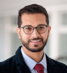 Ramy Ashraf Zeineldin合作论文数Menoufia Univ, Fac Elect Engn, Comp Sci & Engn Dept, Al Minufya, Egypt1Ralf Lobmann合作论文数1
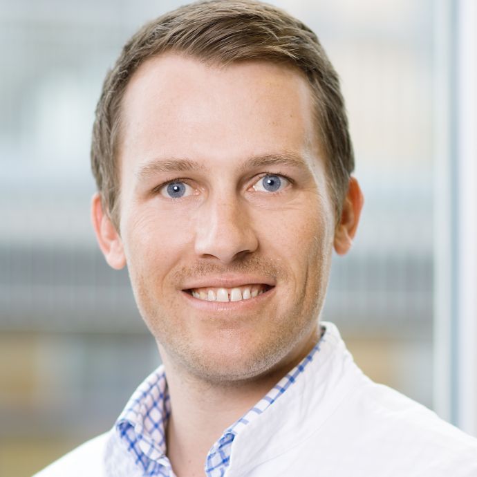 Malte Niklas Bongers合作论文数Universitätsklinikum und Medizinische Fakultät Tübingen;Department for Experimental Radiology, University of Tübingen1
 Andreas Fritsche合作论文数Abteilung Prävention und Therapie des Diabetes Mellitus, Universitätsklinikum und Medizinische Fakultät Tübingen;Instituts für Diabetesforschung und Metabolische Erkrankungen, Helmholtzzentrums München, Universität Tübingen1




