Objective:To investigate the mid-term effectiveness of debridement with prosthesis retention for periprosthetic joint infection (PJI) after total hip arthroplasty (THA) and total knee arthroplasty (TKA), and analyze the influence factors that affected the effectiveness.Methods:A clinical data of 45 cases with PJI after THA (16 cases) and TKA (29 cases) that were treated with debridement with prosthesis retention between January 2011 and January 2015 were collected and analyzed. There were 19 males and 26 females with a mean age of 58.4 years (range, 23-78 years). PJI occurred after primary joint arthroplasty in 40 cases and after revision in 5 cases. The mean time interval between the performance of infection symptoms and the arthroplasty or revision was 15.5 months (range, 0.5-72.0 months). The time interval between the performance of infection symptoms and debridment was 35 days (range, 3-270 days). There were early postoperative infections in 13 cases, acute hematogenous infections in 24 cases, and late chronic infections in 8 cases. X-ray films showed that the location of prosthesis was good. The results of bacilli culture showed that 28 cases were positive and 17 were negative. Twelve cases had sinuses. Length of stay, Hospital for Special Surgery (HSS) score, and Harris score were recorded to evaluate risk factors by using a multivariate logistic regression.Results:The mean length of stay was 22.6 days (range, 5-79 days). All patients were followed up 24-74 months (mean, 52 months). There were 33 cases that retained the prosthesis without further evidence of infection with the success rate was 73.3%. There were significant differences in Harris score and HSS score between pre- and post-operation ( P<0.05). The univariate analysis results showed that the failure of debridement with prosthesis retention had a significant correlation with sinus developing and duration of infection symptoms more than 14 days ( P<0.05). Multivariate logistic regression analysis results showed that sinus developing was an independent risk factor of failure ( P<0.05).Conclusion:Debridement with prosthesis retention plays an important role in treating PJI after THA and TKA. These patients with sinus performing and duration of infection symptoms more than 14 days have higher risk to develop infection again.
Two-stage revision is the standard procedure for periprosthetic knee infection. But when to perform the second-stage is still under debate. We attempt to search the reliable indicators, risk factors, and proper timing for the second-stage revision.
The impact of surgeon handedness on acetabular cup orientation in total hip arthroplasty (THA) is not well studied. The aim of our study is to investigate the difference of cup orientation in bilateral THA performed by right-handed surgeons using posterolateral approach and which cup could be fitter to Lewinneck’s safe zone.
Objective:To study the imaging features of the hip joint by measuring the imaging parameters of spine, pelvis, and hip joint before and after total hip arthroplasty (THA) in patients with ankylosing spondylitis (AS) undergoing THA so as to provide reference for selection of operation methods and prosthesis. Methods:Between January and July 2015, 38 patients (56 hips) with AS underwent primary THA as AS group, and 36 patients (45 hips) with osteonecrosis of the femoral head underwent THA as control group. There was no significant difference in side ( χ2=1.14, P=0.95). The acetabular abduction angle (ABA), acetabular anteversion angle (AVA), center collum diaphyseal (CCD), offset, height from rotation center to lesser trochanter (HRCLT), femoral intertrochanteric distance (FID) were measured by CT three-dimensional morphology. The canal flare index (CFI), cortical thickness index (CTI), pelvic incidence (PI), sacral slope (SS), and pelvic tilt (PT) were measured by X-ray film before operation. The AVA, ABA, and the filling ratio were measured on the postoperative X-ray film. Results:There was no significant difference in preoperative AVA and ABA and postoperative ABA between 2 groups ( P>0.05), but significant difference was found in postoperative AVA ( t=6.71, P=0.00). The mean PI, SS, and PT in AS group were 48.37° (range, 41-58°), 5.64°(range, 2-11°), and 12.85° (range, 5-26°), respectively. There was significant difference in CCD, CFI, and CTI between 2 groups ( t=3.63, P=0.04; t=5.12, P=0.02; t=3.91, P=0.04), but offset, HRCLT, and FID all showed no significant difference ( t=0.41, P=0.36; t=0.33, P=0.56; t=0.59, P=0.12). On the basis of the Noble classification, medullary cavity of the femur was rated as chimney type, ordinary type, and champagne flute type in 32, 18, and 6 hips of AS group, and in 4, 28, and 13 hips of control group respectively. Filling ratio of distal segment in AS group was significantly lower than that in control group ( t=5.64, P=0.02), but there was no significant difference in the filling ratio of middle and proximal segments between 2 groups ( t=0.29, P=0.61; t=0.55, P=0.13). Conclusion:Compared with patients having osteonecrosis of the femeral head, there is no significant difference in preoperative AVA and ABA, but postoperative AVA significantly increase in patients with AS. Because AS patients have mainly chimney type medullary cavity of the femur, the filling ratio of middle and distal segment is lower when tapered stems are used, and the filling ratio of anatomic stems is higher.
BACKGROUND Periprosthetic joint infection (PJI) is a complication of total joint arthroplasty (TJA). The leukocyte esterase (LE) strip test and histology are diagnostic methods for PJI. The aims of this study were to determine the sensitivity and specificity of the LE strip test and to compare it with histology in the diagnosis of PJI. MATERIAL AND METHODS Between January and December 2015, 93 patients who underwent TJA with PJI were enrolled in the study. Synovial fluid samples were tested with an LE strip, and three synovial tissue samples from each patient underwent frozen section and formalin-fixed histology. Recent criteria from the Musculoskeletal Infection Society (MSIS) were used for the diagnosis of PJI. RESULTS Ninety-three patients studied included 38 cases of PJI and 55 non-infected cases. Sensitivity and specificity of the LE strip test were 92.1% (95% CI, 77.5-97.9%) and 96.4% (95% CI, 86.4-99.4%), respectively. There was no significant difference in sensitivity (p=0.249) or specificity (p=0.480) between frozen and paraffin sections for histology; the two methods were strongly correlated (φ=0.892). Comparison of the LE test results with histology showed a strong correlation (φ=0.758, and φ=0.840). CONCLUSIONS The findings of this preliminary study have shown that the LE strip test on synovial fluid showed similar sensitivity and specificity as histology for the diagnosis of PJI, indicating that that further controlled clinical studies should be performed to investigate the role of the LE strip test for the early diagnosis of PJI.
Background: Culture-negative periprosthetic joint infection (PJI) is very intractable when dealing with an infected total knee arthroplasty (TKA) patient. Two-stage revision has been proved to be a reliable solution for PJI patients. Whether it is still credible for culture-negative infected patients remains uncertain.Methods: Our group retrospectively reviewed all total knee revision patients from January 2003 to January 2014, 145 PJI patients were diagnosed as infection with the PJI diagnostic criteria and 129 patients were successfully followed. As different treating strategies were utilized, these patients were divided into culture-negative (18 cases, CN) group, culture-positive with one-stage revision group (CP1, 21 cases) and culture-positive with two-stage revision group (CP2, 87 cases) groups. The CN group and CP2 group underwent two-stage revision with antibiotic loaded cement spacers and intravenous antibiotics, CP1 group received one-stage revision. All the culture results and relevant medical records were thoroughly reviewed.Results: The mean follow-up time was 59.5 +/- 32.3 months (range 12-158 months). The culture-negative rate was 14.2%. The overall infection control rate was 92.12%. Infection recurrence was observed in two cases in CP1 group (9.09%), six cases in CP2 group (6.90%) and two cases in CN group (11.1%). The reinfection rate of culture-negative patients and culture-positive patients was 7.34% and 11.1% with no significant difference (p = 0.94). No statistically difference was observed between CP2 group and CN group (p = 0.90). No Spacer fracture or dislocation was observed.Conclusion: With combined or broad spectrum antibiotics, two-stage revision showed comparable outcome when treating culture-negative infected TKA patients at five-year follow-up. (C) 2016 Published by Elsevier B. V. on behalf of The Japanese Orthopaedic Association.
Periprosthetic femoral fracture (PFF) is a complicated complication of both primary and revision hip arthroplasty with an increasing incidence. The present study aimed to summarize the clinical characteristics and identify the risk factors for PFF which would be potentially helpful in the prevention and treatment of PFF.We retrospectively analyzed the clinical data of 89 cases of PFF, and a case-control study was designed to identify the potential risk for intraoperative and postoperative PFF in both primary and revision hip arthroplasty.The overall incidence of PFF was 2.08% (intraoperative: 1.77%, postoperative: 0.30%, revision: 13.60%, and primary: 0.97%). The most commonly used treatment strategy was fixation with cerclage wire or band for intraoperative PFF, whereas long stem revision with plate or cortical allograft strut fixation was the main treatment strategy for postoperative PFF. The risk factors for intraoperative PFF in primary total hip arthroplasty (THA) included the diagnosis of development dysplasia of the hip (DDH) (odds ratio [OR] = 5.01, 95%CI, 1.218-20.563, P=0.03) and CBR ≥ 0.49 (OR = 3.34, 95%CI, 1.138-9.784, P = 0.03). The increased age was associated with increased incidence of postoperative PFF in primary THA (OR = 1.09, 95%CI, 1.001-1.194, P = 0.04). As for the intraoperative PFF in revision THA, we found that receiving multiple operations before revision (OR = 2.45, 95%CI, 1.06-5.66, P = 0.04), revisions due to prosthetic joint infection (OR = 6.72, 95%CI, 1.007-44.832, P = 0.04), the presence of cementless implant before revision (OR = 13.54, 95%CI, 3.103-59.08, P = 0.001), and femoral deformity (OR = 8.03, 95%CI, 1.656-38.966, P = 0.01) were all risk factors.Screening for high-risk patients, preoperative templating, and detailed discharge instructions may be the potential strategies to reduce the incidence of PFF. The treatment of PFFs should take into account Vancouver classification system, patient's characteristics as well as the experience of the operating surgeon.
目的 探讨白细胞酯酶(leukocyte esterase,LE)试纸条在关节假体周围感染(periprosthetic joint infection,PJI)诊断中的应用.方法 选取2014年10月-2015年7月本院63例髋、膝关节置换术后需行关节腔穿刺鉴别是否感染且穿刺后有可利用关节液者入组本实验.选用临床常见的两种试纸条Combur10Test(R)M Roche(德国)和AUTION Sticks 10PA Arkray(日本),对63例患者进行测定.每例患者每次取1滴关节液滴入试纸白细胞酯酶小格,于2~3 min后分别判读.试纸变为深紫色(比色卡最后一格)判定为阳性,其余颜色判定为阴性.混有血液的关节液,通过离心机离心处理后,取上清液滴定试纸并判读.同时依据肌肉与骨骼感染协会(Muscularskeletal Infection Society,MSIS)诊断标准判断假体周围是否感染.将判断结果与LE试纸测试结果对比,分别计算敏感性、特异性、阳性预测值、阴性预测值及相应95% CI.结果 63例中,依据MSIS标准诊断为PJI 35例,非感染28例.两种LE试纸条测定结果无差异,均出现假阳性1例,假阴性3例;敏感性均为91.4%(95% CI:75.8%~97.8%),特异性均为96.4%(95% CI:79.8%~99.8%),阳性预测值均为97.0%(95% CI:82.5%~99.8%),阴性预测值均为90.0%(95% CI:72.3% ~ 97.4%).结论LE试纸应用于关节假体周围感染的诊断有很高的敏感性和特异性,是一种相对准确且经济、简便、快捷的检测方法.其中AUTION Sticks 10PA Arkray推荐应用于PJI的诊断.尽管Combur10Test(R)M Roche结果与AUTION Sticks 10PA Arkray试纸结果一致,但试纸条覆盖的过滤膜较厚,有时会干扰肉眼直接判读.
OBJECTIVE To evaluate the application and effectiveness of bilateral total hip arthroplasty and total knee arthroplasty in the treatment of severe inflammatory arthropathies. METHODS Between September 2008 and September 2015, 31 patients with severe inflammatory arthropathies were treated with bilateral total hip arthroplasty and total knee arthroplasty. Of 31 cases, 22 were male and 9 were female with an average age of 30 years (range, 20 to 41 years); there were 15 cases of rheumatoid arthritis and 16 cases of ankylosing spondylitis with an average onset age of 14 years (range, 5-28 years); all 4 ankylosed joints were observed in 11 cases, 3 ankylosed joints in 2 cases, 2 ankylosed joints in 6 cases, 1 ankylosed joint in 1 case, and no ankylosed joint in 11 cases. Before operation, the hip range of motion (ROM) value was (17.82±28.18)°, and the knee ROM value score was (26.45±30.18)°; the hip Harris score was 29.64±11.58, and the hospital for special surgery (HSS) score was 27.07±11.04. The patients were grouped and compared in accordance with etiology and ankylosed joint. RESULTS One-stage arthroplasty was performed in 1 case, two-stage arthroplasty in 22 cases, three-stage arthroplasty in 7 cases, and four-stage arthroplasty in 1 case. The total operation time was 325-776 minutes; the total blood loss was 900-3 900 mL; the total transfusion volume was 2 220-8 070 mL; and the total hospitalization time was 21-65 days. The patients were followed up 12-94 months (mean, 51 months). The hip and knee ROM values, Harris score and HSS score at last follow-up were significantly improved when compared with preoperative ones (P<0.05). The subjective satisfaction degree was good in 16 cases, moderate in 10 cases, and poor in 5 cases. Periprosthetic infection occurred in 2 cases (3 knees), joint stiffness in 3 cases (6 knees), joint instability in 1 case (1 knee), leg length discrepancy of >2 cm in 2 cases, and flexion deformity of 10° in 1 case (1 knee). The hip and knee ROM values, Harris score and HSS score showed no significant difference between patients with ankylosing spondylitis and patients rheumatoid arthritis at last follow-up (P>0.05). The hip and knee ROM values of the patients with ankylosed joint were significantly lower than those of patients with no ankylosed joint (P<0.05); the Harris score and HSS score of the patients with ankylosed joint were lower than those of patients with no ankylosed joint, but no significant difference was found (P>0.05). CONCLUSIONS A combination of bilateral hip and knee arthroplasty is an efficient treatment for severe lower extremities deformity, arthralgia and poor quality of life caused by inflammatory arthropathies. However, the postoperative periprosthetic infection and stiffness of knee are important complications influencing the effectiveness of operation.
Patient-specific instrumentation (PSI) technology has been developed to improve alignment when implanting total knee arthroplasty (TKA) and is a new focus in the orthopaedic community. Current controversial data concerning PSI are discussed. A systematic review to compare PSI with conventional instrumentation and assess the radiographic outcomes was performed. Electronic databases (including PubMed, Medline, Embase, the Cochrane Library and the Science Citation Index database) and conference proceedings from 1950 to 2014 in the English language were searched. Data, including relevant patient characteristics, sample size, radiographic method, PSI system manufacturer and outliers of implant positioning and alignment on radiography were independently extracted from all eligible studies by two of the authors. A total of 2739 TKAs were included (1410 performed with PSI and 1329 with conventional instrumentation). There were more TCA outliers (malalignment >3°) and tibial slope outliers (malalignment >3°) in the PSI group than in the conventional group. The other radiographic outcomes assessed, including coronal, sagittal or rotational alignment outliers did not differ between the two groups. With regard to radiographic outcomes, our findings indicate that PSI technology is not superior in reducing outliers of component alignment.
OBJECTIVETo investigate the clinical characteristic differences of cementless total hip arthroplasty (THA) between with and without subtrochanteric femoral shortening osteostomy in Crowe type IV developmental dysplasia of the hip (DDH).METHODSBetween January 2006 and March 2012, 21 patients (21 hips) with Crowe type IV DDH who underwent primary THA were enrolled according to inclusion criteria. According to whether subtrochanteric femoral shortening osteostomy was performed during THA or not, the patients were divided into 2 groups: THA with osteostomy group (n = 9) and THA without osteotomy group (n = 12). There was no significant difference in gender, age, body mass index, and hip Harris score between 2 groups (P > 0.05) except leg length discrepancy (t = -3.170, P = 0.005). The operation time, blood loss, postoperative drainage, complications, and radiography data were compared to evaluate the clinical characteristics.RESULTSThe operation time, blood loss, and postoperative drainage of osteotomy group were all significantly greater than those of no osteotomy group (P < 0.05). All patients achieved primary healing of incision; 1 patient (1 hip) had transient sciatic nerve symptom in osteotomy group. The average follow-up time was 53 months (range, 28-88 months). The X-ray films showed good fracture healing at 3-6 months after operation in osteostomy group. No prosthetic loosening or dislocation was found. The hip Harris score was 90.67 ± 4.06 in osteostomy group and 92.17 ± 3.27 in no osteostomy group, showing no significant difference between 2 groups (t = -0.938, P = 0.360). The leg length discrepancy was (0.22 ± 0.26) cm in osteostomy group and (0.18 ± 0.27) cm in no osteostomy group, showing no significant.difference (t = 107.000, P = 0.546). The leg length discrepancy was found in 6 patients of osteotomy group and 5 patients of no osteotomy group. One patient complained of thigh pain in osteotomy group; 2 patients had slight limp (Trendelenburg +) in no osteotomy group.CONCLUSIONTHA can improve joint function and increase limb length in the treatment of Crowe type IV DDH. Subtrochanteric shortening osteotomy is an effective treatment which can be performed according to preoperative template measurement, leg length shortening, and the soft tissue tension.
OBJECTIVE:To provide the evidence for diagnosis and treatment of the complication by describing the distribution and drug sensitivity of pathogens in patients with prosthetic joint infection (PJI) after primary total knee arthroplasty (TKA).METHODS:Between January 2003 and June 2013, 65 cases (65 knees) with PJI after primary TKA were treated. There were 28 males and 37 females with an average age of 63.2 years (range, 37-80 years). The median interval between PJI and primary TKA was 2.8 years (range, 2 weeks to 11 years), including 29 left knees and 36 right knees. Prosthesis loosening could be found in 27 cases by X-ray examination. The average value of C-reactive protein and erythrocyte sedimentation rate was 37.4 mg/L (range, 12.5-197.0 mg/L) and 63.2 mm/1 h (range, 29.3-73.8 mm/l h) respectively. Preoperative and intraoperative synovial fluid as well as intraoperative tissue samples should be submitted for aerobic and anaerobic culture. The four types of infections were made according to the Tsukayama et al. classification standards.RESULTS:The patients were all diagnosed as having PJI. There were 5 (7.69%) type I infections, 4 (6.15%) type IIA, 8 (12.31%) type IIB, 3 (4.62%) type III, and 45 (69.23%) type IV according to the Tsukayama et al. classification standard. Bacterial culture results were negative in 12 cases and positive in 53 cases, the main pathogen was Gram-positive cocci (39/53). The most common organism identified was Coagulase-negative Staphylococcus (24/53) followed by Staphylococcus Aureus (12/53). Resistant bacterium accounted for 61.11% (22/36) of Staphylococcus. These bacterium were all sensitive to vancomycin, linezolid, meropenem, and fluconazole; and highly resistant to erythrocin, penicillin, and cefoxitin. The main pathogenic bacteria of Coagulase-negative Staphylococcus and Staphylococcus aureus had highest resistant rate to penicillin.CONCLUSION:Gram-positive cocci is the main pathogen in patients with PJI after primary TKA, which is highly resistant to penicillin and macrolides. Antibiotic treatment of this complication should be based on the result of drug sensitivity test, vancomycin and linezolid may be used before the result of drug sensitivity test. It is important to pay attention to rare and multiple resistant bacteria.
 Jiying Chen (陈继营)合作论文数Chinese PLA General Hospital11
 Ming Ni (倪明)合作论文数Chinese PLA General Hospital7
 XingGui Song (宋兴桂)合作论文数6Xin Li (李昕)合作论文数5
 Chi Xu (徐驰)合作论文数4Zhendong Zhang合作论文数4QingMeng Zhang (张庆猛)合作论文数4Xiang Li (李想)合作论文数4
 Wei Chai (柴伟)合作论文数Chinese PLA General Hospital4Fan Yang合作论文数3XiangPeng Kong (孔祥朋)合作论文数2JunLei Song (宋俊雷)合作论文数2
 Rui Li (李锐)合作论文数Chinese PLA General Hospital2Yinqiao Du (杜银桥)合作论文数1Qi Zhuo合作论文数1Yucong Li合作论文数1Bo Wu (吴博)合作论文数1BaoZhan Yu (于宝占)合作论文数1YunJian Yang (杨云建)合作论文数1
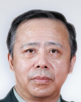 Libo Hao (郝立波)合作论文数Chinese PLA General Hospital1
 Jun Fu (符军)合作论文数State Key Laboratory of Microbial Technology, Shandong University1
 Fenglong Sun (孙凤龙)合作论文数Beijing Rehabilitation Hospital of Capital Medical University1
 Yonggang Zhou (周勇刚)合作论文数Chinese PLA General Hospital1




