Introduction: Laparoscopic inguinal hernia repair (LIHR) is one of the most common surgical procedures performed worldwide, associated with a roughly 10 % rate of complications, most commonly iatrogenic injury to blood vessels, sometimes necessitating conversion to open surgery. Fluorescence-guided laparoscopic surgery using indocyanine green fluorescence angiography (ICG-FA) facilitates the precise identification of numerous anatomical structures, especially vascular, reducing their risk of iatrogenic injury. We present the first published case and video demonstrating LIHR with ICG-FA to prevent intra-operative vascular injury. Presentation of case: A 46-year-old, otherwise-healthy male with a right inguinal hernia underwent fluorescenceguided LIHR using ICG-FA. Before peritoneal dissection, 2 ml ICG was administered intravenously, followed by 10 ml physiological solution. The surgical field was then illuminated using the Stryker fluorescence system. Once vascular structures were located, the sac was dissected. After reversing the peritoneum, but before placing the extraperitoneal mesh, another dose of ICG was administered intravenously to aid in safely securing the mesh. Both times after ICG injection, both the iliac artery and spermatic arteries were clearly visible throughout their course in the surgical field within 45 s. The hernia was repaired successfully with no complications. Discussion: ICG-FA appears to facilitate inguinal hernia repair by enabling real-time visualization of anatomical structures, theoretically reducing the risk of complications, particularly vascular injuries. It is particularly helpful identifying the inguinal area's highly-vascular 'triangle of doom'. Conclusions: Further studies are warranted to evaluate short- and the long-term outcomes and cost-effectiveness of ICG-fluorescence angiography during laparoscopic inguinal hernia repair.
Background: laparoscopic cholecystectomy is one of the most common procedures performed in general surgery. The anatomical differences of the gallbladder and the presence of factors related to inflammation can cause technical issues during surgery. Objective: the aim of the present study was to determine the value of C-reactive protein (CRP) and erythrocyte sedimentation rate (ESR) as predictors of difficult cholecystectomy and to evaluate their application during presurgical planning within a university residency program. Material and methods: we conducted a retrospective and analytical study in a tertiary university hospital. A total of 104 adult patients undergoing laparoscopic cholecystectomy due to symptomatic cholelithiasis between January and July 2019 were included. The patients were categorized into two groups: difficult cholecystectomy and non-difficult cholecystectomy. Results: there were statistically significant differences in ESR and CRP values between both groups (p < 0.001). Sensitivity of ESR was 100%, specificity was 45%, with a PPV of 40% and NPV of 100%. Sen- sitivity of CRP was 87.5%, specificity was 86.3%, with a PPV of 70% and NPV of 95%. Both parameters were elevated in 14 of 16 difficult cholecystectomies and in 2 of 44 non-difficult cholecystectomies. Sensitivity of CRP was 87.5%, specificity was 86.3%, with a PPV of 70% and NPV of 95%. Conclusion: measuring ESR and CRP has proved to be a reliable method to predict difficult chole- cystectomies due to symptomatic cholelithiasis. This could be applied for surgical planning within a university residency program.
Background: Incisionless near-infrared fluorescent cholangiography (NIFC) is emerging as a promising tool to enhance the visualization of extrahepatic biliary structures during laparoscopic cholecystectomies. Methods: We conducted a single-blind, randomized, 2-arm trial comparing the efficacy of NIFC (n = 321) versus white light (WL) alone (n = 318) during laparoscopic cholecystectomy. Using the KARL STORZ Image1 S imaging system with OPAL1 technology for NIR/ICG imaging, we evaluated the detection rate for 7 biliary structures-cystic duct (CD), right hepatic duct (RHD), common hepatic duct, common bile duct, cystic common bile duct junction, cystic gallbladder junction (CGJ), and accessory ducts-before and after surgical dissection. Secondary calculations included multivariable analysis for predictors of structure visualization and comparing intergroup biliary duct injury rates. Results: Predissection detection rates were significantly superior in the NIFC group for all 7 biliary structures, ranging from 9.1% versus 2.9% to 66.6% versus 36.6% for the RHD and CD, respectively, with odds ratios ranging from 2.3 (95% CI 1.6-3.2) for theCGJ to 3.6 (1.6-9.3) for the RHD. After dissection, similar intergroup differences were observed for all structures except CD and CGJ, for which no differences were observed. Significant odds ratios ranged from 2.4 (1.7-3.5) for the common hepatic duct to 3.3 (1.3-10.4) for accessory ducts. Increased bodymass index was associated with reduced detection of most structures in both groups, especially before dissection. Only 2 patients, both in the WL group, sustained a biliary duct injury. Conclusions: In a randomized controlled trial, NIFC was statistically superior to WL alone visualizing extrahepatic biliary structures during laparoscopic cholecystectomy.
Pancreatic cancer is the fourth cause of death related to malignant tumors in Argentina. The Adenocarcinoma is the most frequent histological type, being able to originate from a precursor lesion, such as intraductal papillary mucinous neoplasms. The objective was to describe a case of surgical treatment of pancreatic adenocarcinoma and concomitant Intraductal papillary mucinous neoplasm in an 80-year-old male patient. This association represents an infrequent case that has less loco-regional commitment, better global survival and, therefore, better prognosis than primary adenocarcinomas.
Sarotto, Luis E. MD, FACS; Dip, Fernando MD; Nahmod, Mario; Alle, Lisandro MD; Carles, Gonzalo; Todeschini, Hernan; Rosenthal, Raul J. MD, FACS, FASMBS Author Information
Dip, Fernando MD; Sarotto, Luis MD, PhD; Todeschini, Hernan; Ferraina, Pedro A. MD, FACS; Roy, Mayank MD; Rosenthal, Raul J. MD, FACS; Nahmod, Mario Author Information
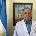 Luis E Sarotto合作论文数6
 Raul J. Rosenthal合作论文数Bariatric and Metabolic Institute, Weston Hospital, Cleveland Clinic Florida4
 Fernando Dip合作论文数4
 Lisandro Alle合作论文数3Nahmod Mario合作论文数3Kevin P White合作论文数2
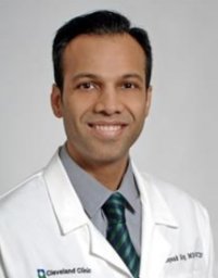 Roy Mayank合作论文数2Thomas Carus合作论文数1
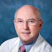 Edward Phillips合作论文数University of Southern California, Los Angeles;Cancer Institute, Cedars-Sinai Medical Center;Department of Surgery, Cedars-Sinai Medical Center1
 Luigi Boni合作论文数Department of Surgical Sciences, University of Insubria1
 Norihiro Kokudo合作论文数National Center for Global Health and Medicine1Pedro A. Ferraina合作论文数1Emanuelle Lomenzo合作论文数1Pedro Ferraina合作论文数1Conrad Simpfendorfer合作论文数1
 Kaja Ludwig合作论文数1Takeaki Ishizawa合作论文数Graduate School of Medicine, University of Tokyo1Sylke Schneider合作论文数1




